. 2012 Nov;137(3):259-70.
doi: 10.1111/imm.12005.

Ribavirin modulates the conversion of human CD4(+) CD25(-) T cell to CD4(+) CD25(+) FOXP3(+) T cell via suppressing interleukin-10-producing regulatory T cell

Affiliations

Ribavirin modulates the conversion of human CD4(+) CD25(-) T cell to CD4(+) CD25(+) FOXP3(+) T cell via suppressing interleukin-10-producing regulatory T cell

Tamaki Kobayashi et al. Immunology. 2012 Nov.

Abstract

Because regulatory T (Treg) cells play an important role in modulating the immune system response against both endogenous and exogenous antigens, their control is critical to establish immunotherapy against autoimmune disorders, chronic viral infections and tumours. Ribavirin (RBV), an antiviral reagent used with interferon, is known to polarize the T helper (Th) 1/2 cell balance toward Th1 cells. Although the immunoregulatory mechanisms of RBV are not fully understood, it has been expected that RBV would affect T reg cells to modulate the Th1/2 cell balance. To confirm this hypothesis, we investigated whether RBV modulates the inhibitory activity of human peripheral CD4(+) CD25(+) CD127(-) T cells in vitro. CD4(+) CD25(+) CD127(-) T cells pre-incubated with RBV lose their ability to inhibit the proliferation of CD4(+) CD25(-) T cells. Expression of Forkhead box P3 (FOXP3) in CD4(+) CD25(-) T cells was down-modulated when they were incubated with CD4(+) CD25(+) CD127(-) T cells pre-incubated with RBV without down-modulating CD45RO on their surface. In addition, transwell assays and cytokine-neutralizing assays revealed that this effect depended mainly on the inhibition of interleukin-10 (IL-10) produced from CD4(+) CD25(+) CD127(-) T cells. These results indicated that RBV might inhibit the conversion of CD4(+) CD25(-) FOXP3(-) naive T cells into CD4(+) CD25(+) FOXP3(+) adaptive Treg cells by down-modulating the IL-10-producing Treg 1 cells to prevent these effector T cells from entering anergy and to maintain Th1 cell activity. Taken together, our findings suggest that RBV would be useful for both elimination of long-term viral infections such as hepatitis C virus infection and for up-regulation of tumour-specific cellular immune responses to prevent carcinogenesis, especially hepatocellular carcinoma.


Figures

Figure 1
Figure 1
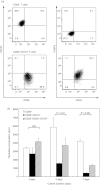
Characteristics of CD25 or CD25+ CD127 CD4+ T cells used in this study. Peripheral blood mononuclear cells (PBMCs) were incubated with magnetic bead-conjugated antibodies without anti-CD4 monoclonal antibodies (mAbs), and the pass-through fraction was collected as CD4+ T cells. Subsequently, the CD4+ T-cell-containing fraction was incubated with FITC-conjugated anti-human CD25 together with Alexa-Fluor647-conjugated anti-human CD127 mAbs, and the CD25 and CD25+ CD127 fractions were obtained using FACSort. (a) Flow cytometric analysis was performed to confirm cell surface molecule expression on the isolated T cells. The expression of cell surface CD25, CD127 and intracellular FOXP3 is shown. The results for CD4+ CD25 T cells are shown in the upper two panels, and those for CD4+ CD25+ CD127 T cells are shown in the lower two panels. The purity of CD25+ CD127 T cells used in this study was approximately 95% (lower right panel). (b) The inhibitory activity of CD4+ CD25+ CD127 T cells against the proliferation of CD4+ CD25 T cells was confirmed; 2 × 10⁴ CD4+ CD25 (empty column) and CD4+ CD25+ CD127 (filled column) T cells were re-stimulated in 96-well U-bottomed culture plates with pB human anti-CD3 mAb 0·05 μg/ml in the presence of 2 × 10⁵ allogeneic irradiated PBMCs. Cell proliferation of CD4+ CD25 and CD4+ CD25+ CD127 T cells was determined 3, 5 and 7 days after stimulation in [3H]thymidine incorporation assays. The cell proliferation of the mixed culture of both cells (shaded column) was also measured. Bonferroni's multiple-comparison test was used to determine the significance of differences between groups.
Figure 2
Figure 2
Effects of ribavirin (RBV) on the characteristics of CD4+ CD25+ CD127 T cells. (a) The effect of RBV against cell viability of the isolated cells was confirmed. Each of the isolated cells was incubated with RBV 0 or 500 ng/ml for 48 hr without stimulation. The dead cells were confirmed as PI-stained cells in flow cytometry and their percentage is indicated. (b) The effects of RBV against the phenotype of the isolated cells were analysed. The isolated CD4+ CD25 and CD4+ CD25+ CD127 T cells were incubated with RBV 0 or 500 ng/ml for 48 hr. The change in FOXP3 expression in CD4+ CD25+ CD127 T cells is indicated in the upper two panels. The expression of inducible co-stimulatory molecule (ICOS) on the surface of the isolated cells is indicated in the lower two panels. ICOS expression of the unstained control (hatched line), on the cells without RBV (bold line), and that with RBV (shaded line) is indicated. (c) The effects of RBV alone against the proliferation of CD4+ CD25 and CD4+ CD25+ CD127 T cells were analysed. Each type of cells was pre-incubated with RBV 0–500 ng/ml for 48 hr and stimulated with 0·05 μg/ml human anti-CD3 mAbs in the presence of irradiated allogeneic PBMCs for 7 days and cell proliferation was confirmed. Student's t-test was used to determine the significance of differences between groups (c, left panel). The inhibitory activity of CD4+ CD25+ CD127 T-cells preincubated with RBV 0–500 ng/ml was evaluated. 104 CD4+ CD25 and CD4+ CD25+ CD127 T-cells preincubated with RBV 0–500 ng/ml were mixed and stimulated with a low dose (0·05 μg/ml, c, centre panel) and maximal dose (5·0 μg/ml, c, right panel) of human anti-CD3 monoclonal antibody in the presence of irradiated allogeneic peripheral blood mononuclear cells for 7 days, and the proliferation of these mixed cells was determined. Bonferroni's multiple-comparison test was used to determine the significance of differences between groups. Each experiment was performed five times, and the results are shown as mean and SD.
Figure 3
Figure 3
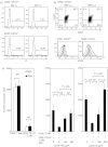
Effects of ribavirin (RBV) on the ability of CD4+ CD25+ CD127 T cells to convert CD4+ CD25 T cells into CD4+ CD25+ FOXP3+ T cells. Intracellular FOXP3 was stained to confirm whether RBV modulated FOXP3 expression in CD4+ CD25 T cells after stimulation for 7 days with CD4+ CD25+ CD127 T cells with or without RBV pre-incubation. Cell surface CD25 and intracellular FOXP3 were stained on CD4+ CD25 T cells (a, upper-left panel), CD4+ CD25 T cells pre-incubated with RBV 500 ng/ml (upper right), CD4+ CD25+ CD127 T cells (centre left), CD4+ CD25+ CD127 T cells pre-incubated with RBV (centre right), CD4+ CD25 and CD4+ CD25+ CD127 T cells (lower left), and CD4+ CD25 T cells and CD4+ CD25+ CD127 T cells pre-incubated with RBV (lower right). The experiments were repeated five times, and representative results are shown. (b) Mean counts of CD25+ FOXP3+ T cells were compared when CD25 T cells were incubated with CD25+ CD127 T cells or RBV-pulsed CD25+ CD127 T cells. The mean inhibition rate was 54·394 ± 11·975%. Bonferroni's multiple-comparison test was used to determine the significance of differences between groups. (c) Expressed CD45RA and CD45RO were stained together with intracellular FOXP3 to determine whether CD4+ CD25 T cells entered anergy in association with the expression of FOXP3 or did not respond to re-stimulation in vitro. Left panels indicate the percentage of CD45RA+ and FOXP3+ cells in the indicated T cells. Right panels indicate the percentage of CD45RO+ and FOXP3+ cells.
Figure 4
Figure 4
Changes in the inhibitory activity of the converted CD4+ T cells against freshly-isolated CD4+ CD25 T cells. The inhibitory effect of the primary-stimulated CD4+ CD25 and CD4+ CD25+ CD127 T cells was examined. The mixed cultures of CD4+ CD25 and CD4+ CD25+ CD127 T cells pre-incubated with or without ribavirin (RBV) 500 ng/ml were harvested, mixed with freshly isolated CD4+ CD25 T cells, and additionally incubated with 0·05 μg/ml of human anti-CD3 monoclonal antibodies in the presence of irradiated allogeneic peripheral blood mononuclear cells for 7 days. (a) Expression of CD25. CD127 and intracellular FOXP3 was confirmed. (b) Thymidine incorporation of the cultured cells including the indicated cells was measured. Bonferroni's multiple-comparison test was used to determine the significance of differences between groups.
Figure 5
Figure 5
Ribavirin (RBV) inhibited activities of CD4+ CD25+ CD127 T cells by interfering with humoral elements released from CD4+ CD25+ CD127 T cells in a cell contact-dependent fashion. Transwell assays were performed to determine whether direct cell contact was required for the inhibitory activity of CD4+ CD25+ CD127 T cells with or without RBV pre-incubation. CD4+ CD25+ CD127 T cells with or without RBV pre-incubation were plated in the upper chamber, and CD4+ CD25 T cells were plated in the lower chamber. Cells were stimulated with soluble anti-CD3 monoclonal antibody 20 μg/ml in the presence of irradiated allogeneic peripheral blood mononuclear cells. After 7 days of stimulation, the upper chamber was removed, and cell proliferation of the CD4+ CD25 T cells in the lower chamber was determined. Thymidine incorporation assays were performed to determine proliferation of the indicated cell fractions. Bonferroni's multiple-comparison test was used to determine the significance of differences between groups.
Figure 6
Figure 6
Inhibitory effects of ribavirin (RBV) against the production of inhibitory cytokines released from CD4+ CD25 or CD4+ CD25+ CD127 T cells. (a) To confirm the key cytokine that RBV affects to inhibit the activity of CD25+ CD127 T cells, levels of interleukin-10 (IL-10) and transforming growth factor-β1 (TGF-β1) produced by CD4+ CD25 or CD4+ CD25+ CD127 T cells stimulated with human anti-CD3 monoclonal antibodies (mAbs) 5 ng/ml in the presence or absence of RBV were measured using ELISA. Student's t-test was used to determine the significance of differences between groups. (b) Neutralization of either IL-10 or TGF-β1 using their neutralizing antibodies was performed to determine which was the key cytokine for the inhibitory activity of CD4+ CD25+ CD127 T cells. CD4+ CD25 T cells were stimulated with the same number of CD4+ CD25+ CD12 T cells with pB anti-CD3 mAb 0·05 μg/ml in the presence or absence of either anti-IL-10 or anti-TGF-β1 mAbs for 7 days. Cell proliferation indicated below the graph was then determined in thymidine incorporation assays. Bonferroni's multiple-comparison test was used to determine the significance of differences between groups.

Similar articles

Cited by

References

    1. Bettini M, Vignali DA. Regulatory T cells and inhibitory cytokines in autoimmunity. Curr Opin Immunol. 2009;21:612–8. - PMC - PubMed
    1. Bui JD, Schreiber RD. Cancer immunosurveillance, immunoediting and inflammation: independent or interdependent processes? Curr Opin Immunol. 2007;19:203–8. - PubMed
    1. Rosen HR, Miner C, Sasaki AW, Lewinsohn DM, Conrad AJ, Bakke A, Bouwer HG, Hinrichs DJ. Frequencies of HCV-specific effector CD4+ T cells by flow cytometry: correlation with clinical disease stages. Hepatology. 2002;35:190–8. - PubMed
    1. Rabinovich GA, Gabrilovich D, Sotomayor EM. Immunosuppressive strategies that are mediated by tumor cells. Annu Rev Immunol. 2007;25:267–96. - PMC - PubMed
    1. Osna N, Silonova G, Vilgert N, et al. Chronic hepatitis C: T-helper1/T-helper2 imbalance could cause virus persistence in peripheral blood. Scand J Clin Lab Invest. 1997;57:703–10. - PubMed

MeSH terms