. 2012 Sep 20;120(12):2501-11.
doi: 10.1182/blood-2011-11-394304. Epub 2012 Aug 13.

EPO-mediated expansion of late-stage erythroid progenitors in the bone marrow initiates recovery from sublethal radiation stress

Affiliations

EPO-mediated expansion of late-stage erythroid progenitors in the bone marrow initiates recovery from sublethal radiation stress

Scott A Peslak et al. Blood. .

Abstract

Erythropoiesis is a robust process of cellular expansion and maturation occurring in murine bone marrow and spleen. We previously determined that sublethal irradiation, unlike bleeding or hemolysis, depletes almost all marrow and splenic erythroblasts but leaves peripheral erythrocytes intact. To better understand the erythroid stress response, we analyzed progenitor, precursor, and peripheral blood compartments of mice post-4 Gy total body irradiation. Erythroid recovery initiates with rapid expansion of late-stage erythroid progenitors-day 3 burst-forming units and colony-forming units, associated with markedly increased plasma erythropoietin (EPO). Although initial expansion of late-stage erythroid progenitors is dependent on EPO, this cellular compartment becomes sharply down-regulated despite elevated EPO levels. Loss of EPO-responsive progenitors is associated temporally with a wave of maturing erythroid precursors in marrow and with emergence of circulating erythroid progenitors and subsequent reestablishment of splenic erythropoiesis. These circulating progenitors selectively engraft and mature in irradiated spleen after short-term transplantation, supporting the concept that bone marrow erythroid progenitors migrate to spleen. We conclude that sublethal radiation is a unique model of endogenous stress erythropoiesis, with specific injury to the extravascular erythron, expansion and maturation of EPO-responsive late-stage progenitors exclusively in marrow, and subsequent reseeding of extramedullary sites.


Figures
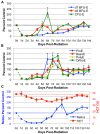
Figure 1
Figure 1
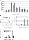
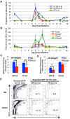
Kinetics of erythroid recovery in the 3 compartments of the erythron after sublethal 4 Gy TBI. (A) Erythroid progenitor (d7 BFU-E, d3 BFU-E, CFU-E) kinetics in the bone marrow post–4 Gy TBI. Recovery initiates in the d3 BFU-E progenitor at 4 days after radiation followed by rapid expansion of CFU-E at 5 to 6 days after TBI. (B) Erythroblast precursor (ProE, BasoE, PolyE, OrthoE) kinetics in the bone marrow post–4 Gy TBI. Erythroid precursor recovery follows expansion of late-stage erythroid progenitors. Progenitors and precursors are normalized per femur and expressed as a percent of unirradiated control marrow. (C) Hematocrit (HCT) and reticulocyte (Retic) levels in the peripheral blood post–4 Gy TBI. Circulating red cell recovery occurs by 8 to 10 days in reticulocytes and by 14 days in RBC. Reticulocytes are calculated as absolute reticulocyte index (percent Retic × total RBC and Retic number × %HCT) and expressed as a percent of unirradiated control levels. RBC levels are expressed as percent HCT. Dotted lines represent unirradiated control levels. Dashed data points at 1 to 2 days after radiation represent erythroid injury data previously published and shown here for clarity. Error bars represent SEM of at least 3 experiments, and 3 or more independently assayed mice were used to determine each data point.
Figure 2
Figure 2
Endogenous EPO induction by anemia is required for expansion of d3 BFU-E and CFU-E during recovery from 4 Gy TBI. (A) Endogenous plasma EPO levels, expressed in pg/mL, as determined by ELISA at 0 to 14 days post–4 Gy TBI. EPO levels increase 13-fold at 4 days post–4 Gy TBI and remain at high levels through 6 days after radiation. (B) Hematocrit levels post–4 Gy TBI ± transfusion of washed RBC at 2 days and 4 days after radiation. Transfusion of irradiated mice prevents radiation-induced anemia. (C) Plasma EPO levels post–4 Gy TBI ± transfusion. Transfusion blocks the induction of EPO normally seen at 4 days and 6 days after radiation. For all experiments, plasma EPO levels and HCT were performed in triplicate with 3 independent blood samples. For each EPO and HCT determination, plasma was isolated from a single terminally bled mouse. All mice were euthanized mid-morning. (D) Erythroid bone marrow progenitor expansion at 6 days after radiation ± transfusion, normalized per femur, and expressed as a percent of unirradiated control marrow. The transfusion-induced block of EPO induction abrogates d3 BFU-E and CFU-E expansion but has no effect on d7 BFU-E during erythroid recovery from 4 Gy TBI. Dotted lines represent unirradiated control levels. Error bars represent SEM of at least 3 experiments, and 3 or more independently assayed mice were used to determine each data point. Statistical analyses were performed using a 2-tailed Student t test (*P < .05; **P < .01; ***P < .001; significantly different from 4 Gy TBI untransfused mice at matched time points).
Figure 3
Figure 3
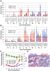
Exogenous EPO is sufficient to initiate late-stage erythroid progenitor expansion and accelerate erythroid recovery after sublethal irradiation. (A) Erythroid progenitor recovery kinetics in the bone marrow ± intraperitoneal injection of 1000 IU/kg EPO at 1 hour post–4 Gy TBI. Recovery of d3 BFU-E is advanced by 2 to 3 days in EPO-injected mice (red bars) compared with mock-treated mice (blue bars) and leads to accelerated CFU-E recovery. (B) Erythroid bone marrow precursor recovery kinetics ± IP EPO injection at 1 hour after TBI. Erythroid precursors in EPO-injected mice (red bars) undergo a wave of recovery 2 to 3 days sooner than mock-treated mice (blue bars) with kinetics that mirror endogenous recovery. Erythroid progenitors and precursors are normalized per femur and expressed as a percent of unirradiated control marrow. (C) Circulating red cell recovery ± EPO injection at 1 hour post–4 Gy TBI. Advanced reticulocyte recovery beginning at 5 days after radiation (red line) leads to partial HCT normalization by 6 days after TBI (green line) in EPO-treated mice. Reticulocytes are calculated as absolute reticulocyte index (% Retic × total RBC and Retic number × %HCT) and expressed as a percent of unirradiated control levels. RBC levels are expressed as percent HCT. Dotted lines represent unirradiated control levels. Error bars represent SEM of at least 3 experiments, and 3 or more independently assayed mice were used to determine each data point. Statistical analyses were performed using a 2-tailed Student t test, (*P < .05; **P < .01; ***P < .001; significantly different from 4 Gy TBI mock-treated mice at matched timepoints). (D) Representative histologic sections of bone marrow ± EPO 4 days post–4 Gy TBI (H&E; 20-micron bars). EPO treatment leads to increased cellularity and decreased vascular dilation compared with mock-treated samples. Images were captured with a Nikon Digital Sight Ds-Fi1 camera using Nikon NIS-Elements software on a Nikon Eclipse 80i upright microscope using a 20× objective.
Figure 4
Figure 4
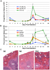
Splenic erythroid progenitor and precursor recovery post–4 Gy TBI. (A) Erythroid progenitor kinetics in the spleen post–4 Gy TBI. Splenic erythroid progenitor recovery does not begin until 7 to 8 days after radiation and peaks at 9 days after TBI. (B) Splenic erythroid precursor kinetics post–4 Gy TBI. Erythroid precursor recovery begins by 8 to 9 days in spleen and remains at high levels, especially in later precursors, at 13 days after TBI. Erythroid progenitors and precursors are expressed as the total number of each cell type per spleen. Error bars represent SEM of at least 3 experiments, and 3 or more independently assayed mice were used to determine each data point. (C) Representative sections of unirradiated spleen and spleen at 6 and 10 days post–4 Gy TBI (H&E staining; 50-micron bars). The low level of steady-state erythropoiesis in spleen is rapidly lost and remains absent at 6 days after radiation; robust erythropoiesis occurs exclusively in the red pulp by 10 days post–4 Gy TBI. White arrows represent areas of erythroid activity in the spleen; rp indicates red pulp; and wp, white pulp. Images were captured with a Nikon Digital Sight Ds-Fi1 camera using Nikon NIS-Elements software on a Nikon Eclipse 80i upright microscope using a 10× objective.
Figure 5
Figure 5
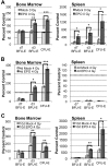
EPO-responsive progenitors are not present in the spleen until after their recovery in the bone marrow. (A) Erythroid progenitor kinetics in bone marrow and spleen 2 days after IP injection of 1000 IU/kg EPO in unirradiated mice. At steady-state, EPO-responsive progenitors are present in both bone marrow and spleen and rapidly expand in response to EPO stimulation. (B) Erythroid progenitor kinetics in bone marrow and spleen at 6 days after radiation ± IP EPO injection at 4 days post–4 Gy TBI. Late-stage erythroid progenitors expand in bone marrow but do not expand in spleen, indicating that EPO-responsive progenitors are not present in spleen during the period of rapid marrow recovery. (C) Erythroid progenitor kinetics in bone marrow and spleen at 12 days after radiation ± IP EPO injection at 10 days post–4 Gy TBI. EPO responsive progenitors are present in spleen by 10 days after radiation, consistent with delayed initiation of splenic expansion. Progenitors are normalized per femur or spleen and expressed as a percent of unirradiated control. Dotted lines represent unirradiated control levels in all graphs. Error bars represent SEM of at least 3 experiments, and 3 or more independently assayed mice were used to determine each data point. Statistical analyses were performed using a 2-tailed Student t test (*P < .05; **P < .01; ***P < .001; significantly different from mock-treated mice at matched time points).
Figure 6
Figure 6
A transient wave of erythroid progenitors circulates in the bloodstream at 6 to 8 days post–4 Gy TBI and selectively engrafts spleen. (A) Erythroid progenitor kinetics in the peripheral blood post–4 Gy TBI. Erythroid progenitors (predominantly d7 BFU-E) are completely lost from the bloodstream after irradiation. A transient wave of erythroid progenitors emerges into the bloodstream between 6 and 8 days after radiation. (B) Erythroid precursor kinetics in the peripheral blood post–4 Gy TBI. Erythroid precursors are also lost after irradiation and transiently emerge into the bloodstream at 6 to 9 days after radiation. Erythroid progenitors and precursors are expressed as the total number of each subpopulation per 300 μL whole blood. (C) Median fluorescence intensity of surface α4-integrin levels on BFU-E and CFU-E obtained from the bone marrow (BM; blue) and circulating blood (red) of mice at 6 days post–4 Gy TBI. α4-integrin levels are significantly decreased on both BFU-E and CFU-E in the bloodstream compared with bone marrow erythroid progenitors at 6 days after radiation. (D) Median fluorescence intensity of surface α5-integrin levels on BFU-E and CFU-E obtained from the bone marrow (BM; blue) and circulating blood (red) of mice at 6 days post–4 Gy TBI. Error bars represent SEM of 3 or more independently assayed mice for each data point. Statistical analyses were performed using a 2-tailed Student t test (**P < .01; ***P < .001; significantly different from 6 day post-TBI BM progenitors at matched time points). (E) Flow cytometric analysis of erythroid progenitor engraftment in irradiated recipient BM and spleen at 12 hours after intravenous injection of Sca1CD16/32 lineage-depleted donor UBC-GFP hematopoietic progenitors from BM and peripheral blood (PB) isolated at 6 days post–4 Gy TBI. Progenitors from bone marrow of UBC-GFP mice at 6 days post–4 Gy TBI can successfully engraft and mature into the bone marrow and spleen of 6.5 day post-TBI recipient mice (center column). In contrast, progenitors isolated from the peripheral blood of UBC-GFP at 6 days post–4 Gy TBI selectively engraft the spleen of recipient mice (right column). GFP Ter119+ recipient BM and spleen cells were used to gate subpopulations of maturing erythroblasts using CD71 and forward scatter characteristics (left column), with R1 representing the most immature and R5 the most mature erythroid subpopulations. Data are shown from 2 independent experiments (shown in black and red), with numbers of GFP+ Ter119+ donor cells present in recipient marrow and spleen per 2 × 10⁶ cells analyzed.
Figure 7
Figure 7
Model for EPO-induced endogenous recovery of the erythron after sublethal radiation stress centered on the expansion, maturation, and migration of bone marrow erythroid progenitors. (A) Sublethal (4 Gy) irradiation causes the near-total loss of erythroid progenitors and precursors in bone marrow, peripheral blood, and spleen leading to the gradual onset of anemia. (B) This anemia induces EPO, which causes the specific, rapid expansion of late-stage erythroid progenitors (d3 BFU-E and CFU-E) in the bone marrow. (C) d3 BFU-E and CFU-E mature into erythroblast precursors and circulating red cells to provide a rapid, short-term erythroid response to acute radiation stress. (D) Simultaneously, early and late-stage erythroid progenitors transiently circulate into the bloodstream. (E) The spleen is subsequently seeded by circulating erythroid progenitors and undergoes robust erythroid reconstitution to augment recovery of the erythron.

Similar articles

Cited by

References

    1. Morley A, Stohlman F., Jr Periodicity during recovery of erythropoiesis following irradiation. Blood. 1969;34(1):96–99. - PubMed
    1. Brady LW, Markoe AM, Ruggieri S, Brodsky I. The effect of sublethal x-irradiation on erythropoiesis in the mouse. Int J Radiat Oncol Biol Phys. 1976;1(5-6):471–479. - PubMed
    1. Okunewick JP, Phillips EL. Extended recovery lag in rat hematopoietic colony forming units following sublethal x-irradiation. J Lab Clin Med. 1972;79(4):550–558. - PubMed
    1. Ben-Ishay Z, Yoffey JM. Ultrastructural studies of erythroblastic islands of rat bone marrow. 3. Effects of sublethal irradiation. Lab Invest. 1974;30(3):320–332. - PubMed
    1. Nakeff A, McLellan WL, Bryan J, Valeriote FA. Response of Megakaryocyte, Erythroid, and Granulocyte-Macrophage Progenitor Cells in Mouse Bone Marrow to Gamma-Irradiation and Cyclophosphamide. In: Baum SJ, Ledney GD, editors. Exp Hematol Today 1979. New York: Springer-Verlag; 1979. pp. 99–104.

Publication types

MeSH terms