Expression profile of flotillin-2 and its pathophysiological role after spinal cord injury
- PMID: 22878913
- PMCID: PMC3545048
- DOI: 10.1007/s12031-012-9873-7
Expression profile of flotillin-2 and its pathophysiological role after spinal cord injury
Abstract
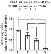
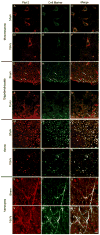
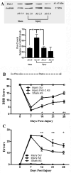
Some receptors that block axonal regeneration or promote cell death after spinal cord injury (SCI) are localized in membrane rafts. Flotillin-2 (Flot-2) is an essential protein associated with the formation of these domains and the clustering of membranal proteins, which may have signaling activities. Our hypothesis is that trauma will change Flot-2 expression and interference of this lipid raft marker will promote functional locomotor recovery after SCI. Analyses were conducted to determine the spatiotemporal profile of Flot-2 expression in adult rats after SCI, using the MASCIS impactor device. Immunoblots showed that SCI produced a significant decrease in the level of Flot-2 at 2 days post-injury (DPI) that increased until 28 DPI. Confocal microscopy revealed Flot-2 expression in neurons, reactive astrocytes and oligodendrocytes specifically associated to myelin structures near or close to the axons of the cord. In the open field test and grid walking assays, to monitor locomotor recovery of injured rats infused intrathecally with Flot-2 antisense oligonucleotides for 28 days showed significant behavioral improvement at 14, 21 and 28 DPI. These findings suggest that Flot-2 has a role in the nonpermissive environment that blocks locomotor recovery after SCI by clustering unfavorable proteins in membrane rafts.
Figures


Similar articles
-
Expression profile and role of EphrinA1 ligand after spinal cord injury.Cell Mol Neurobiol. 2011 Oct;31(7):1057-69. doi: 10.1007/s10571-011-9705-2. Epub 2011 May 21. Cell Mol Neurobiol. 2011. PMID: 21603973 Free PMC article.
-
FasL, Fas, and death-inducing signaling complex (DISC) proteins are recruited to membrane rafts after spinal cord injury.J Neurotrauma. 2007 May;24(5):823-34. doi: 10.1089/neu.2006.0227. J Neurotrauma. 2007. PMID: 17518537
-
Ameliorative Effects of p75NTR-ED-Fc on Axonal Regeneration and Functional Recovery in Spinal Cord-Injured Rats.Mol Neurobiol. 2015 Dec;52(3):1821-1834. doi: 10.1007/s12035-014-8972-6. Epub 2014 Nov 15. Mol Neurobiol. 2015. PMID: 25394381
-
Inhibition of EphA7 up-regulation after spinal cord injury reduces apoptosis and promotes locomotor recovery.J Neurosci Res. 2006 Nov 15;84(7):1438-51. doi: 10.1002/jnr.21048. J Neurosci Res. 2006. PMID: 16983667
-
Promoting axonal myelination for improving neurological recovery in spinal cord injury.J Neurotrauma. 2009 Oct;26(10):1847-56. doi: 10.1089/neu.2008.0551. J Neurotrauma. 2009. PMID: 19785544 Review.
Cited by
-
Neuron-Targeted Caveolin-1 Improves Molecular Signaling, Plasticity, and Behavior Dependent on the Hippocampus in Adult and Aged Mice.Biol Psychiatry. 2017 Jan 15;81(2):101-110. doi: 10.1016/j.biopsych.2015.09.020. Epub 2015 Oct 8. Biol Psychiatry. 2017. PMID: 26592463 Free PMC article.
-
Neurodegeneration and Neuro-Regeneration-Alzheimer's Disease and Stem Cell Therapy.Int J Mol Sci. 2019 Aug 31;20(17):4272. doi: 10.3390/ijms20174272. Int J Mol Sci. 2019. PMID: 31480448 Free PMC article. Review.
References
-
- Basso DM, Beattie MS, Bresnahan JC. A sensitive and reliable locomotor rating scale for open field testing in rats. J Neurotrauma. 1995;12:1–21. - PubMed
-
- Bickel PE, Scherer PE, Scnitzer JE, Oh P, Lisanti MP, Lodish HF. Flotillin and Epidermal Surface Antigen Define a New Family of Caveolae-associated Integral Membrane Proteins. J Biol Chem. 1997;272:13793–13802. - PubMed
Publication types
MeSH terms
Substances
Grants and funding
- U54 RR026139/RR/NCRR NIH HHS/United States
- R24 MH048190/MH/NIMH NIH HHS/United States
- R25 GM061838/GM/NIGMS NIH HHS/United States
- U54 MD007587/MD/NIMHD NIH HHS/United States
- S06-GM008224/GM/NIGMS NIH HHS/United States
- R25-GM068138/GM/NIGMS NIH HHS/United States
- G12 RR003051/RR/NCRR NIH HHS/United States
- S06 GM008224/GM/NIGMS NIH HHS/United States
- 1U54RR026139-01A1/RR/NCRR NIH HHS/United States
- G12 MD007600/MD/NIMHD NIH HHS/United States
- G12RR03051/RR/NCRR NIH HHS/United States
- R01 GM068138/GM/NIGMS NIH HHS/United States
- U54 NS039405/NS/NINDS NIH HHS/United States
LinkOut - more resources
Full Text Sources
Medical

