Review
. 2013 Mar;70(5):863-91.
doi: 10.1007/s00018-012-1096-0. Epub 2012 Aug 7.

Structure and function of biotin-dependent carboxylases

Affiliations
Review

Structure and function of biotin-dependent carboxylases

Liang Tong. Cell Mol Life Sci. 2013 Mar.

Abstract

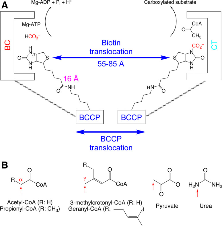
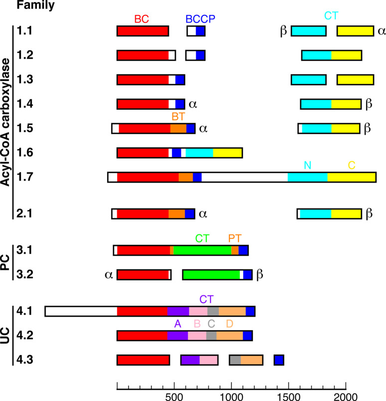
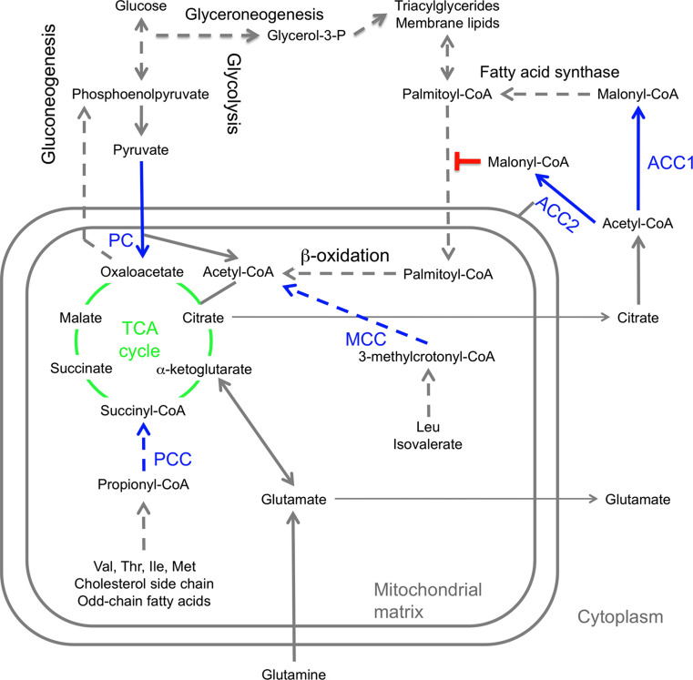
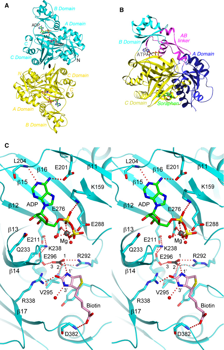
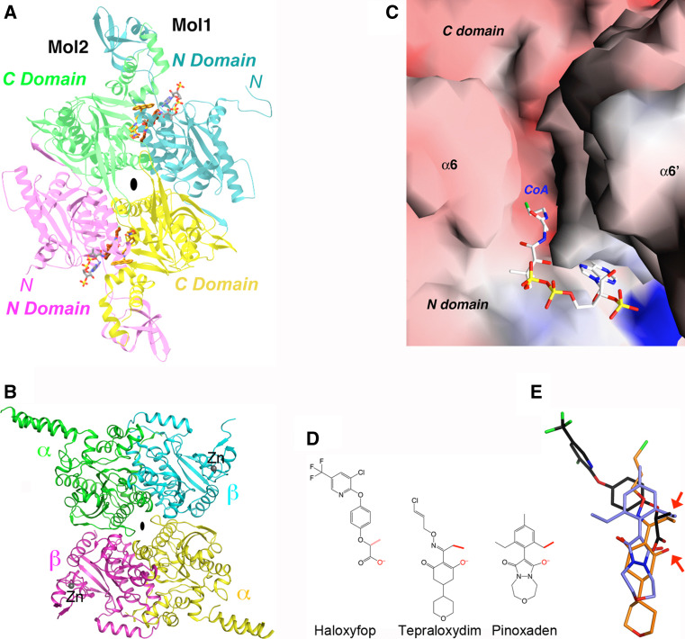
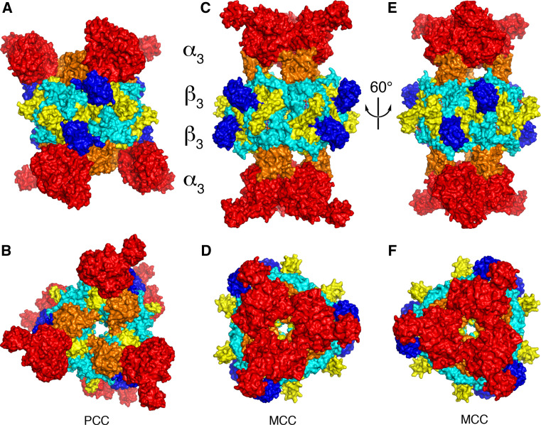
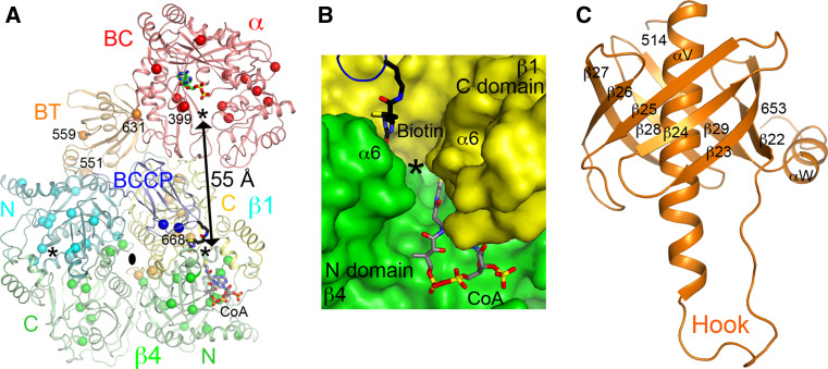
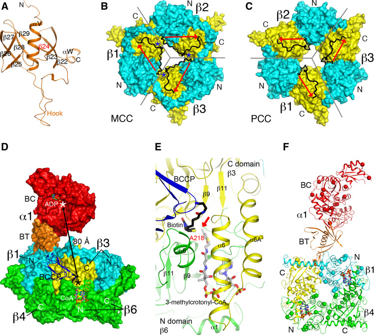
Biotin-dependent carboxylases include acetyl-CoA carboxylase (ACC), propionyl-CoA carboxylase (PCC), 3-methylcrotonyl-CoA carboxylase (MCC), geranyl-CoA carboxylase, pyruvate carboxylase (PC), and urea carboxylase (UC). They contain biotin carboxylase (BC), carboxyltransferase (CT), and biotin-carboxyl carrier protein components. These enzymes are widely distributed in nature and have important functions in fatty acid metabolism, amino acid metabolism, carbohydrate metabolism, polyketide biosynthesis, urea utilization, and other cellular processes. ACCs are also attractive targets for drug discovery against type 2 diabetes, obesity, cancer, microbial infections, and other diseases, and the plastid ACC of grasses is the target of action of three classes of commercial herbicides. Deficiencies in the activities of PCC, MCC, or PC are linked to serious diseases in humans. Our understanding of these enzymes has been greatly enhanced over the past few years by the crystal structures of the holoenzymes of PCC, MCC, PC, and UC. The structures reveal unanticipated features in the architectures of the holoenzymes, including the presence of previously unrecognized domains, and provide a molecular basis for understanding their catalytic mechanism as well as the large collection of disease-causing mutations in PCC, MCC, and PC. This review will summarize the recent advances in our knowledge on the structure and function of these important metabolic enzymes.


Figures

Fig. 1
Fig. 1
The biochemical activity of biotin-dependent carboxylases. a Biotin is carboxylated in the active site of the biotin carboxylase (BC) component, using bicarbonate as the CO2 donor with concomitant ATP hydrolysis. Biotin then translocates to the carboxyltransferase (CT) active site, where the CO2 is transferred to the acceptor (substrate, acetyl-CoA is shown as an example). In the swinging-arm model, biotin itself translocates between the BC and CT active sites, while the biotin-carboxyl carrier protein (BCCP) component remains stationary. The longest distance between the N1′ of biotin and the Cα atom of the covalently linked lysine residue is ~16 Å, giving the swinging arm a maximal reach of ~30 Å. This is significantly shorter than the distances observed in the holoenzymes so far, between 55 and 80 Å. Therefore, the BCCP domain must also translocate during catalysis, and this is known as the swinging-domain model. b The substrates of biotin-dependent carboxylases. The sites of carboxylation are indicated with the red arrows
Fig. 2
Fig. 2
Classification of biotin-dependent carboxylases. These enzymes are classified into four major collections and 13 different families. Domains with sequence homology to each other are shown with the same color. More detailed descriptions of the families can be found in the text, and common examples of acyl-CoA carboxylase family members include E. coli ACC (family 1.1), eukaryotic ACC (family 1.7), PCC (family 1.5), and MCC (family 2.1). The proteins are drawn to size, which is indicated with the scale bar at the bottom (in number of residues)
Fig. 3
Fig. 3
Functions of biotin-dependent carboxylases in mammals. Reactions involving the five biotin-dependent carboxylases are shown in blue. Selected intermediates of the TCA cycle (green) are shown. Glutamine can also be used for anaplerosis, especially in some cancer cells. Dashed arrows indicate pathways with more than one step. In other organisms, the biotin-dependent carboxylases have similar functions (with the exception of ACC2), and they are also involved in additional cellular processes
Fig. 4
Fig. 4
Structural information on the BC component. a Structure of the BC subunit dimer (cyan and yellow) of E. coli ACC in complex with MgADP, bicarbonate, and biotin [154]. The twofold axis of the dimer is indicated with the black oval. b Structure of the BC domain of yeast ACC in complex with the inhibitor soraphen A (green) [161]. The sub-domains of BC are given different colors. The bound position of ATP is also shown to indicate the location of the active site. The view is similar to that for the top monomer in a. c Stereo figure showing detailed interactions between MgADP, bicarbonate, and biotin with the active site of the E. coli BC subunit. Several segments of the protein, including the glycine-rich loop, are omitted for clarity. The structure figures were produced with the program PyMOL (www.pymol.org)
Fig. 5
Fig. 5
Structural information on the CT component of acyl-CoA carboxylases. a Structure of the CT domain dimer of yeast ACC in complex with tepraloxydim (brown) [172]. The N and C domains for monomer 1 are colored in cyan and yellow, respectively, and those for monomer 2 in magenta and green. The bound positions of CoA (gray) [169] and CP-640186 (gold) [174] are also shown. b Structure of the CT subunit of S. aureus ACC [170]. The zinc ions are shown as spheres (dark gray). The view is similar to that for a. c Molecular surface showing the canyon in the active site region of the CT dimer. CoA is recognized by the N domain of one monomer, in the bottom half of the canyon. Biotin is recognized by the C domain of the other monomer, in the top half of the canyon. The side chain of Lys1764 has been omitted in this figure. d Chemical structures of the herbicides haloxyfop (FOP), tepraloxydim (DIM), and pinoxaden. The two anchoring points of interaction with the CT domain are highlighted by the red arrows. e Overlay of the binding modes of haloxyfop (black), tepraloxydim (brown), and pinoxaden (light blue)
Fig. 6
Fig. 6
Striking differences in the overall architecture of the holoenzymes of PCC and MCC. a Crystal structure of the bacterial PCC holoenzyme [22], viewed down the twofold symmetry axis within a β2 dimer. The domains are colored as in Fig. 2. The four layers of the structure are indicated. b Structure of the PCC holoenzyme, viewed down the threefold symmetry axis. c Crystal structure of the P. aeruginosa MCC holoenzyme [23], viewed down the twofold axis within a β2 dimer. d Structure of the MCC holoenzyme, viewed down the threefold axis. e Structure of the MCC holoenzyme, after a ~60° rotation around the vertical axis from c. The view is down the twofold axis relating two β2 dimers. f Structure of the MCC holoenzyme, after a ~60° counterclockwise rotation from panel D
Fig. 7
Fig. 7
The active sites of PCC. a Relationship between the BC and CT active sites (indicated with the asterisks) in the PCC holoenzyme. The CT active site is located at the interface of a β2 dimer, with the β subunit from the bottom layer colored in green. Sites of disease-causing missense mutations are indicated with the spheres. The third asterisk indicates the other active site of the CT dimer. b Molecular surface of the active site region of CT. The observed position of biotin is shown (stick model in black). The position of CoA (gray) is modeled based on that in the structure of the yeast ACC CT domain [169]. c Structure of the BT domain of the bacterial PCC α subunit. The hook region is labeled
Fig. 8
Fig. 8
Structure of P. aeruginosa MCC. a Structure of the BT domain of the P. aeruginosa MCC α subunit. The missing third strand is indicated in red. b Structure of the β6 hexamer of MCC. The subunit boundaries are indicated with the gray lines. The N (cyan) and C (yellow) domains are labeled. The linker between the two domains is shown in black, with the direction given by the red arrow. The linkers from neighboring subunits come very close to each other at one point (blue asterisk), and therefore only a small change is needed to switch to the connectivity seen in the PCC β subunit. c Structure of the β6 hexamer of PCC, shown in the same scheme as panel B. The linker in PCC β runs in the opposite direction compared to MCC β. d Relationship between the BC and CT active sites (indicated with the asterisks) in the MCC holoenzyme. While the BT domain of the α subunit contacts one β2 dimer (β1 and β4), the BCCP domain of that α subunit is actually located in the active site of a different β2 dimer (β3 and β6). e Binding modes of biotin (black) and 3-methylcrotonyl-CoA (gray) to the CT active site of MCC. The position of 3-methylcrotonyl-CoA is modeled based on that of CoA in MCC [23] and crotonyl-CoA in GCDα [30]. The carbon atom to be carboxylated is indicated with the red arrow. f Locations of disease-causing missense mutations in the structure of MCC (indicated with the spheres). Only the β1–β4 dimer is shown
Fig. 9
Fig. 9
Structure of PC. a Crystal structure of SaPC tetramer [21]. The domains of monomer 1 are colored as in Fig. 2, and the other three monomers are in magenta, cyan and yellow. The BC and CT active sites are indicated with the asterisks. The distance between the exo site and the CT active site is also labeled (gray). b Structure of SaPC tetramer, viewed from the bottom layer. c Crystal structure of RePC tetramer [20]. d Structure of RePC tetramer, viewed from the bottom layer. e Structure of the PT domain of SaPC [21]. f Structure of the active site region of the CT domain, in complex with BCCP (blue). The position of biotin in the SaPC structure is shown in black, and that in the HsPC structure in gray [21]. g Molecular surface of the binding site of CoA in SaPC [266]. h Detailed interactions between CoA and the binding site in SaPC. i The activity of various acetyl-CoA analogs in stimulating the catalysis by SaPC [266]
Fig. 10
Fig. 10
Structure of the urea carboxylase (UC) domain of K. lactis urea amidolyase. a The chemical reaction catalyzed by UC, converting urea to allophanate, which is then hydrolyzed to ammonia and bicarbonate. b Crystal structure of K. lactis UC [24]. The domains are labeled, and the active site of BC is indicated with the asterisk. c Interactions of biotin and urea with the CT active site of UC, located at the interface between the B and D domains

Similar articles

Cited by

References

    1. Wakil SJ, Stoops JK, Joshi VC. Fatty acid synthesis and its regulation. Ann. Rev. Biochem. 1983;52:537–579. doi: 10.1146/annurev.bi.52.070183.002541. - DOI - PubMed
    1. Cronan JE, Jr, Waldrop GL. Multi-subunit acetyl-CoA carboxylases. Prog Lipid Res. 2002;41:407–435. doi: 10.1016/S0163-7827(02)00007-3. - DOI - PubMed
    1. Tong L. Acetyl-coenzyme A carboxylase: crucial metabolic enzyme and attractive target for drug discovery. Cell Mol Life Sci. 2005;62:1784–1803. doi: 10.1007/s00018-005-5121-4. - DOI - PMC - PubMed
    1. Jitrapakdee S, St. Maurice M, Rayment I, Cleland WW, Wallace JC, Attwood PV. Structure, mechanism and regulation of pyruvate carboxylase. Biochem J. 2008;413:369–387. doi: 10.1042/BJ20080709. - DOI - PMC - PubMed
    1. Berg IA, Kockelkorn D, Ramos-Vera WH, Say RF, Zarzycki J, Hugler M, Alber BE, Fuchs G. Autotrophic carbon fixation in archaea. Nat Rev Microbiol. 2010;8:447–460. doi: 10.1038/nrmicro2365. - DOI - PubMed

Publication types

MeSH terms

LinkOut - more resources