

Small molecule-assisted, line-independent maintenance of human pluripotent stem cells in defined conditions
- PMID: 22860038
- PMCID: PMC3408405
- DOI: 10.1371/journal.pone.0041958
Small molecule-assisted, line-independent maintenance of human pluripotent stem cells in defined conditions
Abstract
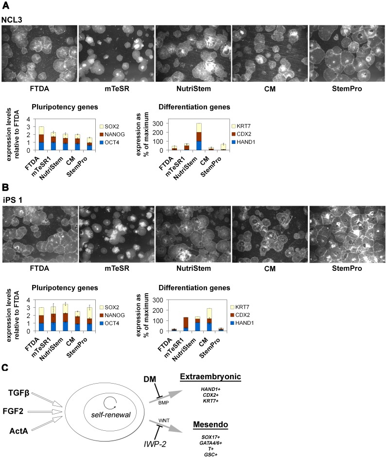
Human pluripotent stem cells (hPSCs) are conventionally grown in a mouse feeder cell-dependent manner. Chemically defined culture conditions are, however, desirable not only for potential medically oriented applications but also for investigating mechanisms of self-renewal and differentiation. In light of the rather high complexity and cost of existing defined hPSC culture systems, we have systematically evaluated over 20 potential media ingredients. Only components that reproducibly gave beneficial effects were ultimately combined to yield a simple and cost-effective formulation termed FTDA. This xeno-free medium is based on mimicking self-renewal factor activities present in mouse embryonic fibroblast-conditioned medium, at minimal dosages. Additionally, small molecule inhibitors of BMP and WNT signaling served to specifically suppress typical types of spontaneous differentiation seen in hPSC cultures. FTDA medium was suitable for the generation of human induced pluripotent stem cells and enabled robust long-term maintenance of diverse hPSC lines including hard-to-grow ones. Comparisons with existing defined media suggested reduced spontaneous differentiation rates in FTDA. Our results imply that using supportive factors at minimal concentrations may still promote robust self-renewal and preserve pluripotency of hPSCs.
Conflict of interest statement
Figures


Similar articles
-
Preparation of mouse embryonic fibroblast cells suitable for culturing human embryonic and induced pluripotent stem cells.J Vis Exp. 2012 Jun 21;(64):3854. doi: 10.3791/3854. J Vis Exp. 2012. PMID: 22760161 Free PMC article.
-
Synergistic effect of medium, matrix, and exogenous factors on the adhesion and growth of human pluripotent stem cells under defined, xeno-free conditions.Stem Cells Dev. 2012 Jul 20;21(11):2036-48. doi: 10.1089/scd.2011.0489. Epub 2012 Jan 26. Stem Cells Dev. 2012. PMID: 22149941
-
Activin A maintains self-renewal and regulates fibroblast growth factor, Wnt, and bone morphogenic protein pathways in human embryonic stem cells.Stem Cells. 2006 Jun;24(6):1476-86. doi: 10.1634/stemcells.2005-0299. Epub 2006 Feb 2. Stem Cells. 2006. PMID: 16456129
-
Engineering biomaterials for feeder-free maintenance of human pluripotent stem cells.Int J Stem Cells. 2012 May;5(1):1-5. doi: 10.15283/ijsc.2012.5.1.1. Int J Stem Cells. 2012. PMID: 24298348 Free PMC article. Review.
-
Progresses and challenges in optimization of human pluripotent stem cell culture.Curr Stem Cell Res Ther. 2010 Sep;5(3):207-14. doi: 10.2174/157488810791824548. Curr Stem Cell Res Ther. 2010. PMID: 20214560 Review.
Cited by
-
Functional high-resolution time-course expression analysis of human embryonic stem cells undergoing cardiac induction.Genom Data. 2016 Sep 28;10:71-74. doi: 10.1016/j.gdata.2016.09.007. eCollection 2016 Dec. Genom Data. 2016. PMID: 27722090 Free PMC article.
-
Using Transcriptomic Analysis to Assess Double-Strand Break Repair Activity: Towards Precise in vivo Genome Editing.Int J Mol Sci. 2020 Feb 18;21(4):1380. doi: 10.3390/ijms21041380. Int J Mol Sci. 2020. PMID: 32085662 Free PMC article.
-
A single-cell and feeder-free culture system for monkey embryonic stem cells.PLoS One. 2014 Feb 5;9(2):e88346. doi: 10.1371/journal.pone.0088346. eCollection 2014. PLoS One. 2014. PMID: 24505480 Free PMC article.
-
Human stem cell-based retina on chip as new translational model for validation of AAV retinal gene therapy vectors.Stem Cell Reports. 2021 Sep 14;16(9):2242-2256. doi: 10.1016/j.stemcr.2021.08.008. Stem Cell Reports. 2021. PMID: 34525384 Free PMC article.
-
A Cleared View on Retinal Organoids.Cells. 2019 Apr 28;8(5):391. doi: 10.3390/cells8050391. Cells. 2019. PMID: 31035373 Free PMC article.
References
-
- Thomson JA, Itskovitz-Eldor J, Shapiro SS, Waknitz MA, Swiergiel JJ, et al. (1998) Embryonic stem cell lines derived from human blastocysts. Science 282: 1145–1147. - PubMed
-
- Reubinoff BE, Pera MF, Fong CY, Trounson A, Bongso A (2000) Embryonic stem cell lines from human blastocysts: somatic differentiation in vitro. Nat Biotechnol 18: 399–404. - PubMed
-
- Amit M, Carpenter MK, Inokuma MS, Chiu CP, Harris CP, et al. (2000) Clonally derived human embryonic stem cell lines maintain pluripotency and proliferative potential for prolonged periods of culture. Dev Biol 227: 271–278. - PubMed
-
- Xu C, Inokuma MS, Denham J, Golds K, Kundu P, et al. (2001) Feeder-free growth of undifferentiated human embryonic stem cells. Nat Biotechnol 19: 971–974. - PubMed
-
- Greber B, Lehrach H, Adjaye J (2007) Fibroblast growth factor 2 modulates transforming growth factor beta signaling in mouse embryonic fibroblasts and human ES cells to support hESC self-renewal. Stem Cells 25: 455–464. - PubMed
MeSH terms
Substances
Grants and funding
LinkOut - more resources
Full Text Sources
Other Literature Sources
Research Materials
