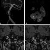
Intracranial atherosclerotic plaque enhancement in patients with ischemic stroke
- PMID: 22859280
- PMCID: PMC7965103
- DOI: 10.3174/ajnr.A3209
Intracranial atherosclerotic plaque enhancement in patients with ischemic stroke
Abstract
Background and purpose: Inflammation of an atherosclerotic plaque is a well-known risk factor in the development of ischemic stroke and myocardial infarction. MR imaging is capable of characterizing inflammation by assessing plaque enhancement in both extracranial carotid arteries and coronary arteries. Our goal was to determine whether enhancing intracranial atherosclerotic plaque was present in the vessel supplying the territory of infarction by using high-resolution vessel wall MR imaging.
Materials and methods: High-resolution vessel wall 3T MR imaging studies performed in 29 patients with ischemic stroke and intracranial vascular stenoses were reviewed for presence and strength of plaque enhancement.
Results: Sixteen patients were studied during the acute phase (<4 weeks from acute stroke), 5 patients in the subacute phase (4-12 weeks), and 8 patients in the chronic phase (>12 weeks) of the ischemic injury. In all of the acute phase patients, atherosclerotic plaque in the vessel supplying the stroke territory demonstrated strong enhancement. There was a trend of decreasing enhancement as the time of imaging relative to the ischemic event increased.
Conclusions: Strong pathologic enhancement of intracranial atherosclerotic plaque was seen in all patients imaged within 4 weeks of ischemic stroke in the vessel supplying the stroke territory. The strength and presence of enhancement of the atherosclerotic plaque decreased with increasing time after the ischemic event. These findings suggest a relationship between enhancing intracranial atherosclerotic plaque and acute ischemic stroke.
Figures


Similar articles
-
Gadolinium Enhancement in Intracranial Atherosclerotic Plaque and Ischemic Stroke: A Systematic Review and Meta-Analysis.J Am Heart Assoc. 2016 Aug 15;5(8):e003816. doi: 10.1161/JAHA.116.003816. J Am Heart Assoc. 2016. PMID: 27528408 Free PMC article. Review.
-
Previous Statin Use and High-Resolution Magnetic Resonance Imaging Characteristics of Intracranial Atherosclerotic Plaque: The Intensive Statin Treatment in Acute Ischemic Stroke Patients With Intracranial Atherosclerosis Study.Stroke. 2016 Jul;47(7):1789-96. doi: 10.1161/STROKEAHA.116.013495. Epub 2016 Jun 14. Stroke. 2016. PMID: 27301946
-
Intracranial plaque enhancement in patients with cerebrovascular events on high-spatial-resolution MR images.Radiology. 2014 May;271(2):534-42. doi: 10.1148/radiol.13122812. Epub 2014 Jan 16. Radiology. 2014. PMID: 24475850 Free PMC article.
-
Higher Plaque Burden of Middle Cerebral Artery Is Associated With Recurrent Ischemic Stroke: A Quantitative Magnetic Resonance Imaging Study.Stroke. 2020 Feb;51(2):659-662. doi: 10.1161/STROKEAHA.119.028405. Epub 2019 Dec 20. Stroke. 2020. PMID: 31856694
-
Vessel wall magnetic resonance imaging of symptomatic middle cerebral artery atherosclerosis: A systematic review and meta-analysis.Clin Imaging. 2022 Oct;90:90-96. doi: 10.1016/j.clinimag.2022.08.001. Epub 2022 Aug 4. Clin Imaging. 2022. PMID: 35952437 Review.
Cited by
-
Recurrent stroke risk in intracranial atherosclerotic disease.Front Neurol. 2022 Sep 1;13:1001609. doi: 10.3389/fneur.2022.1001609. eCollection 2022. Front Neurol. 2022. PMID: 36119685 Free PMC article. Review.
-
Vessel Wall Imaging of Cerebrovascular Disorders.Curr Treat Options Cardiovasc Med. 2019 Nov 14;21(11):65. doi: 10.1007/s11936-019-0782-8. Curr Treat Options Cardiovasc Med. 2019. PMID: 31728661 Review.
-
Correlation of the characteristics of symptomatic intracranial atherosclerotic plaques with stroke types and risk of stroke recurrence: a cohort study.Ann Transl Med. 2022 Jun;10(12):658. doi: 10.21037/atm-22-2586. Ann Transl Med. 2022. PMID: 35845483 Free PMC article.
-
Imaging the intracranial atherosclerotic vessel wall using 7T MRI: initial comparison with histopathology.AJNR Am J Neuroradiol. 2015 Apr;36(4):694-701. doi: 10.3174/ajnr.A4178. Epub 2014 Dec 4. AJNR Am J Neuroradiol. 2015. PMID: 25477359 Free PMC article.
-
Presence of Vessel Wall Hyperintensity in Unruptured Arteriovenous Malformations on Vessel Wall Magnetic Resonance Imaging: Pilot Study of AVM Vessel Wall "Enhancement".Front Neurosci. 2021 Jul 21;15:697432. doi: 10.3389/fnins.2021.697432. eCollection 2021. Front Neurosci. 2021. PMID: 34366779 Free PMC article.
References
-
- Willerson JT, Ridker PM. Inflammation as a cardiovascular risk factor. Circulation 2004;109:II2–10 - PubMed
-
- Wasserman BA. Advanced contrast-enhanced MRI for looking beyond the lumen to predict stroke: building a risk profile for carotid plaque. Stroke 2010;41:S12–16 - PubMed
-
- Yuan C, Mitsumori LM, Beach KW, et al. . Carotid atherosclerotic plaque: noninvasive MR characterization and identification of vulnerable lesions. Radiology 2001;221:285–99 - PubMed
-
- Wasserman BA, Wityk RJ, Trout HH, III, et al. . Low-grade carotid stenosis: looking beyond the lumen with MRI. Stroke 2005;36:2504–13 - PubMed
MeSH terms
LinkOut - more resources
Full Text Sources
Medical
