. 2012 Aug 1;32(31):10758-66.
doi: 10.1523/JNEUROSCI.0565-12.2012.

Role of rhodopsin and arrestin phosphorylation in retinal degeneration of Drosophila

Affiliations

Role of rhodopsin and arrestin phosphorylation in retinal degeneration of Drosophila

Inga Kristaponyte et al. J Neurosci. .

Abstract

Arrestins belong to a family of multifunctional adaptor proteins that regulate internalization of diverse receptors including G-protein-coupled receptors (GPCRs). Defects associated with endocytosis of GPCRs have been linked to human diseases. We used enhanced green fluorescent protein-tagged arrestin 2 (Arr2) to monitor the turnover of the major rhodopsin (Rh1) in live Drosophila. We demonstrate that during degeneration of norpA(P24) photoreceptors the loss of Rh1 is parallel to the disappearance of rhabdomeres, the specialized visual organelle that houses Rh1. The cause of degeneration in norpA(P24) is the failure to activate CaMKII (Ca(2+)/calmodulin-dependent protein kinase II) and retinal degeneration C (RDGC) because of a loss of light-dependent Ca(2+) entry. A lack of activation in CaMKII, which phosphorylates Arr2, leads to hypophosphorylated Arr2, while a lack of activation of RDGC, which dephosphorylates Rh1, results in hyperphosphorylated Rh1. We investigated how reversible phosphorylation of Rh1 and Arr2 contributes to photoreceptor degeneration. To uncover the consequence underlying a lack of CaMKII activation, we characterized ala(1) flies in which CaMKII was suppressed by an inhibitory peptide, and showed that morphology of rhabdomeres was not affected. In contrast, we found that expression of phosphorylation-deficient Rh1s, which either lack the C terminus or contain Ala substitution in the phosphorylation sites, was able to prevent degeneration of norpA(P24) photoreceptors. This suppression is not due to a loss of Arr2 interaction. Importantly, co-expression of these modified Rh1s offered protective effects, which greatly delayed photoreceptor degeneration. Together, we conclude that phosphorylation of Rh1 is the major determinant that orchestrates its internalization leading to retinal degeneration.


Figures
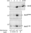
Figure 1.
Figure 1.
Association and dissociation between Rh1 and modified Arr2 in vitro. 35S-Arr2 (wt) binding and release from membranes containing either activated (blue-light-treated, B) or inactivated Rh1 (orange-light-treated, O) were analyzed by SDS/PAGE followed by autoradiography (top). Modified Arr2s containing Asp substitution at residue 366 mimicking phosphorylated Ser (Arr2S366D, middle) or missing the C terminus (Arr21–356, bottom) display similar binding specificity toward activated Rh1 (lane 3), like wt Arr2. Moreover, dissociation of modified Arr2 from inactivated Rh1 (lane 2) is similar to that of wt Arr2. Binding assays were performed using membranes from arr21 that contain 10% of endogenous Arr2 and membranes from ninaEI17 were used for the negative control. A representative autoradiograph is shown.
Figure 2.
Figure 2.
Interaction between activated Rh1 and wt Arr2 or Arr2S366D in vivo. The interaction between activated Rh1 and Arr2 was investigated by fluorescence microscopy in live flies. A, The distribution of wt Arr2-eGFP is predominantly in the rhabdomere of R1–6 photoreceptors. B, The subcellular localization of Arr2S366D-eGFP is similar to that of wt Arr2-eGFP. C, The rhabdomeric distribution of wt Arr2-eGFP is greatly diminished in the ninaEI17 background lacking endogenous Rh1. D, In isolated ommatidia, wt Arr2-eGFP fusion proteins are highly concentrated in rhabdomeres, but display uniform distribution in the absence of Rh1 (ninaE) (E). F, The steady-state level of both Arr2-eGFP fusion proteins is ∼12.1 ± 3.3% (n = 3) of endogenous Arr2 by Western blotting. Three-day-old flies of various genotypes were used for the microscopic and Western analyses.
Figure 3.
Figure 3.
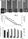
Progression of retinal degeneration in live retinas of norpAP24 photoreceptors. A, Degeneration was characterized by the age-dependent deterioration of rhabdomeres in R1–6 photoreceptors. Rhabdomeres are visualized by wt Arr2-eGFP that binds to activated Rh1. An almost complete loss of wt Arr2-eGFP is evident at 8–9 d posteclosion. Insets depict fdpp. B, Decline of the mean cross-sectional area of rhabdomeres was compared with the intensity of Arr2-eGFP-labeled rhabdomeres from multiple ommatidia (“fluorescent dpp,” see insets in A for depiction of dpp). The age-dependent reduction of dpp and rhabdomere exhibit a similar time course. Data were normalized to 2-d-old norpAP24 and represent means ± SEM (n = 4). C, The age-dependent decrease of the Rh1 level. Rh1, Arr2, and INAD were measured by Western blotting and expressed as a percentage of the 2-d-old flies of the same genotype. The Rh1 content was gradually diminished, while the Arr2 level remained constant in degenerating norpAP24 flies. The level of INAD, a membrane-associated cytosolic protein, served as a positive control. INAD remains constant but starts to decline only at the later stages of degeneration in norpA flies. Values represent means ± SEM (n = 4), *p < 0.05, **p < 0.01.
Figure 4.
Figure 4.
Suppression of CaMKII alone does not promote retinal degeneration. Morphology of rhabdomeres in ala1 flies overexpressing an inhibitory peptide for CaMKII was monitored by wt Arr2-eGFP for up to 4 weeks posteclosion. Ten-day-old wt control (A), 10-d-old ala1 (B), and 28-d-old ala1 (C). D, Comparison of rhabdomere area, intensity of fdpp, and the Rh1 level in ala1 flies. Data were normalized as a percentage of the 2-d-old flies of the same genotype and represent as mean ± SEM (n = 3). *p < 0.05. At 4 weeks posteclosion, ala1 flies displayed a significant reduction in both rhabdomere area and intensity of fluorescent dpp but not in the Rh1 level, when compared with 2-d-old ala1 flies. All flies tested were red eyed. In red-eyed background, the shape of rhabdomeres usually appears more elongated, when compared with that of white-eyed flies (see Fig. 2).
Figure 5.
Figure 5.
Arr2 binds to phosphorylation-deficient Rh1 in vitro and in vivo. A, Interaction between Arr2 and phosphorylation-deficient Rh1 in vitro by membrane binding assays. 35S-Arr2 binding to either Rh1Δ356 or Rh1CT S>A was similar to 35S-Arr2 binding to wt Rh1, whereas binding was greatly reduced when membranes prepared from ninaEI17 were used. A representative autoradiograph depicting the bound 35S-Arr2 is shown (top) and the results from three independent experiments are quantitated, normalized to binding to wt Rh1, and shown as means ± SEM in the histogram. *p < 0.05. B, The in vivo interaction between wt Arr2-eGFP and modified Rh1 lacking the C terminus, Rh1Δ356, or the putative phosphorylation sites, Rh1CT S>A (C), was examined by fluorescence microscopy in 5-d-old live flies. Shown is a representative image from each interaction.
Figure 6.
Figure 6.
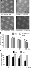
Expression of phosphorylation-deficient Rh1 prevents degeneration of norpAP24 photoreceptors while co-expression greatly delays degeneration. Shown are retinal morphology monitored by wt Arr2-eGFP in norpAP24 flies expressing either Rh1Δ356 (A) or Rh1CT S>A (B) at 20 d posteclosion. Co-expression of Rh1CT S>A greatly delayed degeneration (C, at 21 d; D, at 28 d). Comparison of rhabdomere areas and intensities of fdpp (E) and Rh1 levels (F) of norpAP24 flies co-expressing wt and Rh1CT S>A. Data were normalized as a percentage of the 2-d-old flies of the same genotype and represent as mean ± SEM (n = 4). *p < 0.05, **p < 0.01.
Figure 7.
Figure 7.
A hypothetical mechanism of enhanced internalization and degradation of phosphorylated Rh1 leading to retinal degeneration of norpA photoreceptors. In norpA photoreceptors, the lack of Ca2+ influx renders Rh1 phosphatase RDGC inactive resulting in an increased level of phosphorylated and activated Rh1 (Rh1*). Phosphorylated Rh1 forms stable complexes with Arr2 that promotes the endocytosis of Rh1*. Following internalization, Rh1* may be ubiquitinated and subsequently is trafficked to lysosomes for degradation. A reduction of Rh1 that is critical for the maintenance and function of photoreceptors results in loss of rhabdomeres and eventually, degeneration of photoreceptors. Filled circles denote phosphorylation at the C terminus of Rh1, and filled triangles denote ubiquitination of Rh1.

Similar articles

Cited by

References

    1. Alloway PG, Dolph PJ. A role for the light-dependent phosphorylation of visual arrestin. Proc Natl Acad Sci U S A. 1999;96:6072–6077. - PMC - PubMed
    1. Alloway PG, Howard L, Dolph PJ. The formation of stable rhodopsin-arrestin complexes induces apoptosis and photoreceptor cell degeneration. Neuron. 2000;28:129–138. - PubMed
    1. Byk T, Bar-Yaacov M, Doza YN, Minke B, Selinger Z. Regulatory arrestin cycle secures the fidelity and maintenance of the fly photoreceptor cell. Proc Natl Acad Sci U S A. 1993;90:1907–1911. - PMC - PubMed
    1. Chen J, Shi G, Concepcion FA, Xie G, Oprian D, Chen J. Stable rhodopsin/arrestin complex leads to retinal degeneration in a transgenic mouse model of autosomal dominant retinitis pigmentosa. J Neurosci. 2006;26:11929–11937. - PMC - PubMed
    1. Chinchore Y, Mitra A, Dolph PJ. Accumulation of rhodopsin in late endosomes triggers photoreceptor cell degeneration. PLoS Genet. 2009;5:e1000377. - PMC - PubMed

Publication types

MeSH terms

LinkOut - more resources