

Spatiotemporal dynamics of adenovirus membrane rupture and endosomal escape
- PMID: 22855481
- PMCID: PMC3457294
- DOI: 10.1128/JVI.01428-12
Spatiotemporal dynamics of adenovirus membrane rupture and endosomal escape
Abstract
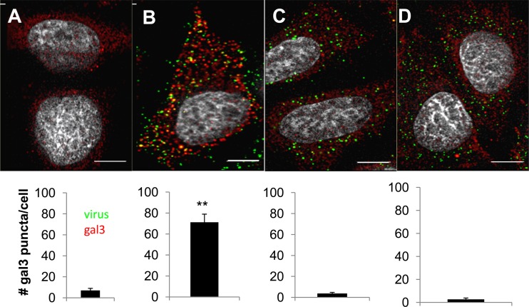
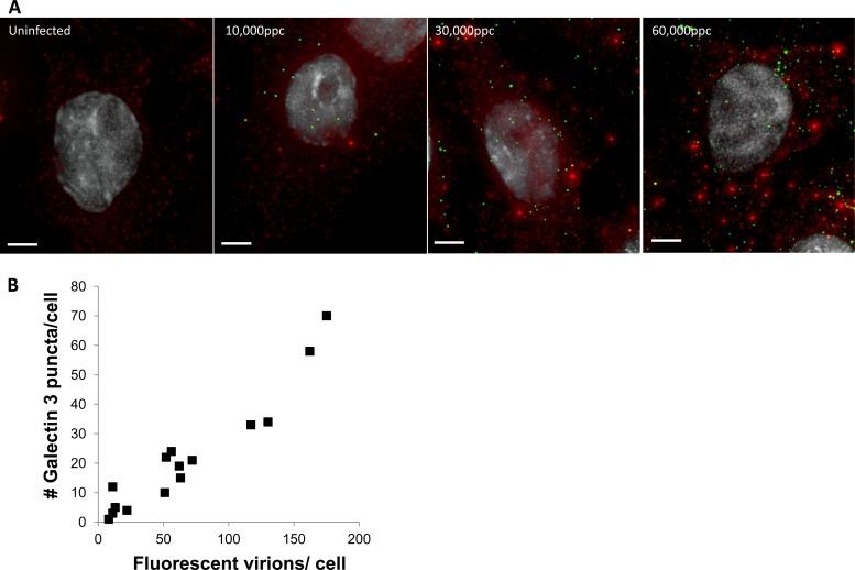
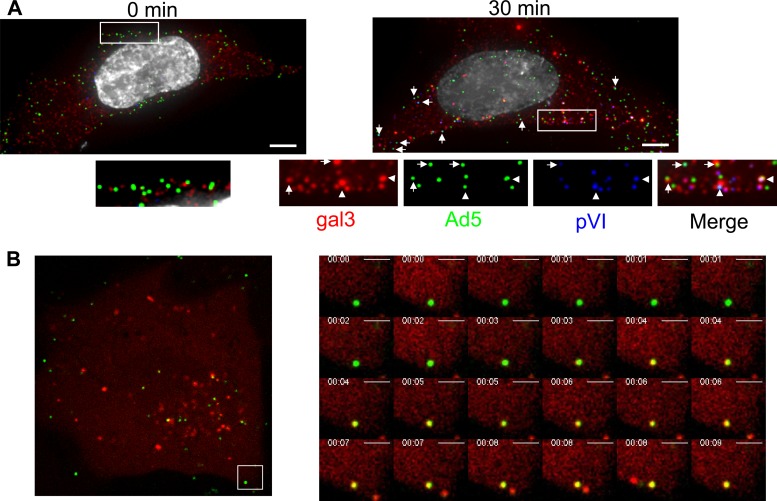
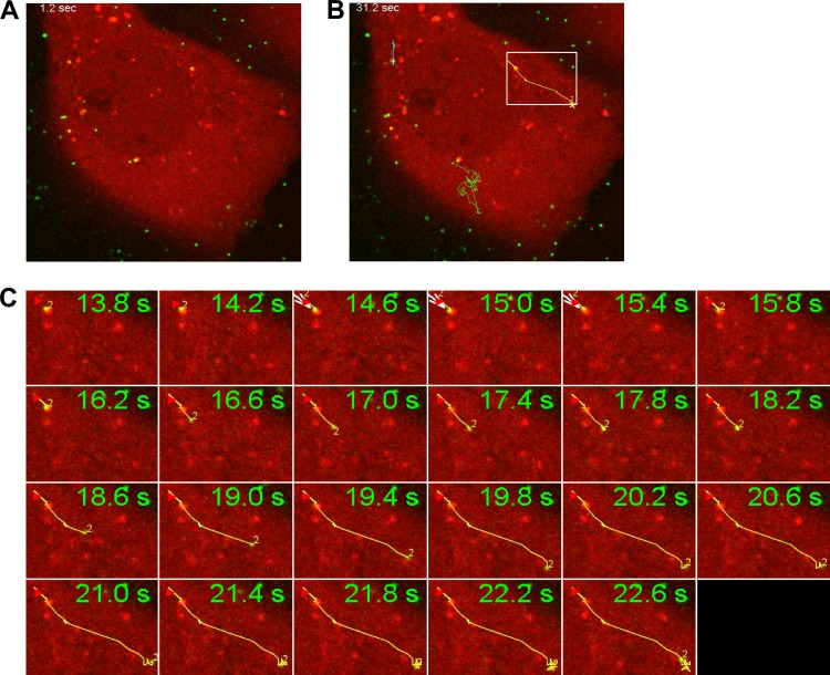
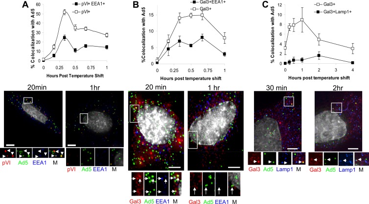
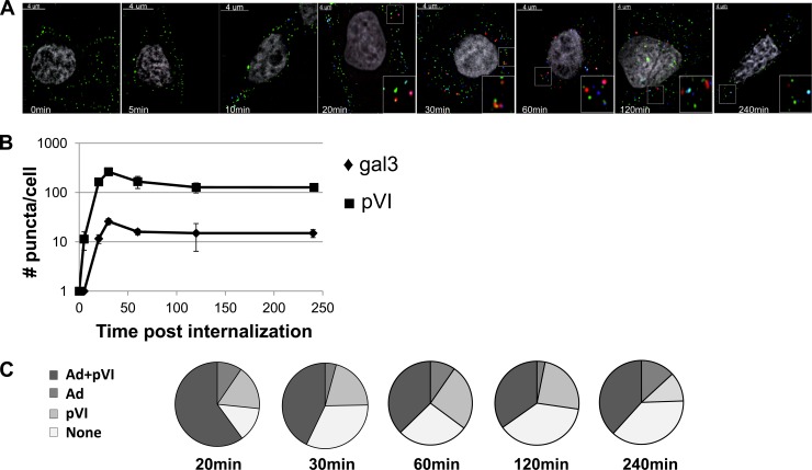
A key step in adenovirus cell entry is viral penetration of cellular membranes to gain access to the cytoplasm and deliver the genome to the nucleus. Yet little is known about this important event in the adenoviral life cycle. Using the cytosolic protein galectin-3 (gal3) as a marker of membrane rupture with both live- and fixed-cell imaging, we demonstrate that in the majority of instances, exposure of pVI and recruitment of gal3 to ruptured membranes occur early at or near the cell surface and occur minimally in EEA-1-positive (EEA-1(+)) early endosomes or LAMP-1(+) late endosomes/lysosomes. Live-cell imaging of Ad5 egress from gal3(+) endosomes occurs most frequently from perinuclear locations. While the Ad5 capsid is observed escaping from gal3(+) endosomes, pVI appears to remain associated with the gal3(+) ruptured endosomes. Thus, Ad5 membrane rupture and endosomal escape appear to be both spatially and temporally distinct events.
Figures
Similar articles
-
Adenovirus serotype 7 retention in a late endosomal compartment prior to cytosol escape is modulated by fiber protein.J Virol. 2001 Feb;75(3):1387-400. doi: 10.1128/JVI.75.3.1387-1400.2001. J Virol. 2001. PMID: 11152512 Free PMC article.
-
HOPS-dependent endosomal fusion required for efficient cytosolic delivery of therapeutic peptides and small proteins.Proc Natl Acad Sci U S A. 2019 Jan 8;116(2):512-521. doi: 10.1073/pnas.1812044116. Proc Natl Acad Sci U S A. 2019. PMID: 30610181 Free PMC article.
-
Transferrin receptor recycling in the absence of perinuclear recycling endosomes.J Cell Biol. 2002 Mar 4;156(5):797-804. doi: 10.1083/jcb.20111048. Epub 2002 Mar 4. J Cell Biol. 2002. PMID: 11877458 Free PMC article.
-
Endocytosis of adenovirus and adenovirus capsid proteins.Adv Drug Deliv Rev. 2003 Nov 14;55(11):1485-96. doi: 10.1016/j.addr.2003.07.010. Adv Drug Deliv Rev. 2003. PMID: 14597142 Review.
-
Dynamics of galectin-3 in the nucleus and cytoplasm.Biochim Biophys Acta. 2010 Feb;1800(2):181-9. doi: 10.1016/j.bbagen.2009.07.005. Epub 2009 Jul 16. Biochim Biophys Acta. 2010. PMID: 19616076 Free PMC article. Review.
Cited by
-
Cellular self-defense: how cell-autonomous immunity protects against pathogens.Science. 2013 May 10;340(6133):701-6. doi: 10.1126/science.1233028. Science. 2013. PMID: 23661752 Free PMC article. Review.
-
Adenovirus membrane penetration: Tickling the tail of a sleeping dragon.Virology. 2015 May;479-480:591-9. doi: 10.1016/j.virol.2015.03.006. Epub 2015 Mar 19. Virology. 2015. PMID: 25798531 Free PMC article. Review.
-
The Revolving Door of Adenovirus Cell Entry: Not All Pathways Are Equal.Pharmaceutics. 2021 Sep 29;13(10):1585. doi: 10.3390/pharmaceutics13101585. Pharmaceutics. 2021. PMID: 34683878 Free PMC article. Review.
-
CRM1 Promotes Capsid Disassembly and Nuclear Envelope Translocation of Adenovirus Independently of Its Export Function.J Virol. 2022 Feb 9;96(3):e0127321. doi: 10.1128/JVI.01273-21. Epub 2021 Nov 10. J Virol. 2022. PMID: 34757845 Free PMC article.
-
Spatially and temporally defined lysosomal leakage facilitates mitotic chromosome segregation.Nat Commun. 2020 Jan 13;11(1):229. doi: 10.1038/s41467-019-14009-0. Nat Commun. 2020. PMID: 31932607 Free PMC article.
References
-
- Barondes SH, et al. 1994. Galectins: a family of animal beta-galactoside-binding lectins. Cell 76:597–598 - PubMed
Publication types
MeSH terms
Substances
Grants and funding
LinkOut - more resources
Full Text Sources
Other Literature Sources
Miscellaneous
