. 2012 Nov;20(11):2087-97.
doi: 10.1038/mt.2012.144. Epub 2012 Jul 31.

Terminal differentiation of cardiac and skeletal myocytes induces permissivity to AAV transduction by relieving inhibition imposed by DNA damage response proteins

Affiliations

Terminal differentiation of cardiac and skeletal myocytes induces permissivity to AAV transduction by relieving inhibition imposed by DNA damage response proteins

Jasmina Lovric et al. Mol Ther. 2012 Nov.

Abstract

Gene therapy vectors based on the adeno-associated virus (AAV) are extremely efficient for gene transfer into post-mitotic cells of heart, muscle, brain, and retina. The reason for their exquisite tropism for these cells has long remained elusive. Here, we show that upon terminal differentiation, cardiac and skeletal myocytes downregulate proteins of the DNA damage response (DDR) and that this markedly induces permissivity to AAV transduction. We observed that expression of members of the MRN complex (Mre11, Rad50, Nbs1), which bind the incoming AAV genomes, faded in cardiomyocytes at ~2 weeks after birth, as well as upon myoblast differentiation in vitro; in both cases, withdrawal of the cells from the cell cycle coincided with increased AAV permissivity. Treatment of proliferating cells with short-interfering RNAs (siRNAs) against the MRN proteins, or with microRNA-24, which is normally upregulated upon terminal differentiation and negatively controls the Nbs1 levels, significantly increased permissivity to AAV transduction. Consistently, delivery of these small RNAs to the juvenile liver concomitant with AAV markedly improved in vivo hepatocyte transduction. Collectively, these findings support the conclusion that cellular DDR proteins inhibit AAV transduction and that terminal cell differentiation relieves this restriction.


Figures

Figure 1
Figure 1
Efficiency of myocardial transduction with adeno-associated virus (AAV) vectors at different times after birth. (a) Schematic representation of the experimental workflow. AAV9-LacZ vectors were administered intraperitoneal (i.p.) in 1-, 7-, and 21-day-old mice; in all cases, analysis was performed 7 days after injection. (b) Histochemical staining for β-galactosidase expression—whole heart sections (upper panels) and enlargements (lower panels; Bar: 100 µm)—after AAV administration for the indicated time intervals (days). (c) Quantification of β-galactosidase expression in total heart tissue (n = 6 per group; mean values ± SD) after AAV administration for the indicated time intervals (days). (d) AAV vector DNA copy numbers per ng of genomic DNA (n = 6 per group; mean values ± SD) after AAV administration for the indicated time intervals (days). (e) Ratio between the amount of expressed β-galactosidase and AAV genome copy numbers (n = 6 per group; mean values ± SD) after AAV administration for the indicated time intervals (days).
Figure 2
Figure 2
Permissivity of cultured neonatal cardiomyocytes to adeno-associated virus (AAV) transduction. (a) Representative images of cardiomyocytes cultures at 4, 7, and 10 days after plating. Cells were stained with anti-α-sarcomeric actinin antibody (green) to reveal cardiomyocytes and ethynyl-2′-deoxyuridine (EdU) to assess DNA synthesis. Bar: 100 µm. (b) Percentage of proliferating (EdU+ and Ki67+) cardiomyocytes at day 4, 7, and 10 after plating (mean values ± SD of five independent experiments). (c) Schematic representation of experimental workflow. Neonatal rat cardiomyocytes were transduced with AAV6-EGFP at days 1, 3, and 7 after plating and analyzed 4 days after transduction. (d) Representative images of cardiomyocytes transduced with AAV6-EGFP at the indicated time intervals. Cells were fixed and immunostained with anti-α-sarcomeric actinin antibody (red) to reveal cardiomyocytes. Cell nuclei were stained with Hoechst 33342. (e) Percentage of transduced (GFP+) cardiomyocytes after AAV6-EGFP transduction for the indicated time intervals (mean values ± SD of three independent experiments). (f) Amount of AAV vector DNA genomes per ng of genomic DNA, determined by real-time quantitative PCR, at 4 hours postinfection, after transduction for day 1, 3, and 7 (mean values ± SD of three independent experiments). (g) Effects of knockdown of Mre11, Rad50, and Nbs1 using RNA interference (RNAi). Cardiomyocytes, isolated at day 1 after birth, were treated with the indicated short-interfering RNAs (siRNAs) and later transduced with AAV6-Luciferase. The levels of luciferase expression were tested 48 hours after transduction (mean values ± SD of three independent experiments)/NTS, nontargeting siRNA control. *Statistical significance (P < 0.05). (h) Levels of Mre11, Rad50, and Nbs1 mRNAs at 72 hours after transfection of the corresponding siRNAs, measured by real-time PCR in the same samples as in panel g. The results are shown relative to those measured in cardiomyocytes treated with the NTS control. EGFP, enhanced green fluorescent protein.
Figure 3
Figure 3
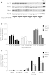
Levels of Mre11/Rad50/Nbs1 (MRN) proteins in the heart at different times after birth and in other organs. (a) Western blot analysis of Mre11, Rad50, and Nbs1 proteins from whole mouse heart samples obtained at different times after birth—days 0 (neonatal), 3, 7, 14, 21, 60 (adult). Representative samples from two mice are shown for each time point. Hsc-70 was used as loading control. (b) Gene expression analysis of proteins of the MRN complex in the mouse heart at different age as determined by real-time PCR. Values (n = 5 per group; mean ± SD) are normalized over glyceraldehyde 3-phosphate dehydrogenase (GAPDH). (c) Gene expression analysis of proteins of the MRN complex in different tissues of 2-month-old mice (adults), evaluated by real-time PCR. Values (n = 5 per group; mean ± SD) are normalized over GAPDH.
Figure 4
Figure 4
Permissivity of C2C12 cells to adeno-associated virus (AAV) transduction markedly increases during cell differentiation. (a) Western blot of Mre11, Rad50, and Nbs1 proteins during differentiation of C2C12 cells. C2C12 myoblasts were incubated in differentiation medium (DM) for 0, 2, 4, 6, 10, and 12 days to induce differentiation and formation of myotubes. Myosin heavy chain (MHC) was used as a differentiation marker and β-tubulin as a loading control. (b) Gene expression analysis of Mre11, Rad50, and Nbs1 by real-time PCR during the C2C12 differentiation process. The graphs represent the change in gene expression over GAPDH (mean values ± SD of three independent experiments). (c) Schematic representation of the experimental setup for analysis of AAV transduction of C2C12 cells. Cultures of nondifferentiated (ND) or differentiated cells (D, 4 days in differentiation medium) were transduced with AAV vectors expressing enhanced green fluorescent protein (EGFP) or luciferase at different multiplicities of infection (MOIs); analysis was performed 4 days after transduction. (d) Representative images of nondifferentiated (ND) and differentiated (D) C2C12 cells transduced with AAV6-EGFP at an MOI of 1 × 10⁵ vg. Four days after transduction cells were fixed and immunostained with anti-α-sarcomeric actinin antibody (red) to reveal formation of differentiated myotubes. Cell nuclei were stained with DAPI. The enlarged merged figures are shown in the rightmost panels. Bars: 200 µm. (e) Quantification of luciferase activity per µg of total protein in nondifferentiated (ND) and differentiated (D) C2C12 cultures transduced with AAV2-Luciferase at the MOI of 1 × 10⁴, 5 × 10⁴, and 1 × 10⁵ vg. Graphs represent mean values ± SD of three independent experiments. (f) Amount of AAV vector genomes internalized by ND and D C2C12 cells at 4 hours postinfection, determined by real-time quantitative PCR (mean values ± SD of three independent experiments).
Figure 5
Figure 5
Levels of expression of microRNAs 24, 23a, and 27a. (a) Expression of miR-24, miR-23a, and miR-27a in mouse heart at different times after birth (days 0 (neonatal), 3, 7, 14, 21, 60 (adult)) as determined by real-time PCR. Graphs represent the change in gene expression over miR-191, shown as mean values ± SD; n = 5 per group. (b) Expression of miR-24, miR-23a, and miR-27a during the differentiation of C2C12 cells (0, 4, and 10 days in differentiation medium (DM)) as determined by real-time PCR. Graphs represent mean values ± SD of three independent samples, normalized to miR-191.
Figure 6
Figure 6
Expression of miR-24 precursor leads to downregulation of Nbs1 and enhances permissivity to adeno-associated virus (AAV) in vitro. (a) Mre11, Rad50, and Nbs1 protein levels in HeLa cells 72 hours after transfection with precursors of miR-24, 23a, and 27a, at a final concentration of 50 nmol/l. β-Tubulin was used as a control for protein levels. (b) Mre11, Rad50, and Nbs1 protein levels in HeLa cells 72 hours after transfection with 25, 50, and 100 nmol/l miR-24 precursor. E2F-1 was used as control of miR-24 efficacy, since the E2F family members are known targets of miR-24. (c) Quantification of immunoblots showing the relative expression of Mre11, Rad50, Nbs1, and E2F-1 in HeLa cells treated with the indicated amounts of miR-24 precursor for 72 hours. The results show mean values ± SD of three independent experiments. (d) Luciferase activity of lysates from HeLa cells transfected with miR-24, miR-23a, or miR-27a precursors (100 nmol/l) or control cells (white bars) after transduction with AAV2-Luciferase at three different multiplicities of infection (MOIs) (1 × 10³, 5 × 10³, and 1 × 10⁴ vg per cell). The graph shows mean values ± SD of three independent experiments. (e) Representative images of HeLa cells transfected with miR-24 precursor (25, 50, 100 nmol/l) or nontargeting short-interfering RNA (siRNA) (NTS; 50 and 100 nmol/l) for 48 hours and then transduced with AAV2-EGFP (MOI 1 × 10⁴). Images were taken 24 hours after AAV transduction. Control cells represent nontransfected cells transduced with AAV2-EGFP. Bar: 100 µm. (f) Analysis of ATP production, as a measurement of cell viability of HeLa cells transfected with miR-24 precursor at the indicated concentrations. The experimental conditions were as in panel e (mean ± SD of three independent experiments). EGFP, enhanced green fluorescent protein.
Figure 7
Figure 7
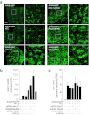
In vivo delivery of short-interfering RNAs against Mre11/Rad50/Nbs1 (MRN) components and of miR-24 precursor increases adeno-associated virus (AAV) transduction of juvenile mice livers. (a) Representative images of liver sections at day 10 after portal vein injection of AAV8-ApoE/hAAT-EGFP together with siRNAs against Mre11, Rad50, and Nbs1 or the miR-24 precursor, as indicated. For each treatment, the panel on the right side shows a magnification of the squared area shown on the left panel. Bar: 1 mm. (b) Quantification of the levels of the enhanced green fluorescent protein (EGFP) mRNA in the liver samples as in panel a. Real-time PCR quantifications were normalized over the amounts of cellular GAPDH and expressed as fold-values over samples treated with the nontargeting siRNA (NTS; mean values ± SD; n = 6 per group). The asterisks denote statistical significance (P < 0.05). (c) Quantification of the number of AAV vector genomes in total DNA samples from the liver of animals treated as in panel a (mean values ± SD; n = 6 per group).

Similar articles

Cited by

References

    1. Mingozzi F., and, High KA. Therapeutic in vivo gene transfer for genetic disease using AAV: progress and challenges. Nat Rev Genet. 2011;12:341–355. - PubMed
    1. Wu Z, Asokan A., and, Samulski RJ. Adeno-associated virus serotypes: vector toolkit for human gene therapy. Mol Ther. 2006;14:316–327. - PubMed
    1. Ferrari FK, Samulski T, Shenk T., and, Samulski RJ. Second-strand synthesis is a rate-limiting step for efficient transduction by recombinant adeno-associated virus vectors. J Virol. 1996;70:3227–3234. - PMC - PubMed
    1. Fisher KJ, Gao GP, Weitzman MD, DeMatteo R, Burda JF., and, Wilson JM. Transduction with recombinant adeno-associated virus for gene therapy is limited by leading-strand synthesis. J Virol. 1996;70:520–532. - PMC - PubMed
    1. McCarty DM, Fu H, Monahan PE, Toulson CE, Naik P., and, Samulski RJ. Adeno-associated virus terminal repeat (TR) mutant generates self-complementary vectors to overcome the rate-limiting step to transduction in vivo. Gene Ther. 2003;10:2112–2118. - PubMed

Publication types

MeSH terms

LinkOut - more resources