. 2012;7(7):e41794.
doi: 10.1371/journal.pone.0041794. Epub 2012 Jul 27.

Elevated IKKα accelerates the differentiation of human neuronal progenitor cells and induces MeCP2-dependent BDNF expression

Affiliations

Elevated IKKα accelerates the differentiation of human neuronal progenitor cells and induces MeCP2-dependent BDNF expression

Ali Khoshnan et al. PLoS One. 2012.

Abstract

The IκB kinase α (IKKα) is implicated in the differentiation of epithelial and immune cells. We examined whether IKKα also plays a role in the differentiation and maturation of embryonic human neuronal progenitor cells (NPCs). We find that expression of an extra copy of IKKα (IKKα+) blocks self-renewal and accelerates the differentiation of NPCs. This coincides with reduced expression of the Repressor Element Silencing Transcription Factor/Neuron-Restrictive Silencing Factor (REST/NRSF), which is a prominent inhibitor of neurogenesis, and subsequent induction of the pro-differentiation non-coding RNA, miR-124a. However, the effects of IKKα on REST/NRSF and miR-124a expression are likely to be indirect. IKKα+ neurons display extensive neurite outgrowth and accumulate protein markers of neuronal maturation such as SCG10/stathmin-2, postsynaptic density 95 (PSD95), syntaxin, and methyl-CpG binding protein 2 (MeCP2). Interestingly, IKKα associates with MeCP2 in the nuclei of human neurons and can phosphorylate MeCP2 in vitro. Using chromatin immunoprecipitation assays, we find that IKKα is recruited to the exon-IV brain-derived neurotrophic factor (BDNF) promoter, which is a well-characterized target of MeCP2 activity. Moreover, IKKα induces the transcription of BDNF and knockdown expression of MeCP2 interferes with this event. These studies highlight a role for IKKα in accelerating the differentiation of human NPCs and identify IKKα as a potential regulator of MeCP2 function and BDNF expression.


Conflict of interest statement

Competing Interests: The authors have declared that no competing interests exist.

Figures

Figure 1
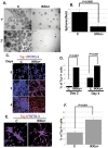
Figure 1. Effects of IKKα on the proliferation of MESC2.10 NPCs.
(A) Elevated IKKα impairs neurosphere formation of MESC2.10 NPCs. The neurosphere assay was carried out as described in Methods. Representative micrographs of primary (1°) (top panels) and secondary (2°) neurospheres (bottom panels) formed by control (C) and IKKα+ NPCs are shown. Assays were done in triplicate. (B) Quantification of neurospheres reveals a significant deficit in the IKKα+ NPCs compared to controls. Six culture wells were counted in each condition and averaged. P value was obtained using student's t-test. (C) Elevated IKKα induces the differentiation of MESC2.10 cells when v-myc expression is repressed. Control and IKKα+ NPCs were cultivated on laminin in the absence (time 0) or the presence of doxycycline for 2 or 4 days. Cells were stained for Tuj-1 expression. Representative micrographs obtained with a confocal microscope are shown. The DNA stain TOTO-3 was used to identify nuclei. (D) The % of Tuj-1 positive cells in day 2 and day 4 cultures is shown. (E) IKKα+ neurospheres undergo spontaneous neuronal differentiation. Day 6 dissociated neurospheres were plated on laminin and stained for Tuj-1 after 24 h. Representative micrographs obtained with a confocal microscope are shown. (F) The % of Tuj-1 positive in day 2 and day 4 cells is shown. For D and F, total and Tuj-1 positive cells were counted in 6 different confocal images and the % positive was calculated. P values were obtained using student's t-test.
Figure 2
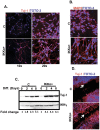
Figure 2. Elevated IKKα promotes the differentiation of MESC2.10 cells.
(A, B) IKKα promotes neurite outgrowth in differentiating NPCs. Control (C) and IKKα+NPCs were differentiated on coverslips for 4 days, fixed and stained with neuronal differentiation markers Tuj-1 (A) and MAP-2 (B). Representative micrographs obtained with a confocal microscope are shown. The DNA stain TOTO-3 was used to identify nuclei. (C) Tuj-1 levels are elevated in differentiating IKKα+ NPCs. Representative western blot results are shown for cytoplasmic lysates staining for Tuj-1 levels at different time points during the differentiation of control and IKKα+ NPCs [diff.(days)]. IKKγ was used as a loading control. Fold-change was obtained by dividing the intensity of Tuj-1 to the corresponding IKKγ, obtained by a Fluorchem 8900 (Alpha Innotech, San Leandro, CA). (D) The scratch assay shows extensive neurite outgrowth in differentiating IKKα+ NPCs. Cultures on the 2nd day of differentiation were wounded by a micropipette tip and further incubated for additional two days. Cells were fixed and stained as above. Arrows point to the areas of neurite extension.
Figure 3
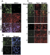
Figure 3. IKKα inhibits the proliferation of early differentiating NPCs.
(A) Elevated IKKα accelerates NPCs commitment to differentiation. BrdU was added on the 4th day of differentiation. 24 h later cells were fixed and stained with a rat anti-BrdU antibody (green) and the neuron-specific marker Tuj-1. Pictures were taken with a confocal microscope. (B) As a further test of proliferation, 4th day cultures were stained with the Ki-67 antibody, which identifies proliferating cells. (C) A time-course (days) of BrdU incorporation reveals the difference in rate of decline in proliferation between the control and the IKKα+ NPCs. Each time point represents 24 h of BrdU incorporation. Samples were processed as in A.
Figure 4
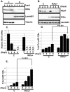
Figure 4. IKKα regulates REST and miR-124a expression.
(A) IKKα accumulates in the nuclei of differentiating MESC2.10 NPCs. Representative Western blot results for levels of endogenous IKKα in the cytoplasm (C) and nuclear (N) fractions of differentiating NPCs (top panel) are shown. IKKα was detected with a mouse anti-IKKα antibody. Nuclear LaminB1 and cytoplasmic tubulin were used as loading controls (middle and bottom panels, respectively). (B) REST protein levels also decline faster in differentiating IKKα+ NPCs compared to differentiating controls. Representative western blot results are shown from nuclear lysates for REST (top panel), IKKα (middle panel) and laminB1 (bottom panel). REST was detected with a mouse anti-REST antibody and Anti-Flag antibody was used to detect IKKα. LaminB1 was used a as loading control. (C) After initiating differentiation, REST mRNA levels decline faster in IKKα+ NPCs than in control cells. Taqman probes were used to quantify the mRNA levels at the days shown. The data are shown relative to the level in proliferating control NPCs. GAPDH mRNA was used for normalization. Triplicate samples were averaged for each point, and the SEMs indicated. N.D., not detected. (D, E) The accumulation of primary (pri-miRNA) and mature miRNA-124a are shown in D and E, respectively. Taqman probes were used for the qPCR. Pri-miRNA was normalized to GAPDH mRNA and mature miRNA was normalized to the small RNA, RNU6. The data are shown relative to the levels in proliferating control NPCs. P values were obtained using student's t-test.
Figure 5
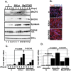
Figure 5. IKKα promotes expression of markers of mature neurons and BDNF.
(A) Elevated IKKα+ enhances neuronal maturation. Cell lysates from various time points (days after inducing differentiation) were examined by western blotting for the levels MeCP2, SCG10, syntaxin, and PSD-95. A lentivirus encoding an shRNA targeting MeCP2 was used to knockdown the expression of MeCP2 in IKKα+ NPCs (labeled as MeCP2KD) (lanes 9–12). IKKγ was used as a loading control. A non-specific band (N.S., below the authentic band) is recognized by the anti-PSD95 antibody. (B) Knockdown of MeCP2 expression does not influence IKKα-induced neuronal differentiation. NPCs were differentiated and examined as described in Fig. 2A. Representative confocal micrographs of 4th day differentiating cultures are shown. (C) IKKα promotes MeCP2-dependent BDNF expression. A time course (days) for BDNF expression during the differentiation of NPCs is shown. Taqman probes were used to quantify mRNA generated from the exon-IV of the BDNF promoter. GAPDH was used for normalization. The data are shown relative to the level in proliferating control (day 0) NPCs. (D) IKKα also promotes MeCP2-dependent BDNF expression following depolarization. Data are shown for cultures after 8th days of differentiation and depolarization with 50 mM KCl for 6 hr immediately preceding harvest. The data are shown relative to the level in non-depolarized control cells. Assays were done in triplicate and P values were obtained using student's t-test.
Figure 6
Figure 6. IKKα associates with MeCP2 and is recruited to the exon-IV BDNF promoter.
(A) Flag-tagged IKKα is recruited to the exon-IV BDNF promoter. ChIP assays were used to immunoprecipitate IKKα/DNA complexes (using anti-Flag antibody) from differentiated IKKα+ and MeCP2KD neurons (day 8). The left panel is ChIP from IKKα+ and the right panel is from MeCP2KD neurons. Non-reactive IgGs were used controls. DNA was amplified by PCR. Products were visualized by agarose gel-electrophoresis and ethidium bromide staining. (B) Western blots were used to assay nuclear lysates for phosphorylation of MeCP2 at Ser421 in 8th day differentiated IKKα+ neurons (middle panel). The top panel shows the total levels of MeCP2 during differentiation (0–8 days). LaminB1 was used as a loading control. (C) IKKα and MeCP2 co-localize in the nuclei of IKKα+ neurons. IKKα+ NPCs were differentiated for 6 days and stained with MeCP2 antibody (green) and an anti-Flag antibody detecting IKKα (red). Representative micrographs obtained with a confocal microscope are shown. (D) MeCP2 co-immunoprecipitates with IKKα. Nuclear lysates from 8th day differentiated IKKα+ neurons were immunoprecipitated with anti-Flag beads (for IKKα) and examined for the presence of MeCP2 with an anti-MeCP2 antibody. A non-immune mouse antibody (C-Ab) was used as a negative control for immunoprecipitation. A strong band for anti-IgG staining is also seen. (E) IKKα phosphorylates MeCP2. Active recombinant IKKα was tested for the ability to phosphorylate MeCP2. The kinase assay was performed as described in Methods with recombinant MeCP2 or GST as substrates. Products were visualized by SDS-PAGE followed by autoradiography.

Similar articles

Cited by

References

    1. Hacker H, Karin M. Regulation and function of IKK and IKK-related kinases. Sci STKE. 2006;357:re13. - PubMed
    1. Li Q, Lu Q, Bottero V, Estepa G, Morrison L, et al. Enhanced NF-kappaB activation and cellular function in macrophages lacking IkappaB kinase 1 (IKK1). Proc Natl Acad Sci U S A. 2005;102:12425–12430. - PMC - PubMed
    1. Lawrence T, Bebien M, Liu GY, Nizet V, Karin M. IKKalpha limits macrophage NF-kappaB activation and contributes to the resolution of inflammation. Nature. 2005;434:1138–1143. - PubMed
    1. Chariot A. The NF-kappaB-independent functions of IKK subunits in immunity and cancer. Trends Cell Biol. 2009;19:404–413. - PubMed
    1. Jepsen K, Solum D, Zhou T, McEvilly RJ, Kim HJ, et al. SMRT-mediated repression of an H3K27 demethylase in progression from neural stem cell to neuron. Nature. 2007;450:415–419. - PubMed

Publication types