A single amino acid change resulting in loss of fluorescence of eGFP in a viral fusion protein confers fitness and growth advantage to the recombinant vesicular stomatitis virus
- PMID: 22832124
- PMCID: PMC3423531
- DOI: 10.1016/j.virol.2012.07.004
A single amino acid change resulting in loss of fluorescence of eGFP in a viral fusion protein confers fitness and growth advantage to the recombinant vesicular stomatitis virus
Abstract
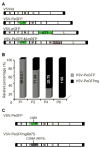
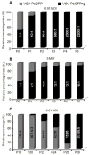
Using a recombinant vesicular stomatitis virus encoding eGFP fused in-frame with an essential viral replication protein, the phosphoprotein P, we show that during passage in culture, the virus mutates the nucleotide C289 within eGFP of the fusion protein PeGFP to A or T, resulting in R97S/C amino acid substitution and loss of fluorescence. The resultant non-fluorescent virus exhibits increased fitness and growth advantage over its fluorescent counterpart. The growth advantage of the non-fluorescent virus appears to be due to increased transcription and replication activities of the PeGFP protein carrying the R97S/C substitution. Further, our results show that the R97S/C mutation occurs prior to accumulation of mutations that can result in loss of expression of the gene inserted at the G-L gene junction. These results suggest that fitness gain is more important for the recombinant virus than elimination of expression of the heterologous gene.
Copyright © 2012 Elsevier Inc. All rights reserved.
Figures


Similar articles
-
Visualization of intracellular transport of vesicular stomatitis virus nucleocapsids in living cells.J Virol. 2006 Jul;80(13):6368-77. doi: 10.1128/JVI.00211-06. J Virol. 2006. PMID: 16775325 Free PMC article.
-
A recombinant vesicular stomatitis virus bearing a lethal mutation in the glycoprotein gene uncovers a second site suppressor that restores fusion.J Virol. 2011 Aug;85(16):8105-15. doi: 10.1128/JVI.00735-11. Epub 2011 Jun 15. J Virol. 2011. PMID: 21680501 Free PMC article.
-
A single amino acid change in the L-polymerase protein of vesicular stomatitis virus completely abolishes viral mRNA cap methylation.J Virol. 2005 Jun;79(12):7327-37. doi: 10.1128/JVI.79.12.7327-7337.2005. J Virol. 2005. PMID: 15919887 Free PMC article.
-
Attenuation of recombinant vesicular stomatitis viruses encoding mutant glycoproteins demonstrate a critical role for maintaining a high pH threshold for membrane fusion in viral fitness.Virology. 1998 Jan 20;240(2):349-58. doi: 10.1006/viro.1997.8921. Virology. 1998. PMID: 9454708
-
Conditional lethal mutants of vesicular stomatitis virus.Curr Top Microbiol Immunol. 1975;69:85-116. doi: 10.1007/978-3-642-50112-8_2. Curr Top Microbiol Immunol. 1975. PMID: 169102 Review. No abstract available.
Cited by
-
Experimental Evolution Generates Novel Oncolytic Vesicular Stomatitis Viruses with Improved Replication in Virus-Resistant Pancreatic Cancer Cells.J Virol. 2020 Jan 17;94(3):e01643-19. doi: 10.1128/JVI.01643-19. Print 2020 Jan 17. J Virol. 2020. PMID: 31694943 Free PMC article.
-
Development and application of reporter-expressing mononegaviruses: current challenges and perspectives.Antiviral Res. 2014 Mar;103:78-87. doi: 10.1016/j.antiviral.2014.01.003. Epub 2014 Jan 23. Antiviral Res. 2014. PMID: 24462694 Free PMC article. Review.
-
Oncolytic viruses: From bench to bedside with a focus on safety.Hum Vaccin Immunother. 2015;11(7):1573-84. doi: 10.1080/21645515.2015.1037058. Hum Vaccin Immunother. 2015. PMID: 25996182 Free PMC article. Review.
-
Applications of Replicating-Competent Reporter-Expressing Viruses in Diagnostic and Molecular Virology.Viruses. 2016 May 6;8(5):127. doi: 10.3390/v8050127. Viruses. 2016. PMID: 27164126 Free PMC article. Review.
-
Deep Sequencing Details the Cross-over Map of Chimeric Genes in Two Porcine Reproductive and Respiratory Syndrome Virus Infectious Clones.Open Virol J. 2017 Jun 30;11:49-58. doi: 10.2174/1874357901711010049. eCollection 2017. Open Virol J. 2017. PMID: 28839504 Free PMC article.
References
-
- Barber GN. VSV-tumor selective replication and protein translation. Oncogene. 2005;24:7710–7719. - PubMed
-
- Dalton KP, Rose JK. Vesicular stomatitis virus glycoprotein containing the entire green fluorescent protein on its cytoplasmic domain is incorporated efficiently into virus particles. Virology. 2001;279:414–421. - PubMed
Publication types
MeSH terms
Substances
Grants and funding
LinkOut - more resources
Full Text Sources

