. 2012 Sep 14;287(38):31948-61.
doi: 10.1074/jbc.M112.348896. Epub 2012 Jul 24.

The extracellular and transmembrane domains of the γC and interleukin (IL)-13 receptor α1 chains, not their cytoplasmic domains, dictate the nature of signaling responses to IL-4 and IL-13

Affiliations

The extracellular and transmembrane domains of the γC and interleukin (IL)-13 receptor α1 chains, not their cytoplasmic domains, dictate the nature of signaling responses to IL-4 and IL-13

Nicola M Heller et al. J Biol Chem. .

Abstract

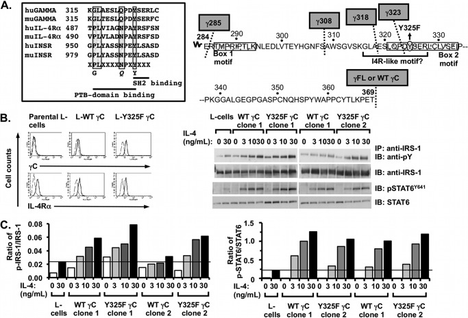
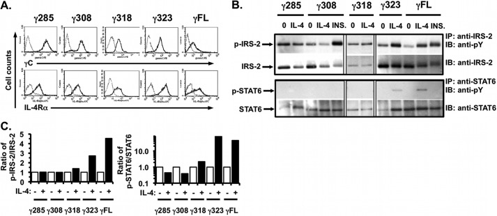
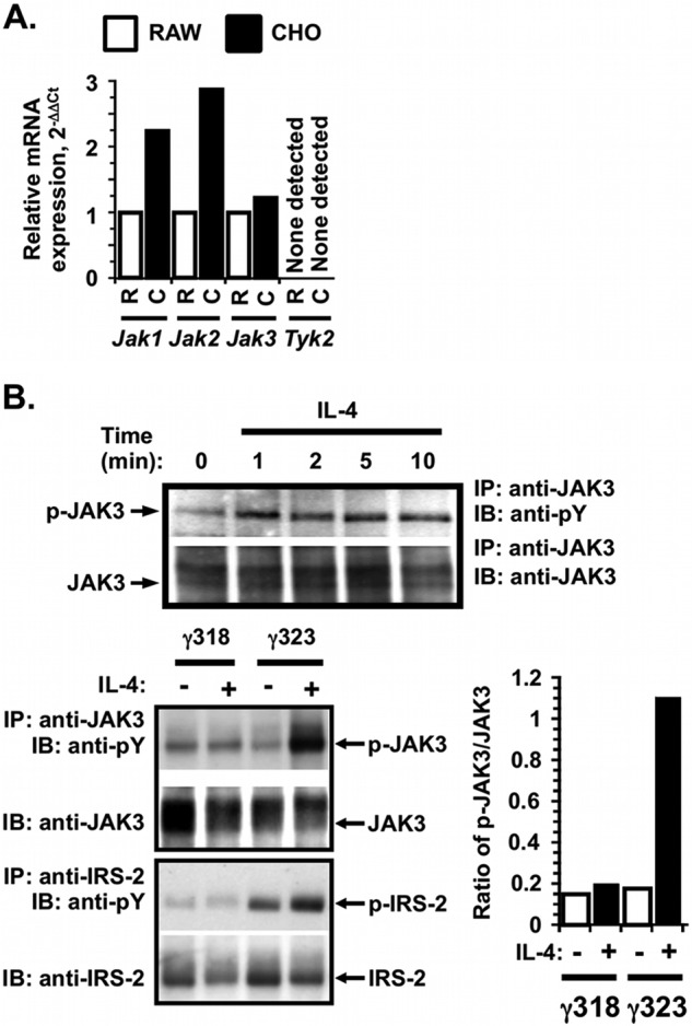
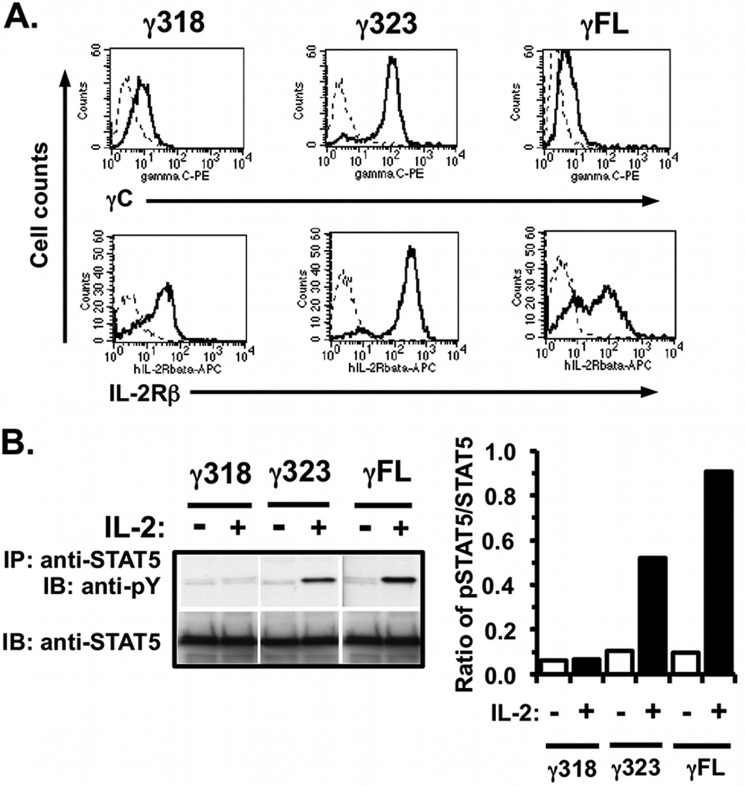
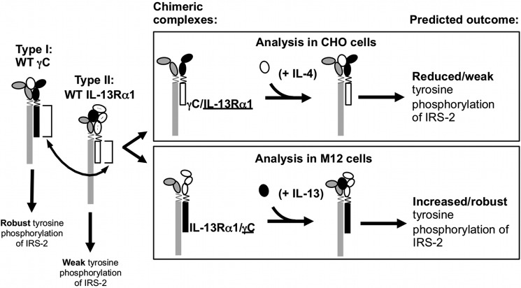
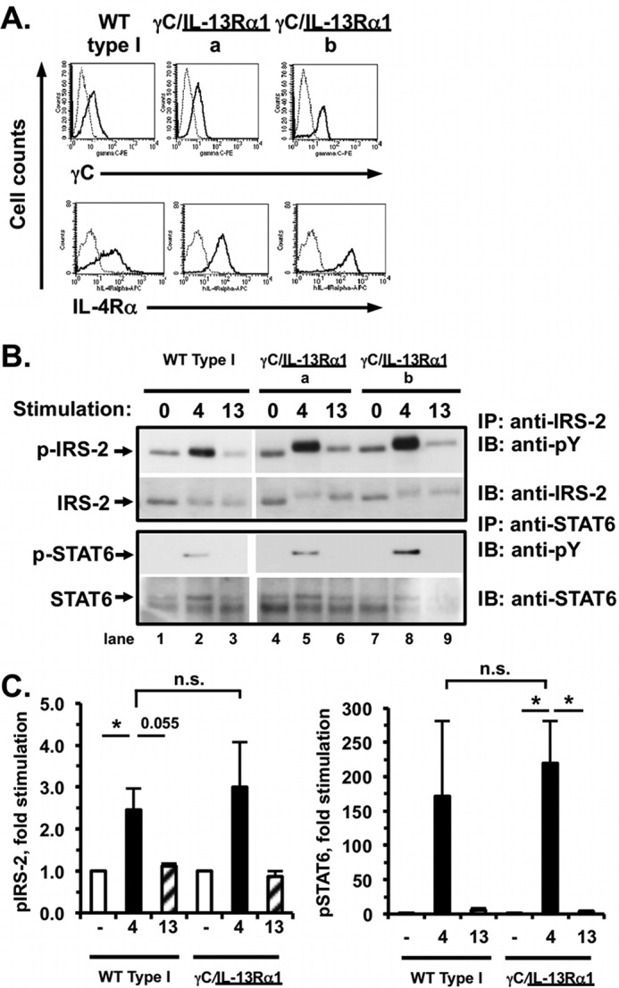
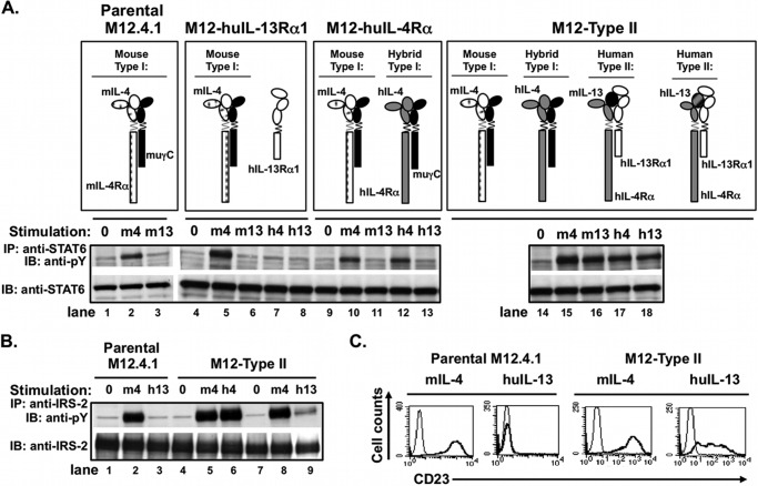
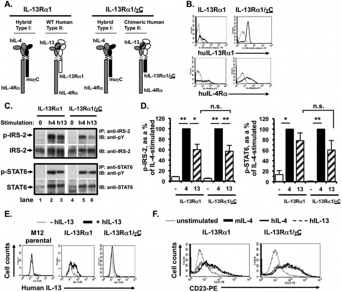
Previously, we demonstrated that the γC subunit of type I IL-4 receptor was required for robust tyrosine phosphorylation of the downstream adapter protein, IRS-2, correlating with the expression of genes (ArgI, Retnla, and Chi3l3) characteristic of alternatively activated macrophages. We located an I4R-like motif (IRS-2 docking sequence) in the γC cytoplasmic domain but not in the IL-13Rα1. Thus, we predicted that the γC tail directed enhanced IRS-2 phosphorylation. To test this, IL-4 signaling responses were examined in a mutant of the key I4R motif tyrosine residue (Y325F) and different γC truncation mutants (γ285, γ308, γ318, γ323, and γFULL LENGTH (FL)) co-expressed in L-cells or CHO cells with wild-type (WT) IL-4Rα. Surprisingly, IRS-1 phosphorylation was not diminished in Y325F L-cell mutants suggesting Tyr-325 was not required for the robust insulin receptor substrate response. IRS-2, STAT6, and JAK3 phosphorylation was observed in CHO cells expressing γ323 and γFL but not in γ318 and γ285 mutants. In addition, when CHO cells expressed γ318, γ323, or γFL with IL-2Rβ, IL-2 induced phospho-STAT5 only in the γ323 and γFL clones. Our data suggest that a smaller (5 amino acid) interval than previously determined is necessary for JAK3 activation/γC-mediated signaling in response to IL-4 and IL-2. Chimeric receptor chains of the γC tail fused to the IL-13Rα1 extracellular and transmembrane domain did not elicit robust IRS-2 phosphorylation in response to IL-13 suggesting that the extracellular/transmembrane domains of the IL-4/IL-13 receptor, not the cytoplasmic domains, control signaling efficiency. Understanding this pathway fully will lead to rational drug design for allergic disease.


Figures

FIGURE 1.
FIGURE 1.
Effect of mutation of Tyr-325 in the putative I4R motif in human γC on IL-4 signaling responses. A, left panel, sequences of human and mouse IL-4Rα and insulin receptor surrounding the I4R motif were aligned with human and mouse γC. The amino acid residues that interact with phosphotyrosine-binding domains and Src homology 2 domains are underlined. The amino acid residues known to be important for binding to IRS-2 are boxed in gray. A, right panel, amino acid sequence within the cytoplasmic domain of human γC is shown. The sites for truncation are indicated with a dotted line and gray boxes. Box 1 and box 2 sequences that bind JAK3 (stippled) and the putative I4R motif (underlined) with the I4R central tyrosine mutant (Y325F) are shown. B, left panel, parental L-cells and L-cell clones stably transfected with wild-type γC (L-WT γC) and the mutant Y325F γC construct (L-Y325F γC) were stained with specific antibodies to human γC and mouse IL-4Rα (heavy black lines) as indicated, and isotype-matched controls (dashed lines) conjugated to PE as described under “Experimental Procedures.” Cells were washed to remove unbound antibody and were analyzed by flow cytometry. B, right panel, WT- or Y325F γC-expressing L-cells were serum-starved for 24 h and then cultured in the presence or absence of increasing concentrations of mouse IL-4 for 30 min. Cell lysates were prepared and immunoprecipitated (IP) with antibodies recognizing IRS-1 or STAT6 as indicated followed by WB with anti-phosphotyrosine (PY) or anti-phospho-STAT6 (Y641). WB membranes were then stripped and re-probed for IRS-1 and STAT6 as appropriate. Representative films were from at least three independent experiments with two independent WT and Y325F γC-expressing L-cell clones per experiment. C, densitometric analysis of the WB films was performed as described under “Experimental Procedures.”
FIGURE 2.
FIGURE 2.
Signaling in response to IL-4 in the γC truncation mutants stably co-expressed with human IL-4Rα in CHO cells. A, FACS analysis for expression of human γC and human IL-4Rα expression in the stable CHO clones of each γC truncation mutant was performed as described (Fig. 1A). B, indicated γC truncation mutant clones were serum-starved for 24 h and then stimulated with human IL-4 (20 ng/ml) or insulin (INS, 20 μg/ml) for 15 min. Tyrosine phosphorylation of IRS-2 and STAT6 was analyzed as described (Fig. 1B). Phosphotyrosine blots were then stripped and re-probed for total IRS-2 and STAT6. Representative films from at least three independent experiments are shown. C, densitometric analysis of the WB films was performed as described under “Experimental Procedures.”
FIGURE 3.
FIGURE 3.
Expression and activation of JAK3 in response to IL-4 in CHO cells expressing γFL, γ323, and γ318. A, total RNA was extracted from CHO (C) cells, and the RAW (R) 264.7 macrophage cell line and cDNA were made. Real time PCR with specific primers for all four JAKs was performed, and the expression of each JAK was calculated using the standard 2−ΔΔCt method, relative to hypoxanthine phosphoribosyltransferase (HPRT). The amount of message found in CHO was expressed as fold change from the amount found in RAW 264.7 ( = 1). B, upper panel, CHO cells expressing human IL-4Rα and γFL were serum-starved prior to stimulation with 10 ng/ml IL-4 for various times. The tyrosine phosphorylation of JAK3 was analyzed by IP and WB as described under “Experimental Procedures.” B, lower left panel, comparison of JAK3 activation in the CHO cells expressing human IL-4Rα and γ318 or γ323. Cells were stimulated as in A for 5 min for JAK3 activation and 15 min for IRS-2 activation. Tyrosine phosphorylation of JAK3 and IRS-2 was analyzed by IP and WB as described in A. Phosphotyrosine blots were then stripped and re-probed for total JAK3 or IRS-2 as appropriate. Representative films from at least three independent experiments are shown. B, lower right panel, densitometric analysis of the JAK3 WB films was performed as described under “Experimental Procedures.”
FIGURE 4.
FIGURE 4.
Signaling in response to IL-2 in the γ318, γ323, truncation mutants and WT γC stably co-expressed with human IL-2Rβ in CHO cells. A, CHO cells were transfected with cDNA encoding human IL-2Rβ and various constructs of human γC (γC FL, γ318, or γ323); stable clones were selected. These cells were evaluated for expression of human γC and IL-2Rβ by FACS analysis as described (Fig. 1A). B, CHO cells expressing human IL-2Rβ and γC truncations were serum-starved for 24 h and then stimulated with human IL-2 (28 ng/ml) for 30 min. Cell lysates were prepared and immunoprecipitated with anti-STAT5 followed by WB with anti-phosphotyrosine (PY). WB membranes were then stripped and re-probed with anti-STAT5. Densitometric analysis of the WB films was performed as described under “Experimental Procedures.” These results are representative of two independent experiments.
FIGURE 5.
FIGURE 5.
Chimeric receptor chains γC/IL-13Rα1 and IL-13Rα1/γC and expected signaling responses in cells co-expressing human IL-4Rα and either of chimeric receptor chains. The cytoplasmic domains of the γC chain (black) or the IL-13Rα1 chain (white) were swapped (arrow) by a two-step overlapping PCR fusion approach as described under “Experimental Procedures.” The cytokine-binding homology regions of the extracellular domains are indicated by the ovals and the transmembrane domain is shown by a zig-zag line. Two chimeric receptor chains were created, γC/IL-13Rα1 and IL-13Rα1/γC (the underline denotes the cytoplasmic domain). The signaling responses of wild-type receptors are shown below the receptor diagram, as well as the predicted signaling responses of receptor complexes containing the respective chimeric receptor chains (boxed).
FIGURE 6.
FIGURE 6.
Signaling responses of CHO cells expressing human IL-4Rα and either γC (WT type I CHO) or chimeric receptor γC/IL-13Rα1 to IL-4 and IL-13. A, CHO cells were transfected with human IL-4Rα and either WT γC or γC/IL-13Rα1, and stable clones were selected. Expression of human γC and IL-4Rα on the surface of the indicated clones was evaluated by FACS as described (Fig. 1A). B, stable CHO cell clones expressing human IL-4Rα and either WT γC (WT Type I) or γC/IL-13Rα1 were serum-starved for 4 h and then either stimulated with human IL-4 (4) or human IL-13 (13, 5 ng/ml) for 15 min or were not stimulated with cytokine (0). Tyrosine phosphorylation of IRS-2 and STAT6 was analyzed as described (Fig. 1B). Phosphotyrosine blots were then stripped and re-probed for total IRS-2 and STAT6. Representative films from four independent experiments with one wild-type clone and two different chimeric clones are shown. C, densitometric analysis of the WB films was performed as described under “Experimental Procedures.” The amount of phosphoprotein is graphed as the fold induced above unstimulated cells. n = 5 (WT type I) and n = 7 (chimeric receptor), *, p < 0.05; n.s., not significant.
FIGURE 7.
FIGURE 7.
Signaling responses of M12 cells expressing either human IL-4Rα, human IL-13Rα1, or both human IL-4Rα and IL-13Rα1 (type II M12) to IL-4 and IL-13. The murine B-cell lymphoma cell line M12.4.1 was transfected with human (hu) IL-13Rα1, huIL-4Rα, or both as indicated, and stable clones were selected. A, activated receptor complexes present in these transfectants following stimulation with mouse or human IL-4 and IL-13 are shown. These cells were deprived of serum for 2 h before treating with murine IL-4 (m4), murine IL-13 (m13), human IL-4 (h4), or human IL-13 (h13) (20 ng/ml) for 30 min. Cell lysates were prepared and precipitated with anti-STAT6 (A) or anti-IRS-2 (B) followed by Western blotting with anti-phosphotyrosine (pY). The blots were stripped and reprobed with anti-STAT6 or anti-IRS-2 as appropriate. C, parental M12 cells and M12 cells stably transfected with both human IL-13Rα1 and human IL-4Rα (M12-type II) were cultured in the presence (thick black line) or absence (thin line) of the indicated cytokine (20 ng/ml) for 24 h, and CD23 expression was evaluated by FACS.
FIGURE 8.
FIGURE 8.
Signaling responses of M12 cells expressing human IL-4Rα and either IL-13Rα1 (type II M12) or chimeric receptor IL-13Rα1/γC to IL-4 and IL-13. A, M12.4.1 cells were transfected with human (hu) IL-4Rα and either huIL-13Rα1 or the huIL-13Rα1/γC chimera. Activated receptor complexes formed following stimulation with human IL-4 and IL-13 are shown. B, stable clones were evaluated for expression of huIL-13Rα1 and huIL-4Rα by FACS. C, indicated cells were serum-starved for 2 h and then stimulated with human IL-4 (h4) or IL-13 (h13) (5 ng/ml) for 15 min. Tyrosine phosphorylation of IRS-2 and STAT6 was analyzed as described above. Phosphotyrosine blots were then stripped and re-probed for total IRS-2 and STAT6. Representative films from two independent experiments using two independent WT and three chimeric type II clones. D, densitometric analysis of the WB films was performed as described under “Experimental Procedures.” The amount of phosphoprotein is expressed as a percentage of the IL-4-stimulated amount. n = 5 (WT type II) and n = 7 (chimeric receptor); **, p < 0.0001; *, p < 0.05. E, human IL-13 binding by the parental M12, M12-type II, and IL-13Rα1/γC-expressing chimeric receptor-expressing clones and cells. Cells were incubated in the presence or absence of human IL-13 on ice for 30 min in buffer containing 0.1% azide. After washing away unbound cytokine, bound IL-13 was measured by incubating the cells with anti-human IL-13-biotin, followed by streptavidin-PE and performing FACS analysis as described under “Experimental Procedures.” A representative set of FACS histograms is shown from two independent experiments performed with two IL-13Rα1/γC-expressing chimeric receptor-expressing clones. F, CD23 induction on the type II M12 cells and chimeric receptor IL-13Rα1/γC-expressing clones. Cells were cultured in the presence or absence (dotted line) of murine IL-4 (thick black line), human IL-4 (thin line), or human IL-13 (dashed line) (5 ng/ml) for 24 h. CD23 expression was determined by FACS analysis as described under “Experimental Procedures.”

Similar articles

Cited by

References

    1. Barnes P. J. (2011) Pathophysiology of allergic inflammation. Immunol. Rev. 242, 31–50 - PubMed
    1. Paul W. E., Zhu J. (2010) How are T(H)2-type immune responses initiated and amplified? Nat. Rev. Immunol. 10, 225–235 - PMC - PubMed
    1. Al-Muhsen S., Johnson J. R., Hamid Q. (2011) Remodeling in asthma. J. Allergy Clin. Immunol. 128, 451–462 - PubMed
    1. Chatila T. A. (2004) Interleukin-4 receptor signaling pathways in asthma pathogenesis. Trends Mol. Med. 10, 493–499 - PubMed
    1. Lowenthal J. W., Castle B. E., Christiansen J., Schreurs J., Rennick D., Arai N., Hoy P., Takebe Y., Howard M. (1988) Expression of high affinity receptors for murine interleukin 4 (BSF-1) on hemopoietic and nonhemopoietic cells. J. Immunol. 140, 456–464 - PubMed

MeSH terms