. 2012 Oct;22(10):1479-501.
doi: 10.1038/cr.2012.110. Epub 2012 Jul 24.

The R-Ras/RIN2/Rab5 complex controls endothelial cell adhesion and morphogenesis via active integrin endocytosis and Rac signaling

Affiliations

The R-Ras/RIN2/Rab5 complex controls endothelial cell adhesion and morphogenesis via active integrin endocytosis and Rac signaling

Chiara Sandri et al. Cell Res. 2012 Oct.

Abstract

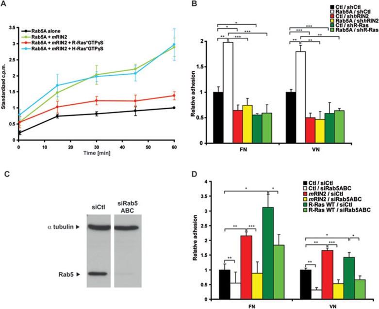
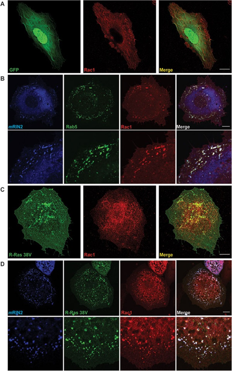
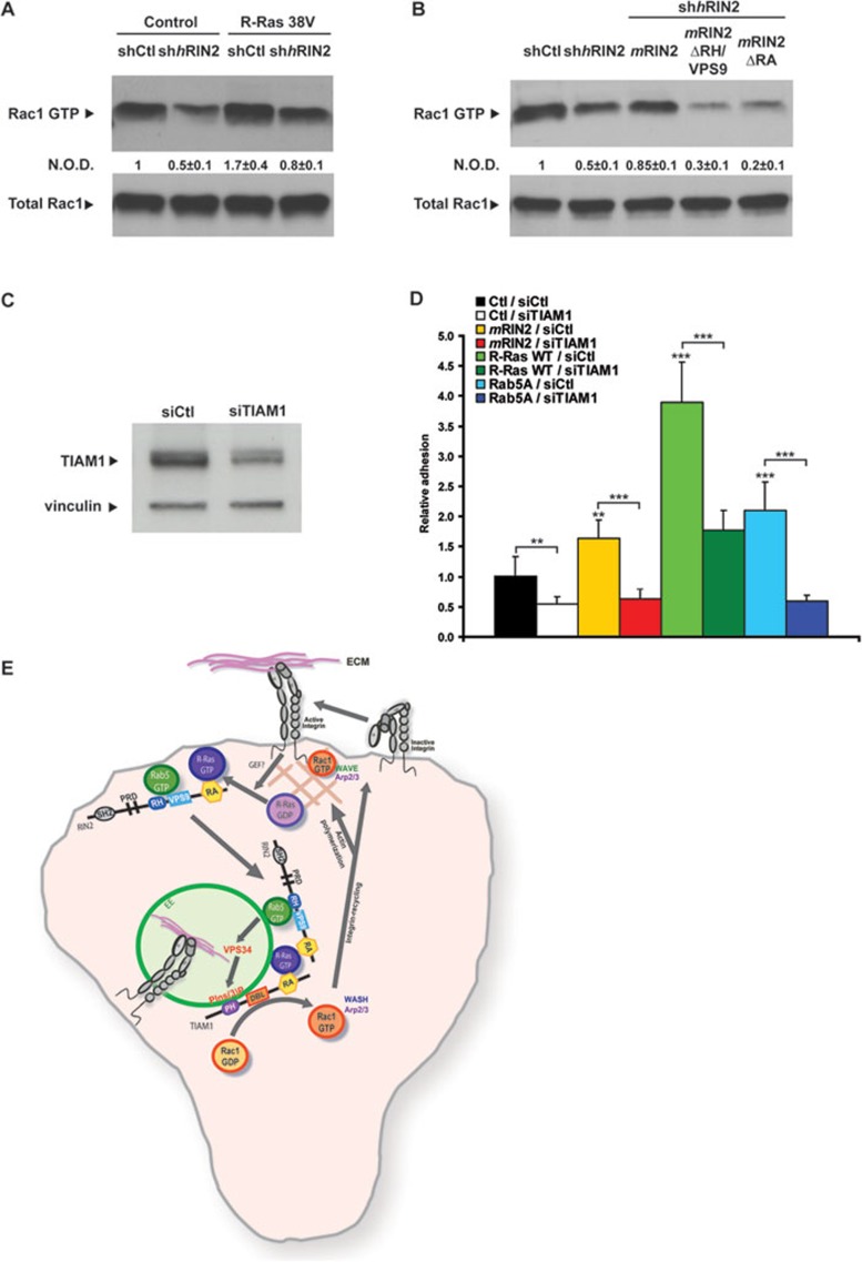
During developmental and tumor angiogenesis, semaphorins regulate blood vessel navigation by signaling through plexin receptors that inhibit the R-Ras subfamily of small GTPases. R-Ras is mainly expressed in vascular cells, where it induces adhesion to the extracellular matrix (ECM) through unknown mechanisms. We identify the Ras and Rab5 interacting protein RIN2 as a key effector that in endothelial cells interacts with and mediates the pro-adhesive and -angiogenic activity of R-Ras. Both R-Ras-GTP and RIN2 localize at nascent ECM adhesion sites associated with lamellipodia. Upon binding, GTP-loaded R-Ras converts RIN2 from a Rab5 guanine nucleotide exchange factor (GEF) to an adaptor that first interacts at high affinity with Rab5-GTP to promote the selective endocytosis of ligand-bound/active β1 integrins and then causes the translocation of R-Ras to early endosomes. Here, the R-Ras/RIN2/Rab5 signaling module activates Rac1-dependent cell adhesion via TIAM1, a Rac GEF that localizes on early endosomes and is stimulated by the interaction with both Ras proteins and the vesicular lipid phosphatidylinositol 3-monophosphate. In conclusion, the ability of R-Ras-GTP to convert RIN2 from a GEF to an adaptor that preferentially binds Rab5-GTP allows the triggering of the endocytosis of ECM-bound/active β1 integrins and the ensuing funneling of R-Ras-GTP toward early endosomes to elicit the pro-adhesive and TIAM1-mediated activation of Rac1.


Figures

Figure 1
Figure 1
R-Ras and RIN2 promote EC-to-ECM binding, adhesion, migration, and vascular morphogenesis; RIN2 mediates the pro-adhesive effects of R-Ras. (A) Representative western blot analysis of R-Ras and β-tubulin protein expression in control (shCtl) and R-Ras-silenced (shhR-Ras) human ECs. (B) Comparison between shCtl and shhR-Ras ECs adhering to FN and VN. A representative of three independent experiments with similar results is shown (mean ± SD; n = 3 samples per experimental condition). (C) Western blot analysis of RIN2 expression in human ECs silenced for RIN2 (sihRIN2) or transfected with control siRNA (siCtl). β-tubulin was used as an internal and loading control. (D) Comparison between siCtl- and sihRIN2-transfected human ECs adhering to FN and VN. A representative of eight (FN) and five (VN) independent experiments with similar results is shown (mean ± SD; n = 3 samples per experimental condition). (E) Comparative analysis of stimulation of cell adhesion to FN and VN by WT and constitutively active R-Ras 38V in either control (shCtl) or RIN2-silenced (shhRIN2) human ECs. Ctl corresponds to ECs transduced with the empty PINCO retroviral vector. A representative of three independent experiments with similar results is shown (mean ± SD; n = 3 samples per experimental condition). (F) FACS analysis of rhodamine-FN binding to siCtl, sihR-Ras, and sihRIN2 ECs. A representative of three independent experiments with similar results is shown (mean ± SD; n = 4 samples per experimental condition). (G, H) Comparison between siCtl and sihR-Ras (G) or sihRIN2 (H) EC migration rate (μm/h) in wound healing assays. A representative of three independent experiments with similar results is shown (mean ± SD; n = 3 samples per experimental condition). (I-L) Comparison of the number of branching points/mm2 of vascular networks formed by siCtl and sihR-Ras (I) or sihRIN2 (L) ECs plated on growth factor-reduced Matrigel matrix. A representative of three independent experiments with similar results is shown (mean ± SD; n = 3 samples per experimental condition). (M) Representative pictures of vascular networks formed by siCtl, sihR-Ras, sihRIN2 ECs plated on growth factor-reduced Matrigel matrix. Scale bars, 500 μm. *P < 0.05, **P < 0.01, ***P < 0.001.
Figure 2
Figure 2
RIN2 colocalizes with active R-Ras in NAs, FCs and Rab5-positive early endosomes. (A) Fluorescent confocal microscopy analysis of ECs transfected with mRIN2 (left panels, red) and GFP-R-Ras 38V (central panels, green) indicates that mRIN2 and GFP-R-Ras 38V colocalize in peripheral NAs (arrowheads), and vesicles (dashed arrows) as shown in merge (right panels). Lower panels are magnifications of the corresponding upper panels. (B) Fluorescent confocal microcopy reveals that in ECs transfected with mRIN2 (left panels, red) and GFP-vinculin (central panels, green), mRIN2 colocalizes (right panels) in full with peripheral NAs (external arrowheads), in part with the external portion of FCs maturing into FAs (internal arrowheads), but not with elongated FAs (arrows). Lower panels are magnifications of the corresponding upper panels. (C) Fluorescent confocal analysis of ECs transfected with mRIN2 (left panels, red) and GFP-Rab5A (central panels, green) unveils that mRIN2 and Rab5A colocalize in early endosomes as shown in merge (right panels). A minor amount of GFP-Rab5A is present at the cell periphery as well, where it colocalizes with HA-RIN2 in NAs. Panels are representative of more than 25 cells in four independent experiments. Scale bars, 10 μm.
Figure 3
Figure 3
R-Ras controls active β1 integrin endocytosis via RIN2 in ECs. (A-D) Comparison of the amounts of total (A, C) or active (B, D) β1 integrins on the plasma membrane of siCtl versus sihR-Ras (A, B) or sihRIN2 (C, D), as evaluated by surface biotinylation and capture ELISA assays. Values are mean ± SD from more than three independent experiments (n = 6; P ≥ 0.2 and differences are not statistically significant). (E-H) Time-course analysis of the amounts of total (E, G) or active (F, H) β1 integrins endocytosed in siCtl versus sihR-Ras (E, F) or sihRIN2 (G, H), as evaluated by integrin internalization assay and capture ELISA assay. Values are mean ± SD from more three independent experiments (n = 6). In E and G, P ≥ 0.3 and differences are not statistically significant. (I) Time-course analysis of the amounts of active β1 integrins endocytosed in shCtl versus shhRIN2 ECs overexpressing or not R-Ras WT or 38V, as evaluated by integrin internalization assay and capture ELISA assay. Values are mean ± SD from more than three independent experiments (n = 6). *P < 0.05, **P < 0.01, ***P < 0.001.
Figure 4
Figure 4
Stimulation of EC adhesion to ECM proteins by RIN2 depends on its Rab5- and Ras-binding domains and endogenous RIN2 associates with both R-Ras and Rab5. (A) Schematic representation of full-length mRIN2 together with its Rab5- and Ras-binding defective deletion mutants mRIN2 ΔRH/VPS9 and mRIN2 ΔRA. (B) Western blot analysis of exogenously transduced and silencing-resistant HA-tagged mRIN2 constructs and endogenous β-tubulin expression in ECs first infected with PINCO retrovirus carrying or not (Ctl) mRIN2 constructs and then with a shhRIN2 lentivirus. (C) Comparison of WT full-length mRIN2, mRIN2 ΔRH/VPS9, and mRIN2 ΔRA efficiency in rescuing the defective adhesion of shhRIN2 ECs to FN and VN. A representative of three independent experiments with similar results is shown (mean ± SD; n = 3 samples per experimental condition). (D) Comparative analysis of the modulation of cell adhesion to FN and VN by overexpression of either full-length mRIN2 or mRIN2 ΔRH/VPS9 and mRIN2 ΔRA deletion mutants in human ECs. Ctl corresponds to ECs transduced with the empty PINCO retroviral vector. A representative of three independent experiments with similar results is shown (mean ± SD; n = 3 samples per experimental condition). (E) Immunoprecipitation (IP) of endogenous hRIN2 followed by western blotting with anti-R-Ras, anti-Rab5, or anti-RIN2 antibodies. In ECs, RIN2 associates with both R-Ras and Rab5. An EC lysate was employed as positive control or immunoprecipitated with irrelevant IgG for negative control purposes. **P < 0.01, ***P < 0.001.
Figure 5
Figure 5
RIN2 promotes the recruitment of R-Ras to Rab5-positive early endosomes. Fluorescent confocal microscopy analysis of ECs transfected with GFP-Rab5A (central left panels, green), mCherry-R-Ras 38V (central right panels, red) and HA-mRIN2 (A) or HA-mRIN2 RH/VPS9 (B) or HA-mRIN2 RA (C) (left panels, blue). Right panels correspond to merged image. (A) HA-mRIN2, GFP-Rab5A, and mCherry-R-Ras 38V colocalize on early endosomes. (B) HA-mRIN2 RH/VPS9 and mCherry-R-Ras 38V colocalize in the perinuclear vesicular compartment, but they are excluded from the peripheral Rab5-labeled early endosomes. (C) HA-mRIN2 RA colocalizes with GFP-Rab5A in peripheral early endosomes, whereas mCherry-R-Ras 38V is excluded from these compartments. Lower panels are magnifications of the corresponding upper panels. (D) Quantification of colocalization signal between: (1) mRIN2 constructs and GFP-Rab5; (2) mRIN2 constructs and mCherry-R-Ras 38V; (3) mCherry-R-Ras 38V and GFP-Rab5 in ECs simultaneously transfected with different mRIN2 constructs (mean ± SD; n = 25 cells per experimental condition). Scale bars, 10 μm. ***P < 0.001.
Figure 6
Figure 6
R-Ras, RIN2, and Rab5 constitute a new signaling module that controls the adhesion of ECs to the ECM. (A) The GEF activity of mRIN2 on Rab5A is measured by a nucleotide exchange assay from unlabeled prebound GDP to [3H]-GDP on GST-Rab5A. The rate of [3H]-GDP association is presented as a correlation of measured counts per minute at each time point for each sample divided by the counts per minute after 60 min for the control sample Rab5A alone (“standardized c.p.m.”). The GEF activity of RIN2 on Rab5A is inhibited by the presence of R-Ras*GTPγS, but not H-Ras*GTPγS. Error bars correspond to the standard error of the mean. (B) Comparative analysis of the modulation of cell adhesion to FN and VN in control (shCtl) or hRIN2 (shhRIN2)- or R-Ras (shR-Ras)-silenced human ECs overexpressing Rab5A or empty PINCO retroviral vector (Ctl). Error bars correspond to standard deviation. A representative of three independent experiments with similar results is shown (mean ± SD; n = 3 samples per experimental condition). (C) Western blot analysis of Rab5 expression in human ECs silenced for Rab5A, B, C (sihRab5ABC) or transfected with control siRNA (siCtl). β-tubulin was used as an internal and loading control. (D) Comparative analysis of the modulation of cell adhesion to FN and VN by overexpression of mRIN2 or R-Ras WT in control (siCtl) or hRab5ABC (sihRab5ABC)-silenced human ECs. Ctl corresponds to ECs transfected with the empty PINCO retroviral vector. A representative of three independent experiments with similar results is shown (mean ± SD; n = 3 samples per experimental condition). *P < 0.05, **P < 0.01, ***P < 0.001.
Figure 7
Figure 7
RIN2, R-Ras and Rac1 colocalize on Rab5-positive early endosomes. (A) Fluorescent confocal microscopy analysis of ECs transfected with GFP (left panel, green) and stained for endogenous Rac1 (central panel, red) shows how Rac1 is quite homogeneously distributed throughout the cell. Right panel corresponds to merged image. (B) Fluorescent confocal microscopy analysis of ECs transfected with HA-mRIN2 (left panels, blue) and GFP-Rab5A (central left panels, green) and stained for Rac1 (central right panels, red) shows that HA-mRIN2 and Rac1 colocalize in vesicular compartment marked by GFP-Rab5A. Right panels correspond to merged images. Lower panels are magnifications of the corresponding upper panels. (C) Fluorescent confocal microscopy analysis of ECs transfected with GFP-R-Ras 38V (left panel, green) and stained for endogenous Rac1 (central panel, red) shows a partial colocalization of GFP-R-Ras 38V and Rac1 in bona fide vesicular structures. Right panel corresponds to merged image. (D) Fluorescent confocal microscopy analysis of ECs transfected with HA-mRIN2 (left panels, blue) and GFP-R-Ras 38V (central left panels, green) and stained for Rac1 (central right panels, red) shows the colocalization of HA-mRIN2, GFP-R-Ras 38V and Rac1 in vesicular compartments. Right panels correspond to merged images. Lower panels are magnifications of the corresponding upper panels. Panels are representative of more than 25 cells in three independent experiments. Scale bars, 10 μm.
Figure 8
Figure 8
The R-Ras/RIN2/Rab5 module controls Rac1 activation and EC adhesion via TIAM1. (A) Pull-down assay of active Rac1 in ECs overexpressing constitutively active R-Ras 38V or the control empty PINCO retroviral vector and silenced for hRIN2 (shhRIN2) or infected with control shRNA (shCtl). Total Rac1, detected in the input fractions, is used to calculate the normalized optical density (N.O.D.) of active Rac1. A representative of three independent pull-down assays with similar results is shown. Bands were quantified and N.O.D.s were calculated relative to control (Values are means ± SD; n = 3 separate assays). (B) The ability of mRIN2 or mRIN2 RH/VPS9 or mRIN2 RA to rescue the Rac1 activation defect in control (shCtl) or RIN2-silenced (shhRIN2) ECs was tested by pull-down assay. Total Rac1, detected in the input fractions, is used to calculate the N.O.D. of active Rac1. A representative of three independent pull-down assays with similar results is shown. Bands were quantified and N.O.D.s were calculated relative to control (Values are means ± SD; n = 3 separate assays). (C) Western blot analysis of TIAM1 expression in HeLa silenced for TIAM1 (siTIAM1) or transfected with control siRNA (siCtl). Vinculin was used as an internal and loading control. (D) Comparative analysis of the modulation of cell adhesion to FN by overexpression of mRIN2 or R-Ras WT or Rab5A in control (siCtl) or TIAM1 (siTIAM1)-silenced HeLa. Ctl corresponds to HeLa transfected with the empty PINCO retroviral vector. A representative of two independent experiments with similar results is shown (mean ± SD; n = 6-9 samples per experimental condition). (E) Model of how the R-Ras/RIN2/Rab5 complex controls active integrin endocytosis and signals the TIAM1-dependent activation of Rac1 and EC adhesion to the ECM. At the leading edge of motile ECs, ECM-bound/active integrins trigger GEF-mediated GTP loading of R-Ras. At NAs, active R-Ras interacts with the RA domain of RIN2, inhibits its Rab5 GEF activity, and transforms RIN2 into an adaptor that binds GTP-Rab5 via its RH/VPS9 domain, thus localizing this Rab GTPase at the plasma membrane. Here, the R-Ras/RIN2/Rab5 complex drives the endocytosis of ECM-bound/active integrins and the ensuing translocation of R-Ras and RIN2 proteins to Rab5-positive early endosomes (EE). Also TIAM1, a protein that presents a Ras-binding domain, is recruited to Rab5-positive early endosomes; indeed, TIAM1 possesses a PH domain that binds PIns(3)P, which is highly enriched at the endosomal surface and positively regulates TIAM1 GEF activity,. Upon its double binding to PIns(3)P and R-Ras, TIAM1 gets completely activated and acts as a GEF for Rac1. Active Rac1 can promote, in turn, integrin-dependent cell adhesion to the ECM through several mechanisms. Indeed, the Arf6-dependent delivery of Rac1-GTP to the plasma membrane can drive WAVE-Arp2/3-dependent actin polymerization that mediates the formation of new NAs. Furthermore, Rac1-dependent endosomal activation of the WASH-Arp2/3 complex can support the recycling of internalized integrins to the plasma membrane, a process that is central to directional cell migration. **P < 0.01, ***P < 0.001.

Comment in

Similar articles

Cited by

References

    1. Fraisl P, Mazzone M, Schmidt T, Carmeliet P. Regulation of angiogenesis by oxygen and metabolism. Dev Cell. 2009;16:167–179. - PubMed
    1. Jones EA, le Noble F, Eichmann A. What determines blood vessel structure? Genetic prespecification vs. hemodynamics. Physiology (Bethesda) 2006;21:388–395. - PubMed
    1. Adams RH, Alitalo K. Molecular regulation of angiogenesis and lymphangiogenesis. Nat Rev Mol Cell Biol. 2007;8:464–478. - PubMed
    1. Jain RK. Lessons from multidisciplinary translational trials on anti-angiogenic therapy of cancer. Nat Rev Cancer. 2008;8:309–316. - PubMed
    1. Jain RK. Normalization of tumor vasculature: an emerging concept in antiangiogenic therapy. Science. 2005;307:58–62. - PubMed

Publication types

MeSH terms