

SLAT/Def6 plays a critical role in the pathogenic process of experimental autoimmune uveitis (EAU)
- PMID: 22815639
- PMCID: PMC3398495
SLAT/Def6 plays a critical role in the pathogenic process of experimental autoimmune uveitis (EAU)
Abstract
Purpose: SWAP 70-like adaptor of T cells (SLAT; aka Def6) is a recently discovered guanine nucleotide exchange factor for Rho guanosine triphosphate (GTP)ases that has been previously shown to play a role in cluster of differentiation(CD)4+ T cell activation, T-helper (Th)1/Th2/Th17 differentiation and development of experimental autoimmune encephalomyelitis. Here, we investigated the role of SLAT/Def6 in the development of experimental autoimmune uveitis (EAU), an animal model for several uveitic conditions in humans.
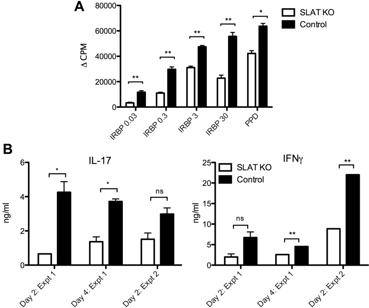
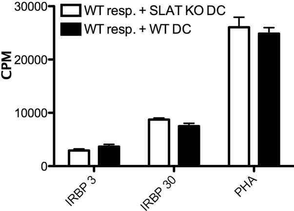
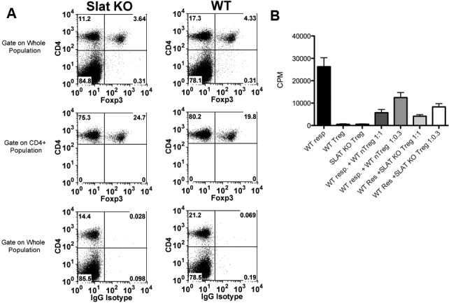
Methods: SLAT/Def6 deficient ("KO") mice and C57BL/6 controls were immunized with interphotoreceptor retinoid-binding protein (IRBP), along with pertussis toxin. The development of ocular inflammation was determined by both fundoscopy and histological examination. Lymphoid cells from draining lymph nodes were cultured with IRBP to measure lymphocyte proliferation and release of cytokines. Purified dendritic cells were tested for their capacity to present antigen to responding lymphocytes. In addition, the lymphoid cells were tested for the expression of forkhead box P3 (FoxP3), using conventional methods, and the activity of T-regulatory cells was determined by their capacity to inhibit in vitro proliferative responses. Serum anti -IRBP antibody levels were measured by enzyme-linked immunosorbant assay (ELISA). quantitative polymerase chain reaction (qPCR) was used to determine the transcript levels of cytokines in inflamed eyes.
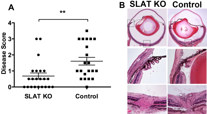
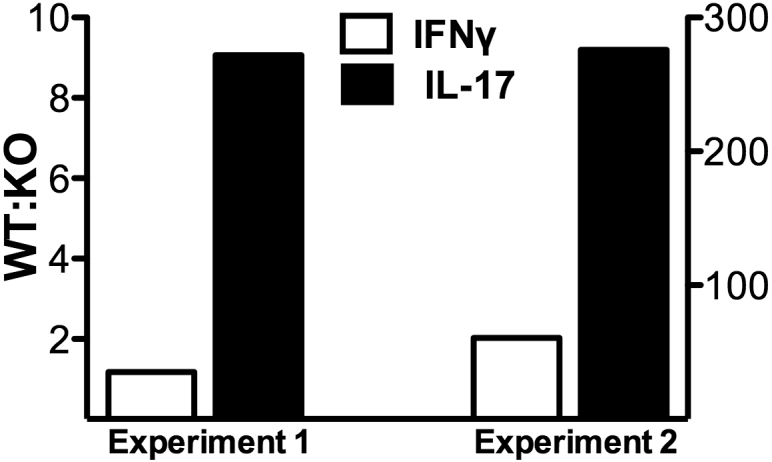
Results: SLAT/Def6 KO mice had significantly reduced EAU compared to controls. Cells isolated from draining lymph nodes of SLAT/Def6 KO mice exhibited impaired proliferation and production of Th1 and Th17 signature cytokines (interferon [IFN]-γ and interleukin [IL]-17, respectively) when compared with cells isolated from control mice. qPCR of inflamed eyes detected similar levels of IFN-γ transcript in control and SLAT/Def6 KO mice, whereas the IL-17 transcript levels in eyes of the SLAT/Def6 KO mice were lower than in eyes of the controls. The SLAT/Def6 KO mice resembled their wild type (WT) controls, however, in the levels of their serum antibody against IRBP, the antigen presenting capacity of their dendritic cells, the proportion of cells expressing Foxp3 and the immunosuppressive activity of their T-regulatory cells.
Conclusions: SLAT/Def6 KO mice exhibit reduced capacity to develop ocular inflammation and cellular activity when immunized with IRBP. Our study provides new data showing that SLAT/Def6 plays a major role in the T cell-mediated autoimmune processes that bring about the inflammatory eye disease, EAU.
Figures

Similar articles
-
Blockade of interleukin-6 signaling suppresses not only th17 but also interphotoreceptor retinoid binding protein-specific Th1 by promoting regulatory T cells in experimental autoimmune uveoretinitis.Invest Ophthalmol Vis Sci. 2011 May 17;52(6):3264-71. doi: 10.1167/iovs.10-6272. Invest Ophthalmol Vis Sci. 2011. PMID: 21330657
-
Differentiation of Th1 and Th2 cells in lymph nodes and spleens of mice during experimental autoimmune uveoretinitis.Jpn J Ophthalmol. 2001 Sep-Oct;45(5):463-9. doi: 10.1016/s0021-5155(01)00369-0. Jpn J Ophthalmol. 2001. PMID: 11583666
-
[Effects of exogenous interleukin-2, interleukin-23 on differentiation of IRBP 1-20-specific T cells toward Th1 and Th17 and comparison of the pathogenicity of IRBP 1-20-specific T cells].Zhonghua Yan Ke Za Zhi. 2013 Mar;49(3):224-9. Zhonghua Yan Ke Za Zhi. 2013. PMID: 23866703 Chinese.
-
Autoimmunity in the immune privileged eye: pathogenic and regulatory T cells.Immunol Res. 2008;42(1-3):41-50. doi: 10.1007/s12026-008-8031-3. Immunol Res. 2008. PMID: 18629448 Free PMC article. Review.
-
SWAP-70-like adapter of T cells: a novel Lck-regulated guanine nucleotide exchange factor coordinating actin cytoskeleton reorganization and Ca2+ signaling in T cells.Immunol Rev. 2009 Nov;232(1):319-33. doi: 10.1111/j.1600-065X.2009.00839.x. Immunol Rev. 2009. PMID: 19909373 Free PMC article. Review.
Cited by
-
Leucine-Rich Repeat Kinase 2 (Lrrk2) Deficiency Diminishes the Development of Experimental Autoimmune Uveitis (EAU) and the Adaptive Immune Response.PLoS One. 2015 Jun 11;10(6):e0128906. doi: 10.1371/journal.pone.0128906. eCollection 2015. PLoS One. 2015. PMID: 26067490 Free PMC article.
-
IRF4-Dependent and IRF4-Independent Pathways Contribute to DC Dysfunction in Lupus.PLoS One. 2015 Nov 6;10(11):e0141927. doi: 10.1371/journal.pone.0141927. eCollection 2015. PLoS One. 2015. PMID: 26544714 Free PMC article.
-
Human DEF6 deficiency underlies an immunodeficiency syndrome with systemic autoimmunity and aberrant CTLA-4 homeostasis.Nat Commun. 2019 Jul 15;10(1):3106. doi: 10.1038/s41467-019-10812-x. Nat Commun. 2019. PMID: 31308374 Free PMC article.
-
Regulated Tristetraprolin Overexpression Dampens the Development and Pathogenesis of Experimental Autoimmune Uveitis.Front Immunol. 2021 Jan 25;11:583510. doi: 10.3389/fimmu.2020.583510. eCollection 2020. Front Immunol. 2021. PMID: 33569048 Free PMC article.
References
-
- Pearce G, Angeli V, Randolph GJ, Junt T, von Andrian U, Schnittler H-J, Jessberger R. Signaling protein SWAP-70 is required for efficient B cell homing to lymphoid organs. Nat Immunol. 2006;7:827–34. - PubMed
-
- Tanaka Y, Bi K, Kitamura R, Hong S, Altman Y, Matsumoto A, Tabata H, Lebedeva S, Bushway PJ, Altman A. SWAP-70-like adapter of T cells, an adapter protein that regulates early TCR-initiated signaling in Th2 lineage cells. Immunity. 2003;18:403–14. - PubMed
Publication types
MeSH terms
Substances
Grants and funding
LinkOut - more resources
Full Text Sources
Medical
Molecular Biology Databases
Research Materials