. 2012 Sep;23(17):3348-56.
doi: 10.1091/mbc.E11-12-1020. Epub 2012 Jul 18.

Dnt1 acts as a mitotic inhibitor of the spindle checkpoint protein dma1 in fission yeast

Affiliations

Dnt1 acts as a mitotic inhibitor of the spindle checkpoint protein dma1 in fission yeast

Yamei Wang et al. Mol Biol Cell. 2012 Sep.

Abstract

The Schizosaccharomyces pombe checkpoint protein Dma1 couples mitotic progression with cytokinesis and is important in delaying mitotic exit and cytokinesis when kinetochores are not properly attached to the mitotic spindle. Dma1 is a ubiquitin ligase and potential functional relative of the human tumor suppressor Chfr. Dma1 delays mitotic exit and cytokinesis by ubiquitinating a scaffold protein (Sid4) of the septation initiation network, which, in turn, antagonizes the ability of the Polo-like kinase Plo1 to promote cell division. Here we identify Dnt1 as a Dma1-binding protein. Several lines of evidence indicate that Dnt1 inhibits Dma1 function during metaphase. First, Dnt1 interacts preferentially with Dma1 during metaphase. Second, Dma1 ubiquitin ligase activity and Sid4 ubiquitination are elevated in dnt1 cells. Third, the enhanced mitotic defects in dnt1Δ plo1 double mutants are partially rescued by deletion of dma1(+), suggesting that the defects in dnt1 plo1 double mutants are attributable to excess Dma1 activity. Taken together, these data show that Dnt1 acts to restrain Dma1 activity in early mitosis to allow normal mitotic progression.


Figures

FIGURE 1:
FIGURE 1:
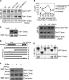
Identification of Dnt1 as a Dma1-binding protein. (A) Results of tandem mass spectrometry analysis of protein mixtures from two independent Dma1-TAP purifications (1st and 2nd) and from one Dnt1-TAP purification. (B) Confirmation of the physical association between Dma1 and Dnt1 in vivo. Lysates were prepared from unsynchronized yeast cells expressing no tags, either Dma1-GFP or Dnt1-13myc, or both Dma1-GFP and Dnt1-13myc. Dma1-GFP was immunoprecipitated, and samples were analyzed by immunoblotting using anti-GFP and anti-Myc antibodies as indicated. (C) Dma1 interacts with Dnt1 by yeast two-hybrid assay. Dma1 was fused with the DNA-binding domain of GAL4 (BD) and Dnt1 with the transcriptional activation domain of GAL4 (AD). S. cerevisiae host strain PJ69-4A was cotransformed with plasmids as indicated, and growth on synthetic defined medium/−Leu, −Trp and synthetic defined medium/−Leu, −Trp, −His, +2 mM 3-aminotriazole is shown (left). As controls, coexpressions of Dma1 and empty AD vector and of empty BD vector with Dnt1 (negative control) or an AD fusion with S. pombe Sid4 (positive control) are shown. The two-hybrid interaction between Dma1 and Sid4 has been shown previously (Guertin et al., 2002b). Mean β-galactosidase activity units from liquid β-galactosidase assay are also shown (right). Error bars, SD from three independent experiments.
FIGURE 2:
FIGURE 2:
Dma1 binds phosphorylated Dnt1 in early mitosis. (A) Dma1 and Dnt1 interact strongly at the prometaphase–metaphase transition. Lysates were prepared from asynchronous dma1-GFP dnt1-13myc cells or cells arrested at different stages of the cell cycle (S phase by HU, G1 phase by cdc10-129, metaphase by nda3-KM311 and mts3-1), and Dma1-GFP was immunoprecipitated and samples analyzed by Western blotting using anti-GFP and anti-Myc antibodies as indicated. The mts3-1 and nda3-KM311 mutations were used to arrest cells at prometaphase–metaphase transition. About 3–4% of total lysates were loaded as input to detect endogenous Dnt1-13myc. (B) Interaction between Dma1 and Dnt1 persists in anaphase. nda3-km311 dma1-3HA-TAP dnt1-13myc cells were first arrested at prometaphase–metaphase transition at 18°C for 6 h and released at 30°C, and samples were collected at the indicated time points. Top, cells with condensed chromosomes (indicating prometaphase–metaphase arrest), binucleate cells (indicating anaphase), or septated cells are quantified over a time course. Bottom, Dma1-3HA-TAP was immunoprecipitated, and samples were analyzed by immunoblotting using anti-hemagglutinin and anti-Myc antibodies as indicated. (C) Dma1-bound Dnt1 can be dephosphorylated in vitro. MBP-Dma1 was produced in bacteria and mixed with protein extracts prepared from mts3-1 dnt1-13myc cells, and the MBP-Dma1 pull-down complexes were treated with or without calf-intestinal alkaline phosphatase and analyzed by immunoblotting with anti-myc antibodies. (D, E) Dma1 and Dnt1 interact via the Dma1 FHA domain in vivo. Schematic diagrams of Dma1 variants fused to GFP are shown in D. Asterisks in D indicate the positions of point mutations (R64A [#3], H88A [#4], and C210;H212A [#6]). In E, mts3-1 dnt1-13myc dma1Δ cells (YDM3274) expressing the different Dma1 versions shown in D from plasmids (pREP42-GFP-dma1+) were arrested in metaphase using mts3-1 temperature shift, and then Dma1-GFP was immunoprecipitated, and samples were analyzed by immunoblotting using anti-GFP and anti-Myc antibodies as indicated. (F) Pretreatment of lysates prepared from mts3-1 dnt1-13myc yeast cells with λ-PPase caused reduced binding of Dnt1-13myc to bacteria-produced MBP-Dma1. Lysates prepared from dnt1-13myc yeast cells were first treated without or with λ-PPase for 75min before being mixed with bacteria-expressed MBP-Dma1 and subsequent precipitation by amylose resin.
FIGURE 3:
FIGURE 3:
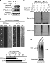
Dnt1 affects Dma1 localization at SPB and its E3 ligase activity but not its protein level. (A) Dma1 protein level is not influenced by the presence or absence of Dnt1. Wild-type or dnt1Δ cells with or without Dma1-13myc were cultured in liquid YE media and were collected and subjected to immunoprecipitation (with anti-myc antibodies) or straight Western assay (with anti-myc antibodies). Blot with anti-TAT1 (α-tubulin) served as a loading control. (B) Dma1-GFP intensity at SPBs is increased in dnt1Δ cells. Dma1-GFP and Sid4-RFP intensities were quantitated in wild-type and dnt1Δ cells, and final values are expressed as GFP/RFP ratio. Arrows indicate SPBs. For each cell quantitated, both SPBs were measured and averaged, for a total of 20 SPBs in 10 cells. Averaged measurements for each cell were then averaged for statistical analysis. Error bars represent the SEM; *p < 0.05. (C) Dma1 autoubiquitination is enhanced in dnt1Δ cells. Dma1-TAP was purified from metaphase-arrested (by mts3-1 mutation) wild-type or dnt1Δ cells. Ubiquitin ligase assays were carried out by mixing various combinations of E1 and E2 enzymes or Dma1-TAP as shown. The reactions were allowed to proceed for 0 or 90 min at room temperature (∼23°C). The Apc11 E3 enzyme is used as a control. Both Dma1 and Apc11 can autoubiquitinate. (D) Sid4 ubiquitination is increased in dnt1Δ cells. Sid4-HBH was purified from asynchronous wild-type or dnt1Δ cells under denatured conditions, and its ubiquitination status was assessed by immunoblotting with an ubiquitin antiserum and streptavidin.
FIGURE 4:
FIGURE 4:
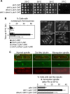
Absence of dnt1+ compromises Plo1 function but is rescued by deletion of dma1+. (A, B) The plo1-24C mutant is sensitive to deletion of dnt1+, and the sensitivity can be alleviated by disruption of Dma1 function. (A) Serial dilutions (10-fold) of the indicated single-, double-, or triple-mutant strains were spotted on YE and incubated at the indicated temperatures. (B) Liquid cultures of the indicated strains were also grown at 25°C and then shifted to 36°C, and percentages of cells with condensed chromosomes/metaphase arrest were quantified after being fixed and stained with DAPI (n = 200; left). Error bars, SD from three independent experiments. Examples of mutant cells incubated at 37°C for 4 h are also shown (right). (C) Enhanced spindle formation defects in dnt1Δ plo1-24C double mutant can be partially rescued by dma1Δ. Liquid cultures of mutant strains carrying GFP-atb2 and Sad1-mCherry were first grown at 25°C and then shifted to 36°C for 4 h; spindle formation was visualized in live cells. Examples of mutant cells showing either normal, dot-like tubulin or a short, monopolar spindle are shown (top). Percentages of cells in each strain with defective spindles were quantified (n = 200; bottom). Error bars, SD from three independent experiments.
FIGURE 5:
FIGURE 5:
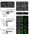
Overexpression of Dma1 in dnt1∆ cells leads to arrest in early mitosis. (A) Examples of nmt1-dma1+ dnt1+ and nmt1-dma1+ dnt1Δ cells after overexpression of Dma1 in EMM liquid media without thiamine for 20 h at 30°C. Cells were fixed and stained with DAPI. In the dnt1+ strain, open arrows and triangles indicate cells with two clustered nuclei in a postmitotic configuration and multinucleate cells, respectively. Asterisks indicate cells with condensed chromosomes in the dnt1Δ strain. Note that the major phenotype in Dma1-overexpressing dnt1+ cells is defective cytokinesis, whereas cells with condensed chromosomes mainly accumulate upon overexpression of Dma1 in dnt1Δ. (B) The functional ring finger (RF) and FHA domains in Dma1 are required for inducing metaphase arrest when Dma1 is overproduced in dnt1Δ cells. The dma1+ open reading frame with deleted RF domain (Dma1ΔRF), two point mutations in RF domain (Dma1C210;H212A), deleted FHA domain (Dma1ΔFHA), or two point mutations in FHA domain (Dma1R64A or Dma1H88A) was integrated into the yeast genome under the full-strength nmt1 promoter and overexpressed in wild-type or dnt1Δ cells. The indicated strains were fixed and DAPI stained after Dma1overexpression was induced as in A, and the frequencies of cells with cytokinetic defects (i.e., binucleate with the nuclei in a postmitotic configuration and multinucleate) and condensed chromosomes were quantified in wild-type cells or in dnt1Δ cells, respectively. We counted n > 200 cells for each strain. Error bars, SD from three independent experiments. (C) Securin Cut2 and mitotic cyclin Cdc13 persist in dnt1Δ cells upon overexpression of Dma1. nmt1-dma1+ dnt1Δ cells carrying Cut2-GFP or Cdc13-GFP were induced for Dma1 overexpression as in A, fixed, and examined for localization of Cut2-GFP or Cdc13-GFP. Asterisks indicate cells with condensed chromosomes. (D) The phenotype of hypercondensed chromosomes in nmt1-dma1+ dnt1Δ cells is dependent on the spindle assembly checkpoint. Dma1 was overexpressed in the indicated strains as in A, and cells with hypercondensed chromosomes determined by DAPI staining were counted (n > 200). Error bars, SD from three independent experiments. (E) Normal spindle formation upon overexpression of Dma1 in wild-type cells. Microtubules were labeled by indirect immunofluorescence with TAT1 antibody in wild-type cells induced for 20 h at 30°C for dma1+ overexpression. Nuclei were stained with DAPI. Left, the nucleus shows condensed chromosomes at metaphase with a fully assembled spindle; right, an early-anaphase nucleus shows an intact spindle. (F) Microtubules were stained in dnt1Δ cells overproducing Dma1. These cells showed no spindle, partial spindles, or monopolar spindles, and the percentages of each type in cells with hypercondensed chromosomes (n > 200) are indicated.
FIGURE 6:
FIGURE 6:
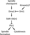
Model for Dnt1 regulation of Dma1 in early mitosis. Dnt1 is phosphorylated by an unknown kinase in prometaphase, which promotes binding of Dnt1 to Dma1 through Dma1's FHA domain. Dnt1–Dma1 interaction inhibits Dma1, which normally targets Sid4 for ubiquitination and subsequently prevents Plo1 from localization to the SPB. While at metaphase–anaphase transition, Dnt1's negative effect on Dma1 may be redirected or terminated by an unknown mechanism.

Similar articles

Cited by

References

    1. Bahler J, Steever AB, Wheatley S, Wang Y, Pringle JR, Gould KL, McCollum D. Role of polo kinase and Mid1p in determining the site of cell division in fission yeast. J Cell Biol. 1998a;143:1603–1616. - PMC - PubMed
    1. Bahler J, Wu JQ, Longtine MS, Shah NG, McKenzie A 3rd, Steever AB, Wach A, Philippsen P, Pringle JR. Heterologous modules for efficient and versatile PCR-based gene targeting in Schizosaccharomyces pombe. Yeast. 1998b;14:943–951. - PubMed
    1. Balasubramanian MK, McCollum D, Gould KL. Cytokinesis in fission yeast Schizosaccharomyces pombe. Methods Enzymol. 1997;283:494–506. - PubMed
    1. Brooks L 3rd, Heimsath EG, Jr, Loring GL, Brenner C. FHA-RING ubiquitin ligases in cell division cycle control. Cell Mol Life Sci. 2008;65:3458–3466. - PMC - PubMed
    1. Carnahan RH, Gould KL. The PCH family protein, Cdc15p, recruits two F-actin nucleation pathways to coordinate cytokinetic actin ring formation in Schizosaccharomyces pombe. J Cell Biol. 2003;162:851–862. - PMC - PubMed

Publication types

MeSH terms

LinkOut - more resources