. 2012 Jul 17:12:293.
doi: 10.1186/1471-2407-12-293.

Prolyl hydroxylase 2 dependent and Von-Hippel-Lindau independent degradation of Hypoxia-inducible factor 1 and 2 alpha by selenium in clear cell renal cell carcinoma leads to tumor growth inhibition

Affiliations

Prolyl hydroxylase 2 dependent and Von-Hippel-Lindau independent degradation of Hypoxia-inducible factor 1 and 2 alpha by selenium in clear cell renal cell carcinoma leads to tumor growth inhibition

Sreenivasulu Chintala et al. BMC Cancer. .

Abstract

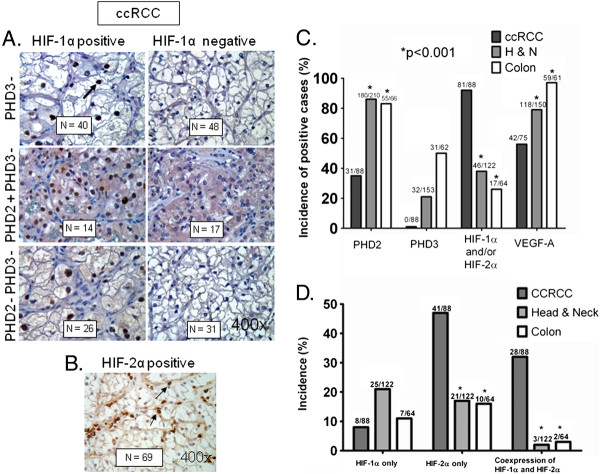
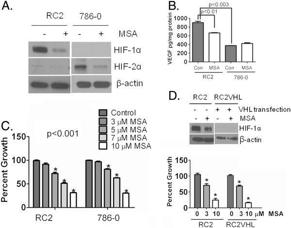
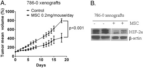
Background: Clear cell renal cell carcinoma (ccRCC) accounts for more than 80% of the cases of renal cell carcinoma. In ccRCC deactivation of Von-Hippel-Lindau (VHL) gene contributes to the constitutive expression of hypoxia inducible factors 1 and 2 alpha (HIF-α), transcriptional regulators of several genes involved in tumor angiogenesis, glycolysis and drug resistance. We have demonstrated inhibition of HIF-1α by Se-Methylselenocysteine (MSC) via stabilization of prolyl hydroxylases 2 and 3 (PHDs) and a significant therapeutic synergy when combined with chemotherapy. This study was initiated to investigate the expression of PHDs, HIF-α, and VEGF-A in selected solid cancers, the mechanism of HIF-α inhibition by MSC, and to document antitumor activity of MSC against human ccRCC xenografts.

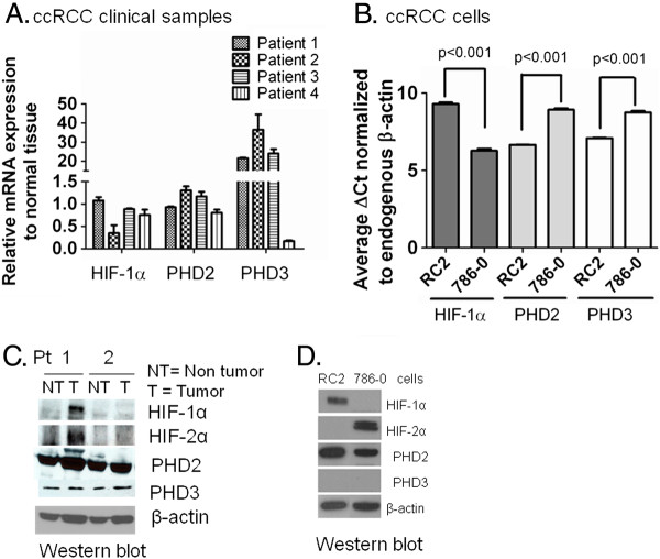
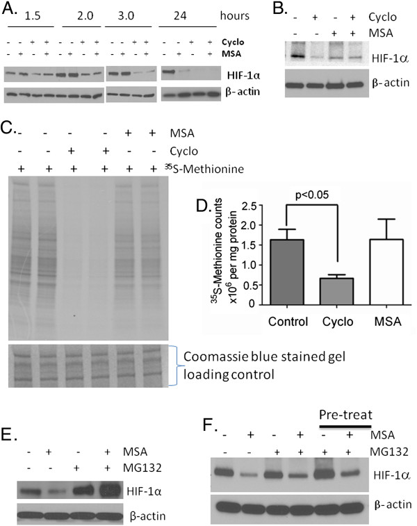
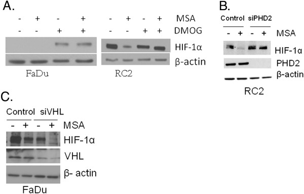
Methods: Tissue microarrays of primary human cancer specimens (ccRCC, head & neck and colon) were utilized to determine the incidence of PHD2/3, HIF-α, and VEGF-A by immunohistochemical methods. To investigate the mechanism(s) of HIF-α inhibition by MSC, VHL mutated ccRCC cells RC2 (HIF-1α positive), 786-0 (HIF-2α positive) and VHL wild type head & neck cancer cells FaDu (HIF-1α) were utilized. PHD2 and VHL gene specific siRNA knockdown and inhibitors of PHD2 and proteasome were used to determine their role in the degradation of HIF-1α by MSC.

Results: We have demonstrated that ccRCC cells express low incidence of PHD2 (32%), undetectable PHD3, high incidence of HIF-α (92%), and low incidence of VEGF-A compared to head & neck and colon cancers. This laboratory was the first to identify MSC as a highly effective inhibitor of constitutively expressed HIF-α in ccRCC tumors. MSC did not inhibit HIF-1α protein synthesis, but facilitated its degradation. The use of gene knockdown and specific inhibitors confirmed that the inhibition of HIF-1α was PHD2 and proteasome dependent and VHL independent. The effects of MSC treatment on HIF-α were associated with significant antitumor activity against ccRCC xenograft.

Conclusions: Our results show the role of PHD2/3 in stable expression of HIF-α in human ccRCC. Furthermore, HIF-1α degradation by MSC is achieved through PHD2 dependent and VHL independent pathway which is unique for HIF-α regulation. These data provide the basis for combining MSC with currently used agents for ccRCC.


Figures

Figure 1
Figure 1
Incidence of PHD2/3, HIF-α, and VEGF-A in ccRCC, head & neck and colon primary cancers. (A) Double immunohistochemical detection of HIF-1α and PHD2/3 in TMAs of ccRCC. Representative photomicrographs (all magnification x400) of HIF-1α positive and HIF-1α negative tumors (upper panel) showing nuclear staining of HIF-1α. Arrow indicates brown nuclear staining. Representative photomicrographs of PHD2 positive and PHD3 negative tumors (pink cytoplasmic staining, middle panel) and PHD2/3 negative tumors showing no cytoplasmic pink staining (lower panel). Numbers shown in the boxes are the positive/negative tumors. (B) Representative photomicrograph (x400) of HIF-2α positive tumors in TMA of ccRCC. Arrows indicate brown nuclear staining. (C) Percent incidence of PHD2, PHD3, HIF-α (HIF-1α and/or HIF-2α), and VEGF-A in ccRCC, head & neck, and colon primary tumor biopsies arranged in TMA. Numbers at the top of column indicate the number of positive cases among all evaluable cases. (D) Incidence of exclusively HIF-1α, HIF-2α and both HIF-1α and HIF-2α positive cases from the primary cancers in TMA. Some of the HIF-1α positive tumors turned out to be positive for HIF-2α and vice versa for HIF-2α positive tumors, which were excluded to count only HIF-1α, HIF-2α, and both HIF-1α and HIF-2α positive cases. The co-expression of HIF-1α and HIF-2α together was also significantly higher in ccRCC as compared to head & neck and colon cancers. Fisher Exact test revealed the statistically significant difference of incidence in ccRCC when compared to head & neck and colon cancers.*P < 0.001.
Figure 2
Figure 2
Expression of HIF-α, PHD2, and PHD3 mRNA and protein in ccRCC clinical specimens and cell lines. (A) Quantitative analysis of HIF-1α, PHD2, and PHD3 mRNA by real-time RT-PCR (qRT-PCR) in ccRCC primary tumors. Expressions were normalized to the matched normal kidney tissue by calculating 2delta-deltaCT values relative to normal kidney reference. Expression of mRNA in individual tumors was shown. B. Expression analysis of HIF-1α, PHD2, and PHD3 in ccRCC cells RC2 and 786–0. Expression was normalized to endogenous β-actin by calculating delta cycle threshold (ΔCt). ΔCt = Ct value of specific gene (HIF-1α, PHD2 and PHD3) - Ct value of β-actin. The lower the ΔCt value the higher the expression of the gene. Experiment was repeated twice with triplicates and p < 0.05 was considered as significant P < 0.001. (C) Detection of HIF-1α, HIF-2α and PHD2/3 in ccRCC primary tumors and their matched normal kidney tissues by western blot analysis. 80 micrograms of protein extract was electrophoresed through Mini-Protean precast 4-20% gradient gel. Expression of β-actin was used as a loading control. (D) PHD3 protein was undetectable in ccRCC cells. Detection of HIF-α and PHD2/3 by western blot analysis in ccRCC cells RC2 and 786–0 cells. β-actin expression was used as a loading control.
Figure 3
Figure 3
Effect of selenium treatment on HIF-1α, HIF-2α, VEGF and cell growth. (A) MSA inhibits both HIF-1α and HIF-2α. RC2 cells expressing HIF-1α and 786–0 cells expressing HIF-2α were treated with and without 10 μM MSA for 24 h; HIF-1α and HIF-2α were detected by western blot. β-actin expression was used as a loading control. (B) MSA down-regulates secreted VEGF in RC2 cells but not in 786–0 cells. Secreted VEGF was measured by ELISA in ccRCC cells. Cells were treated with and without MSA for 24 h and media were used to measure VEGF in RC2 and 786–0 cells and normalized with protein and expressed as pg/mg protein. Experiment was repeated twice with duplicates and P value < 0.05 was considered as significant. (C). HIF-α inhibition by MSA was associated with growth inhibition. Cells were treated with various concentrations MSA (3, 5, 7 and 10 μM) for 24 h. Medium was removed, rinsed and fresh medium was added and allowed to proliferate for 96 h. Cells were fixed and determined the cell survival by SRB assay. Growth inhibition was presented as percent growth inhibition compared to untreated controls. Experiment was repeated twice with 4–5 replicate samples. *p <0.001. (D). VHL transfected RC2 cells which do not express HIF-1α were equally sensitive to MSA like RC2 cells which express HIF-1α . Expression of HIF-1α in RC2 and VHL transfected RC2 VHL cells with and without MSA (upper panel). Cytotoxic effects of MSA in RC2 and RC2 VHL cells (lower panel). Cell survival was determined by SRB assay. *p <0.001.
Figure 4
Figure 4
Effect of MSA on HIF-1α protein synthesis and degradation. (A) HIF-1α synthesis was inhibited by cycloheximide but not by MSA in FaDu cells. FaDu cells were treated with 1 μM MSA and 10 μM cycloheximide alone and in combination for 1.5, 2, 3 and 24 hours at 0.5% oxygen and protein extracts were used to determine HIF-1α by western blot. β-actin expression was used as a loading control. (B) Effect of MSA on HIF-1α protein synthesis in RC2 cells. Cells were treated with cycloheximide or MSA alone and in combination for 8 h. HIF-1α was detected by western blot. β-actin was used as loading control (C) Incorporation of 35 S-Methionine was not affected by MSA in RC2. Cells were treated with cycloheximide or MSA separately in duplicate samples for 5 h and 35 S-Methionine (2.3 μCi/ml) was added at the last 1 h of treatment. Protein extracts (20 μg) were used to separate by SDS polyacrylamide gel electrophoresis and detected the incorporated 35 S-Methionin by autoradiography. Lower panel showing the coomassie blue stained proteins as loading control. (D) Determination of 35 S-Methionine radioactivity counts in cycloheximide or MSA treated RC2 cells. Protein extracts (20 μl) were used to detect 35 S-methionine radioactivity in the cells by counting in Liquid Scientilator Counter. Total counts were calculated in one milligram of protein and presented the number of counts in millions as compared to untreated control cells. P < 0.05 was considered as significant. (E) HIF-1α degradation by MSA is proteasome dependent. FaDu cells which do not express constitutive HIF-1α under normoxic culture conditions were subjected to 0.5% oxygen and treated with 1 μM MSA alone and in combination with 10 μM proteasome inhibitor MG132 for 24 h. Cell extracts were prepared and the expression of HIF-l α was analyzed. Expression of β-actin was used as a loading control. (F) Proteasome independent degradation of HIF-1α in VHL mutated RC2 cells. RC2 cells were treated with MSA and MG132 alone and in combination for 8 h. In a separate experiment, 1 h pre-treatment of MG132 followed by 7 h MSA treatment was performed to see the effect of MSA on HIF-1α. Cells were processed to extract protein and HIF-1α levels were determined. Expression of β-actin was used as a loading control.
Figure 5
Figure 5
Role of PHDs in HIF-1α degradation by MSA. (A) Inhibition of PHDs activity by DMOG reversed the degradation of HIF-1α by MSA in VHL active FaDu, and VHL inactive RC2 cells. Cells were treated with 10 μM MSA and 0.5 mM DMOG alone and in combination and HIF-1α was analyzed by western blot. β-actin expression was used as a loading control. (B) Gene specific silencing of PHD2 in RC2 cells by siRNA prevented the degradation of HIF-1α by MSA. PHD2 siRNA was transfected with lipofectamine 2000 for 24 h. Cells were treated with and without 10 μM MSA for 24 h and HIF-1α was detected by western blot. β-actin expression was used as a loading control. (C) Degradation of HIF-1α by MSA is VHL independent. VHL was inhibited by siRNA in FaDu cells expressing active VHL and treated with MSA to determine the HIF-1α degradation. This experiment was done under 0.5% oxygen level to stabilize HIF-1α in FaDu cells. β-actin expression was used as a loading control.
Figure 6
Figure 6
MSC effect on tumor growth and HIF-2α expression in 786–0 ccRCC xenografts. (A) Inhibition of ccRCC tumor growth by MSC in xenografts. HIF-2α expressing 786–0 cells (10 million) were transplanted into nude mice for establishing the xenografts. Small pieces (~50 mg) of tumor tissues were transplanted subcutaneously into nude mice and treatment began when tumor weighed 200–250 mg. Mice were randomized and divided into two groups, each containing 5 mice. One group was treated with saline and the other group was treated with MSC (0.2 mg/mouse/day; the optimal nontoxic dose) daily for 18 days. Tumor volume was measured daily. (B) HIF-2α is inhibited by the therapeutic dose of MSC in 786–0 xenografts. Tumor xenografts collected after 18 days of the MSC treatments and processed to extract protein and HIF-2α levels were assessed by western blot. β-actin expression was used as a loading control.

Similar articles

Cited by

References

    1. Linehan WM, Srinivasan R, Schmidt LS. The genetic basis of kidney cancer: a metabolic disease. Nature reviews. 2010;7(5):277–285. - PMC - PubMed
    1. US National Institutes of Health. Institute N.C. kidney cancer [online] 2010. http://www.cancer.gov/cancertopics/types/kidney.
    1. Hervouet E, Demont J, Pecina P, Vojtiskova A, Houstek J, Simonnet H, Godinot C. A new role for the von Hippel-Lindau tumor suppressor protein: stimulation of mitochondrial oxidative phosphorylation complex biogenesis. Carcinogenesis. 2005;26(3):531–539. - PubMed
    1. Semenza GL. Targeting HIF-1 for cancer therapy. Nat Rev Cancer. 2003;3(10):721–732. doi: 10.1038/nrc1187. - DOI - PubMed
    1. Chan DA, Kawahara TL, Sutphin PD, Chang HY, Chi JT, Giaccia AJ. Tumor vasculature is regulated by PHD2-mediated angiogenesis and bone marrow-derived cell recruitment. Cancer Cell. 2009;15(6):527–538. doi: 10.1016/j.ccr.2009.04.010. - DOI - PMC - PubMed

Publication types

MeSH terms

Substances