. 2012 Sep;23(17):3420-8.
doi: 10.1091/mbc.E12-02-0107. Epub 2012 Jul 11.

An essential novel component of the noncanonical mitochondrial outer membrane protein import system of trypanosomatids

Affiliations

An essential novel component of the noncanonical mitochondrial outer membrane protein import system of trypanosomatids

Mascha Pusnik et al. Mol Biol Cell. 2012 Sep.

Abstract

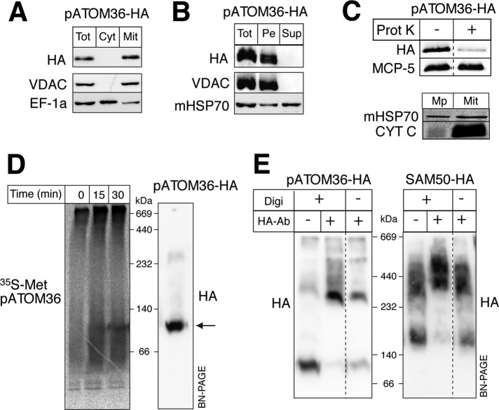
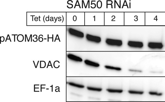
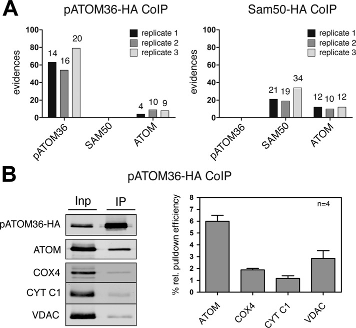
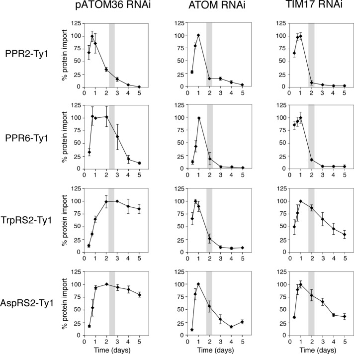
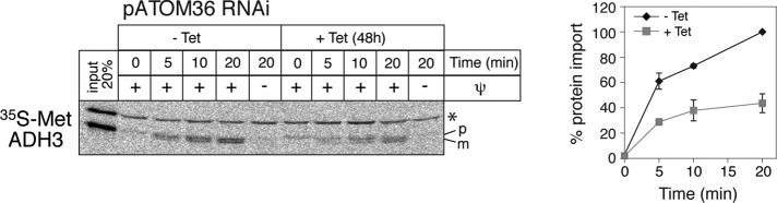
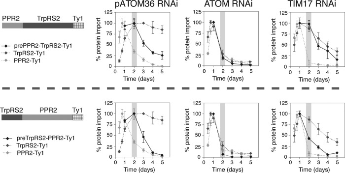
The mitochondrial outer membrane protein Tom40 is the general entry gate for imported proteins in essentially all eukaryotes. Trypanosomatids lack Tom40, however, and use instead a protein termed the archaic translocase of the outer mitochondrial membrane (ATOM). Here we report the discovery of pATOM36, a novel essential component of the trypanosomal outer membrane protein import system that interacts with ATOM. pATOM36 is not related to known Tom proteins from other organisms and mediates the import of matrix proteins. However, there is a group of precursor proteins whose import is independent of pATOM36. Domain-swapping experiments indicate that the N-terminal presequence-containing domain of the substrate proteins at least in part determines the dependence on pATOM36. Secondary structure profiling suggests that pATOM36 is composed largely of α-helices and its assembly into the outer membrane is independent of the sorting and assembly machinery complex. Taken together, these results show that pATOM36 is a novel component associated with the ATOM complex that promotes the import of a subpopulation of proteins into the mitochondrial matrix.


Figures

FIGURE 1:
FIGURE 1:
pATOM36 is an integral protein of the mitochondrial outer membrane. (A) Immunoblots of total (Tot), digitonin-extracted crude cytosolic (Cyt), and mitochondrial (Mit) fractions from a cell line expressing pATOM36 with an HA tag at its C-terminus (pATOM36-HA) probed with anti-HA antibodies (HA). VDAC and elongation factor 1a (EF-1a) serve as mitochondrial outer membrane and cytosolic marker, respectively. (B) Immunoblot of an alkaline carbonate extraction of pATOM36-HA–containing mitochondria. Pe, pellet; Sup, supernatant. The integral membrane protein VDAC and the soluble mHSP70 serve as markers. (C) Top, immunoblot of untreated and proteinase K-treated (50 μg/ml) pATOM36-HA–containing mitochondria. Mitochondrial carrier protein 5 (MCP-5) serves as a loading control. Bottom, immunoblot of isolated mitoplasts (Mp) having a disrupted outer membrane and the intact pATOM36-HA–containing mitochondria (Mit) shown at the top. CYT C and mHSP70 serve as markers for the intermembrane space and the matrix, respectively. (D) Left, import of in vitro–translated, 35S-Met–labeled pATOM36 into isolated mitochondria analyzed by BN-PAGE. Right, immunoblot of pATOM36-HA–containing mitochondrial extract separated on the same BN-PAGE. The main pATOM36 complex is indicated by the arrow. (E) Antibody shift experiments. Left, pATOM36-HA containing digitonin-lysed (+Digi) and intact mitochondria (−Digi) were incubated with polyclonal anti-HA antibodies (+HA, −antibody), separated by BN-PAGE, and analyzed by immunoblots using a monoclonal anti-HA antiserum (HA). Untreated pATOM36-HA (−HA, −antibody) complex is shown on the left as a control. Right, same as shown on the left, but SAM50-HA–expressing mitochondria were used.
FIGURE 2:
FIGURE 2:
In vivo assembly of pATOM36 is independent of SAM50. Levels of pATOM36-HA in total cellular extracts are not affected, whereas the levels of the β-barrel protein VDAC declines during tet induction of SAM50 RNAi.
FIGURE 3:
FIGURE 3:
pATOM36 is a peripheral component of the ATOM complex. (A) A 0.15% digitonin lysate of isolated, HA-tagged pATOM36 (left) and HA-tagged SAM50 containing mitochondria (right) were immunoprecipitated using anti-HA antibodies. The corresponding eluates were analyzed for the presence of pATOM36, SAM50, and ATOM using mass spectrometry. The graphs depict the number of evidences that were detected in the eluate of each experiment. The number of unique peptides identified for each protein is depicted on the top of each column. The experiment was performed in triplicate, and immunoprecipitations using wild-type mitochondria lacking any tagged proteins served as controls. pATOM36, SAM50, nor ATOM was recovered in the eluates of wild-type mitochondria. The only exception was ATOM that was detected by a single peptide/evidence in the first replicates of wild-type immunoprecipitates (data not shown). (B) A 0.15% digitonin lysate of isolated, HA-tagged pATOM36–containing mitochondria was immunoprecipitated using anti-HA antibodies and analyzed by immunoblots. Five percent of the total extract (Inp) and 95% of the bound fraction (IP) were analyzed by immunoblot using a monoclonal anti-HA antibody. The same samples were also analyzed with polyclonal ATOM, VDAC, COX4, and CYT C1 antisera. The experiment shown on the left was performed in quadruplicate and quantitated using the Odyssey infrared imaging system (Li-Cor Biosciences, Lincoln, NE). The means of the relative amounts of ATOM, VDAC, COX4, and CYT C1 that were recovered in the bound fraction were calculated. The pATOM36 that was recovered in the pellet was set to 100%. Standard errors are indicated.
FIGURE 4:
FIGURE 4:
pATOM36 is essential for normal growth of procyclic and bloodstream forms of T. brucei. (A) Growth curve of an uninduced and tet-induced procyclic pATOM36 RNAi cell line. Right, Northern blot of total RNA isolated from uninduced and induced (2 d) RNAi cell lines probed for pATOM36 mRNA. The rRNA region of the ethidium bromide–stained gel (EtBr) was used as a loading control. (B) As in A, but results are for an uninduced and tet-induced pATOM36 RNAi cell line of the bloodstream form.
FIGURE 5:
FIGURE 5:
pATOM36 is required for in vivo import of a subset of matrix proteins. Degradation of newly synthesized tagged matrix proteins during induction of RNAi of pATOM36 (first column), ATOM (second column), and TIM17 (third column) was used as a proxy for inhibition of mitochondrial protein import. The levels of indicated newly synthesized, C-terminally Ty1-tagged import substrates during pATOM36 RNAi were quantified by immunoblot analysis using antitag antibodies and normalized to the levels of cytosolic EF-1a. All experiments were replicated at least three times. The graphs depict the means and the standard errors of the normalized protein levels for each time point. Maximal expression was set to 100% for each tested substrate protein. Gray bar in graphs indicates the time of onset of growth arrest. The two control panels testing import of PPR2-Ty1 and TrpRS2-Ty1 in the ATOM RNAi cell line were published previously (Pusnik et al., 2011).
FIGURE 6:
FIGURE 6:
pATOM36 is required for in vitro import of matrix proteins. Inhibition of in vitro import into isolated mitochondria from induced pATOM36 RNAi cells. In vitro–translated, 35S-Met–labeled alcohol dehydrogenase 3 (ADH3) precursor was imported for the indicated times into mitochondria isolated from uninduced (–Tet) and induced (+Tet) pATOM36 RNAi cell lines and analyzed by SDS–PAGE. The Coomassie-stained gel is shown as a loading control. All import reaction were proteinase treated. Input, 20% of added substrate; ψ, membrane potential. The band indicated by the asterisk is a labeled protein that is template independently synthesized in this batch of reticulocyte lysate. The positions of the precursor (p) and mature (m) forms of the protein are indicated.
FIGURE 7:
FIGURE 7:
pATOM36 function depends on a N-terminal presequence-containing domain of substrate proteins. Degradation of newly synthesized tagged substrate proteins during induction of pATOM36 (first column), ATOM (second column), and TIM17 (third column) RNAi was measured by immunoblots and used as a proxy for inhibition of mitochondrial protein import. Top, Ty1-tagged prePPR2-TrpRS2 consisting of the N-terminal 45 amino acids of PPR2 followed by TrpRS2 lacking the N-terminal 45 amino acids was used as a substrate. Bottom, Ty1-tagged preTrpRS2-PPR2 consisting of the N-terminal 45 amino acids of TrpRS2 followed by PPR2 lacking the N-terminal 45 amino acids was used as a substrate. All experiments were replicated at least three times. The graphs depict the means and the standard errors of the protein levels normalized to EF-1a for each time point. Maximal expression set to 100% for each tested substrate protein. Gray bar in graphs indicates the time of onset of growth arrest. The results for the tagged parent proteins TrpRS-Ty1 and PPR-Ty1 as shown in Figure 3 are shown in gray for comparison.

Similar articles

Cited by

References

    1. Abe Y, Shodai T, Muto T, Mihara K, Torii H, Nishikawa S-I, Endo T, Kohda D. Structural basis of presequence recognition by the mitochondrial protein import receptor Tom20. Cell. 2000;100:551–560. - PubMed
    1. Ahting U, Thieffry M, Engelhardt H, Hegerl R, Neupert W, Nussberger S. Tom40, the pore-forming component of the protein-conducting TOM channel in the outer membrane of mitochondria. J Cell Biol. 2001;153:1151–1160. - PMC - PubMed
    1. Allen R, Egan B, Gabriel K, Beilharz T, Lithgow T. A conserved proline residue is present in the transmembrane-spanning domain of Tom7 and other tail-anchored protein subunits of the TOM translocase. FEBS Lett. 2002;514:347–350. - PubMed
    1. Alsford S, Turner DJ, Obado SO, Sanchez-Flores A, Glover L, Berriman M, Hertz-Fowler C, Horn D. High-throughput phenotyping using parallel sequencing of RNA interference targets in the African trypanosome. Genome Res. 2011;21:915–924. - PMC - PubMed
    1. Baker KP, Schatz G. Mitochondrial proteins essential for viability mediate protein import into yeast mitochondria. Nature. 1991;349:205–208. - PubMed

Publication types

MeSH terms

Substances