. 2012;7(7):e39642.
doi: 10.1371/journal.pone.0039642. Epub 2012 Jul 2.

Interplay of Nkx3.2, Sox9 and Pax3 regulates chondrogenic differentiation of muscle progenitor cells

Affiliations

Interplay of Nkx3.2, Sox9 and Pax3 regulates chondrogenic differentiation of muscle progenitor cells

Dana M Cairns et al. PLoS One. 2012.

Abstract

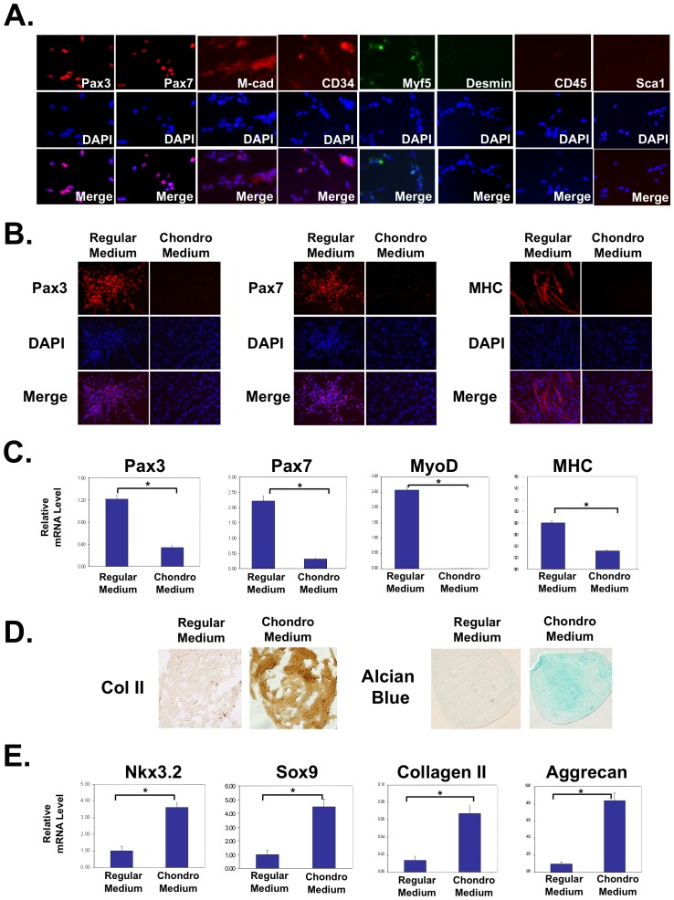
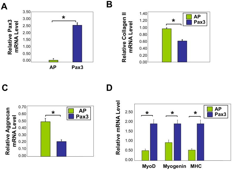
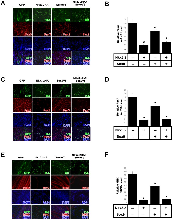
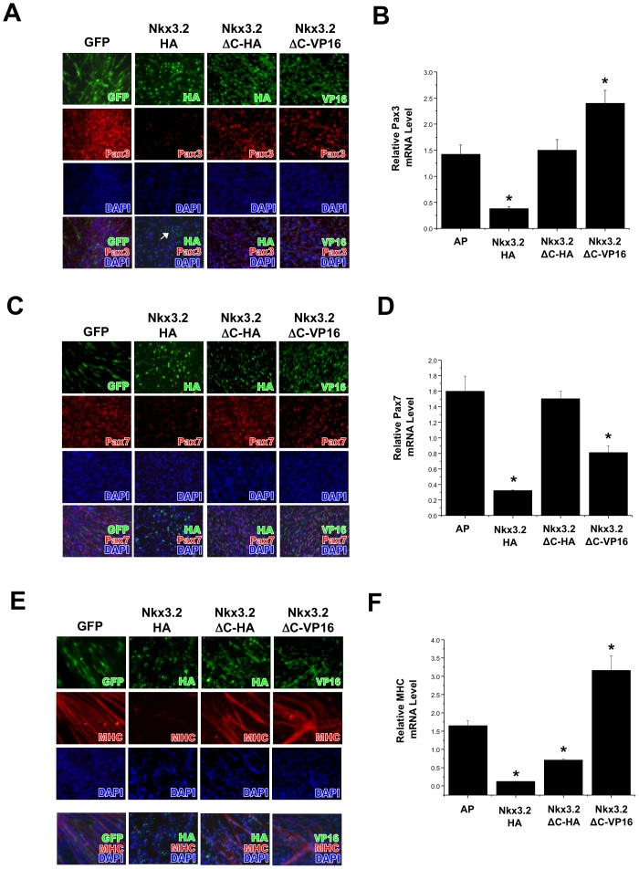
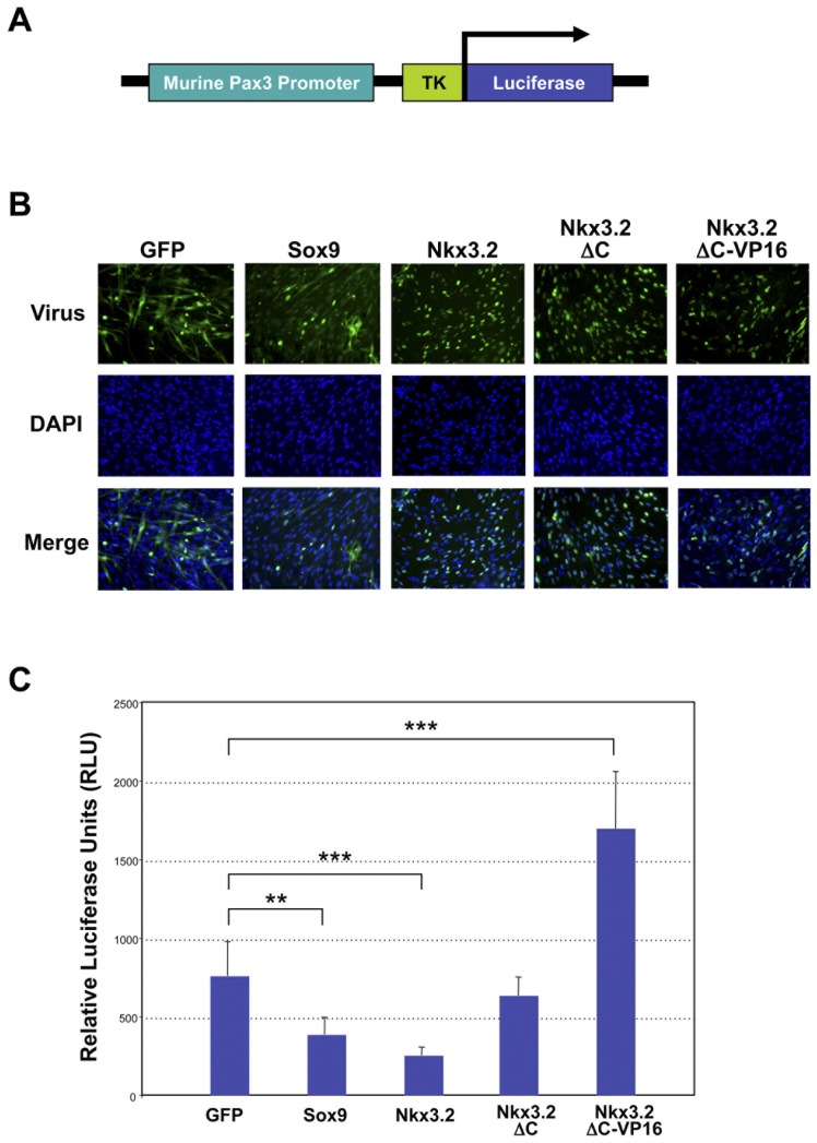
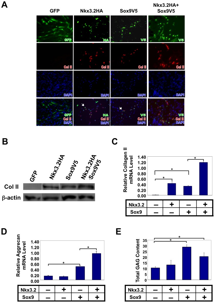
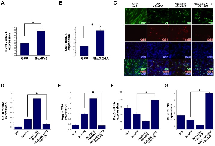
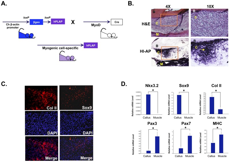
Muscle satellite cells make up a stem cell population that is capable of differentiating into myocytes and contributing to muscle regeneration upon injury. In this work we investigate the mechanism by which these muscle progenitor cells adopt an alternative cell fate, the cartilage fate. We show that chick muscle satellite cells that normally would undergo myogenesis can be converted to express cartilage matrix proteins in vitro when cultured in chondrogenic medium containing TGFß3 or BMP2. In the meantime, the myogenic program is repressed, suggesting that muscle satellite cells have undergone chondrogenic differentiation. Furthermore, ectopic expression of the myogenic factor Pax3 prevents chondrogenesis in these cells, while chondrogenic factors Nkx3.2 and Sox9 act downstream of TGFß or BMP2 to promote this cell fate transition. We found that Nkx3.2 and Sox9 repress the activity of the Pax3 promoter and that Nkx3.2 acts as a transcriptional repressor in this process. Importantly, a reverse function mutant of Nkx3.2 blocks the ability of Sox9 to both inhibit myogenesis and induce chondrogenesis, suggesting that Nkx3.2 is required for Sox9 to promote chondrogenic differentiation in satellite cells. Finally, we found that in an in vivo mouse model of fracture healing where muscle progenitor cells were lineage-traced, Nkx3.2 and Sox9 are significantly upregulated while Pax3 is significantly downregulated in the muscle progenitor cells that give rise to chondrocytes during fracture repair. Thus our in vitro and in vivo analyses suggest that the balance of Pax3, Nkx3.2 and Sox9 may act as a molecular switch during the chondrogenic differentiation of muscle progenitor cells, which may be important for fracture healing.


Conflict of interest statement

Competing Interests: The authors have declared that no competing interests exist.

Figures

Figure 1
Figure 1. Isolated muscle satellite cells can be redirected toward a cartilage phenotype at the expense of the default muscle phenotype.
(A) Immunocytochemistry analysis indicating that isolated chicken muscle progenitor cells at day 0 are over 95% positive for Pax3, Pax7, M-cadherin and CD34 (red staining). They are 33%-positive for Myf5 and are negative for Desmin, CD45 and Sca-1. Percentage positive cells = number of positively stained cells/total cell number (based on DAPI staining). (B) Immunocytochemistry results indicating a dramatic downregulation of Pax3, Pax7, and MHC in response to chondrogenic stimuli. (C) qRT-PCR analysis showing decreased expression of muscle markers Pax3, Pax7, MyoD, and MHC in chondrogenic media as compared to those cultured in regular media. (D) Immunocytochemistry analysis on sectioned micromass cultures showing increased collagen II protein expression upon chondrogenic media treatment. Alcian Blue staining indicates increased glycosaminoglycan levels when cultured in chondrogenic media as compared to those cultured in regular media. (E) qRT-PCR analysis showing increased expression of cartilage markers Nkx3.2, Sox9, collagen II, and aggrecan in chondrogenic media as compared to those cultured in regular media. All PCR results were normalized to GAPDH. “*” denotes p<0.05 in statistical analysis.
Figure 2
Figure 2. Viral infection with Pax3 in muscle satellite cells inhibits chondrogenesis while maintaining muscle gene expression.
qRT-PCR analysis of satellite cells infected with RCAS-A-Pax3 or RCAS-A-AP (alkaline phosphatase, control virus) and cultured in 3D micromasses in chondrogenic media. (A) Pax3 mRNA, (B) Collagen II mRNA, (C) Aggrecan mRNA, and (D) MyoD, Myogenin, MHC mRNA expression. All PCR results were normalized to GAPDH. “*” denotes p<0.05 in statistical analysis.
Figure 3
Figure 3. Nkx3.2 and Sox9 inhibit muscle-specific gene expression in muscle satellite cells.
Muscle satellite cells were infected with RCAS viruses encoding GFP, Nkx3.2HA, Sox9V5 or Nkx3.2HA+Sox9V5. Immunocytochemistry and qRT-PCR analyses were performed. Virus staining images were overlaid with Pax3, Pax7 and MHC images, and DAPI stains cell nuclei. For all qRT-PCR, GAPDH was used for normalization. (A) Pax3 immunostaining results. (B) Pax3 mRNA expression. (C) Pax7 immunostaining results. (D) Pax7 mRNA expression. (E) MHC immunostaining results. (F) MHC mRNA expression. “*” denotes statistically significant differences (p<0.05) relative to control samples.
Figure 4
Figure 4. The C-terminus of Nkx3.2 is required to inhibit muscle cell fate in muscle satellite cells.
Muscle satellite cells were infected with RCAS viruses encoding GFP, Nkx3.2HA, Nkx3.2-ΔCHA or Nkx3.2-ΔC-VP16. Immunocytochemistry and qRT-PCR analyses were performed. Virus staining images were overlaid with Pax3, Pax7 and MHC images, and DAPI stains cell nuclei. For all qRT-PCR, GAPDH was used for normalization. (A) Pax3 immunostaining results. (B) Pax3 mRNA expression. (C) Pax7 immunostaining results. (D) Pax7 mRNA expression. (E) MHC immunostaining results. (F) MHC mRNA expression. “*” denotes statistically significant differences (p<0.05) relative to control samples.
Figure 5
Figure 5. Nkx3.2 and Sox9 inhibit mouse Pax3 promoter activity.
(A) Schematic diagram of the mouse Pax3 promoter luciferase construct. (B) Immunocytochemistry analysis showing equal infection efficiencies of all viruses (GFP, Sox9, Nkx3.2, Nkx3.2-ΔC-HA, Nkx3.2-ΔC-VP16). (C) Luciferase analysis on satellite cells infected with all viruses (GFP, Sox9, Nkx3.2, Nkx3.2-ΔC-HA, Nkx3.2-ΔC-VP16) and transfected with the Pax3 luciferase construct. A control luciferase vector was used for normalization. “**” denotes p<0.01 and “***” denotes p<0.001 in statistical analysis.
Figure 6
Figure 6. Nkx3.2 and Sox9 can induce muscle satellite cells to express cartilage markers collagen II and aggrecan.
Muscle satellite cells were infected with RCAS viruses encoding GFP, Nkx3.2HA, Sox9V5 or Nkx3.2HA+Sox9V5. Immunocytochemistry and qRT-PCR analyses were performed. For all qRT-PCR, GAPDH was used for normalization. (A) Immunocytochemistry analysis of collagen II protein expression in satellite cells. Virus staining images were overlaid with collagen II images, and DAPI stains cell nuclei. (B) Western Blot analysis of collagen II protein expression in satellite cells. ß-actin, internal control. (C) qRT-PCR analysis of collagen II mRNA and (D) Aggrecan mRNA expression. (E) Glycoaminoglycan (GAG) assay. “*” denotes p<0.05 in statistical analysis.
Figure 7
Figure 7. Nkx3.2 is required for Sox9 to activate a cartilage program and to inhibit the muscle program in muscle satellite cells.
For all qRT-PCR, GAPDH was used for normalization. (A) qRT-PCR analysis of Nkx3.2 expression in satellite cells infected with control RCAS-GFP virus or RCAS-Sox9V5 virus. (B) qRT-PCR analysis of Sox9 expression in satellite cells infected with control RCAS-GFP virus or RCAS-Nkx3.2HA virus. (C)–(G) Muscle satellite cells were co-infected with the following combination of viruses: RCAS-A-GFP+RCAS-B-AP (alkaline phosphatase); RCAS-A-AP+RCAS-B-Sox9V5; RCAS-A-Nkx3.2HA+RCAS-B-Sox9V5; RCAS-A-Nkx3.2-ΔC-VP16+RCAS-B-Sox9V5. These infected cells were subject to immunocytochemistry analysis and qRT-PCR. (C) Immunocytochemistry analysis of Collagen II protein expression in satellite cells. Virus staining images were overlaid with Collagen II images, and DAPI stains cell nuclei. (D) Collagen II mRNA expression. (E). Aggrecan mRNA expression. (F). Pax3 mRNA expression. (G). MHC mRNA expression. In A–B, “*” denotes p<0.05 in statistical analysis. In D–G, “*” denotes statistically significant differences (p<0.05) between RCAS-A-AP+RCAS-B-Sox9V5 and RCAS-A-Nkx3.2-ΔC-VP16+RCAS-B-Sox9V5 co-infected samples.
Figure 8
Figure 8. Nkx3.2 and Sox9 are induced in the muscle progenitor cells that contribute to cartilage formation in an in vivo mouse model of fracture healing.
(A) Schematic diagram illustrating the generation of the transgenic mice in which MyoD+ lineage cells are labeled with heat-resistant alkaline phosphatase (hPLAP). (B) H&E histological analysis showing the fracture callus site 1 week post-fracture. Muscle progenitor cells were identified by assaying for heat-resistant alkaline phosphatase (arrow). Left panels, low magnification (4×); right panels, high magnification of the boxed areas from the left panels (10×). B, bone. C, callus, M, muscle. (C) Immunohistochemistry analyses of collagen II and Sox9 expression in the fracture callus. DAPI staining images were overlaid with collagen II and Sox9 staining images. (D) Laser Capture Microscopy (LCM) analysis of muscle progenitor cells in the fracture callus and in the neighboring muscle. qRT-PCR analyses were performed for the following genes: Nkx3.2, Sox9, collagen II, Pax3, Pax7 and MHC. For all qRT-PCR, 18S RNA was used for normalization. “*” denotes p<0.05 in statistical analysis.

Similar articles

Cited by

References

    1. Buckingham M, Bajard L, Chang T, Daubas P, Hadchouel J, et al. The formation of skeletal muscle: from somite to limb. J Anat. 2003;202:59–68. - PMC - PubMed
    1. Kuang S, Kuroda K, Le Grand F, Rudnicki MA. Asymmetric self-renewal and commitment of satellite stem cells in muscle. Cell. 2007;129:999–1010. - PMC - PubMed
    1. Relaix F, Rocancourt D, Mansouri A, Buckingham M. A Pax3/Pax7-dependent population of skeletal muscle progenitor cells. Nature. 2005;435:948–953. - PubMed
    1. Kanisicak O, Mendez JJ, Yamamoto S, Yamamoto M, Goldhamer DJ. Progenitors of skeletal muscle satellite cells express the muscle determination gene, MyoD. Dev Biol. 2009;332:131–141. - PMC - PubMed
    1. Bryson-Richardson RJ, Currie PD. The genetics of vertebrate myogenesis. Nat Rev Genet. 2008;9:632–646. - PubMed

Publication types

MeSH terms