Regulation of hindbrain Pyy expression by acute food deprivation, prolonged caloric restriction, and weight loss surgery in mice
- PMID: 22761162
- PMCID: PMC3468511
- DOI: 10.1152/ajpendo.00033.2012
Regulation of hindbrain Pyy expression by acute food deprivation, prolonged caloric restriction, and weight loss surgery in mice
Abstract
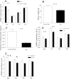
PYY is a gut-derived putative satiety signal released in response to nutrient ingestion and is implicated in the regulation of energy homeostasis. Pyy-expressing neurons have been identified in the hindbrain of river lamprey, rodents, and primates. Despite this high evolutionary conservation, little is known about central PYY neurons. Using in situ hybridization, PYY-Cre;ROSA-EYFP mice, and immunohistochemistry, we identified PYY cell bodies in the gigantocellular reticular nucleus region of the hindbrain. PYY projections were present in the dorsal vagal complex and hypoglossal nucleus. In the hindbrain, Pyy mRNA was present at E9.5, and expression peaked at P2 and then decreased significantly by 70% at adulthood. We found that, in contrast to the circulation, PYY-(1-36) is the predominant isoform in mouse brainstem extracts in the ad libitum-fed state. However, following a 24-h fast, the relative amounts of PYY-(1-36) and PYY-(3-36) isoforms were similar. Interestingly, central Pyy expression showed nutritional regulation and decreased significantly by acute starvation, prolonged caloric restriction, and bariatric surgery (enterogastroanastomosis). Central Pyy expression correlated with body weight loss and circulating leptin and PYY concentrations. Central regulation of energy metabolism is not limited to the hypothalamus but also includes the midbrain and the brainstem. Our findings suggest a role for hindbrain PYY in the regulation of energy homeostasis and provide a starting point for further research on gigantocellular reticular nucleus PYY neurons, which will increase our understanding of the brain stem pathways in the integrated control of appetite and energy metabolism.
Figures


Similar articles
-
Characterization of brainstem peptide YY (PYY) neurons.J Comp Neurol. 2008 Jan 10;506(2):194-210. doi: 10.1002/cne.21543. J Comp Neurol. 2008. PMID: 18022952
-
Diet and gastrointestinal bypass-induced weight loss: the roles of ghrelin and peptide YY.Diabetes. 2011 Mar;60(3):810-8. doi: 10.2337/db10-0566. Epub 2011 Feb 3. Diabetes. 2011. PMID: 21292870 Free PMC article.
-
Peptide YY 3-36 and pancreatic polypeptide differentially regulate hypothalamic neuronal activity in mice in vivo as measured by manganese-enhanced magnetic resonance imaging.J Neuroendocrinol. 2011 Apr;23(4):371-80. doi: 10.1111/j.1365-2826.2011.02111.x. J Neuroendocrinol. 2011. PMID: 21251093
-
News in gut-brain communication: a role of peptide YY (PYY) in human obesity and following bariatric surgery?Eur J Clin Invest. 2005 Jul;35(7):425-30. doi: 10.1111/j.1365-2362.2005.01514.x. Eur J Clin Invest. 2005. PMID: 16008543 Review.
-
The role of PYY in feeding regulation.Regul Pept. 2008 Jan 10;145(1-3):12-6. doi: 10.1016/j.regpep.2007.09.011. Epub 2007 Sep 18. Regul Pept. 2008. PMID: 17920705 Review.
Cited by
-
Peptide YY signaling in the lateral parabrachial nucleus increases food intake through the Y1 receptor.Am J Physiol Endocrinol Metab. 2015 Oct 15;309(8):E759-66. doi: 10.1152/ajpendo.00346.2015. Epub 2015 Sep 1. Am J Physiol Endocrinol Metab. 2015. PMID: 26330345 Free PMC article.
-
Peptide YY3-36 and 5-hydroxytryptamine mediate emesis induction by trichothecene deoxynivalenol (vomitoxin).Toxicol Sci. 2013 May;133(1):186-95. doi: 10.1093/toxsci/kft033. Epub 2013 Mar 1. Toxicol Sci. 2013. PMID: 23457120 Free PMC article.
-
Peripheral activation of the Y2-receptor promotes secretion of GLP-1 and improves glucose tolerance.Mol Metab. 2013 Mar 14;2(3):142-52. doi: 10.1016/j.molmet.2013.03.001. eCollection 2013. Mol Metab. 2013. PMID: 24049729 Free PMC article.
-
Peptide YY: more than just an appetite regulator.Diabetologia. 2014 Sep;57(9):1762-9. doi: 10.1007/s00125-014-3292-y. Epub 2014 Jun 11. Diabetologia. 2014. PMID: 24917132 Review.
-
The homeostatic role of neuropeptide Y in immune function and its impact on mood and behaviour.Acta Physiol (Oxf). 2015 Mar;213(3):603-27. doi: 10.1111/apha.12445. Epub 2015 Jan 9. Acta Physiol (Oxf). 2015. PMID: 25545642 Free PMC article. Review.
References
-
- Abbott CR, Small CJ, Kennedy AR, Neary NM, Sajedi A, Ghatei MA, Bloom SR. Blockade of the neuropeptide Y Y2 receptor with the specific antagonist BIIE0246 attenuates the effect of endogenous and exogenous peptide YY(3–36) on food intake. Brain Res 1043: 139–144, 2005 - PubMed
-
- Adrian TE, Ferri GL, Bacarese-Hamilton AJ, Fuessl HS, Polak JM, Bloom SR. Human distribution and release of a putative new gut hormone, peptide YY. Gastroenterology 89: 1070–1077, 1985 - PubMed
-
- Athanassiadis T, Olsson KA, Kolta A, Westberg KG. Identification of c-Fos immunoreactive brainstem neurons activated during fictive mastication in the rabbit. Exp Brain Res 165: 478–489, 2005 - PubMed
-
- Banks WA, Coon AB, Robinson SM, Moinuddin A, Shultz JM, Nakaoke R, Morley JE. Triglycerides induce leptin resistance at the blood-brain barrier. Diabetes 53: 1253–1260, 2004 - PubMed
-
- Batterham RL, Cohen MA, Ellis SM, Le Roux CW, Withers DJ, Frost GS, Ghatei MA, Bloom SR. Inhibition of food intake in obese subjects by peptide YY3–36. N Engl J Med 349: 941–948, 2003 - PubMed
Publication types
MeSH terms
Substances
Grants and funding
LinkOut - more resources
Full Text Sources
Medical
Molecular Biology Databases
Miscellaneous

