. 2012 Feb 1;2(2):59-69.
doi: 10.4161/bioa.20359.

A tethering complex dimer catalyzes trans-SNARE complex formation in intracellular membrane fusion

Affiliations

A tethering complex dimer catalyzes trans-SNARE complex formation in intracellular membrane fusion

Aditya Kulkarni et al. Bioarchitecture. .

Abstract

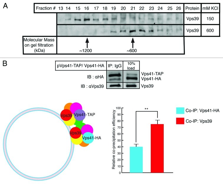
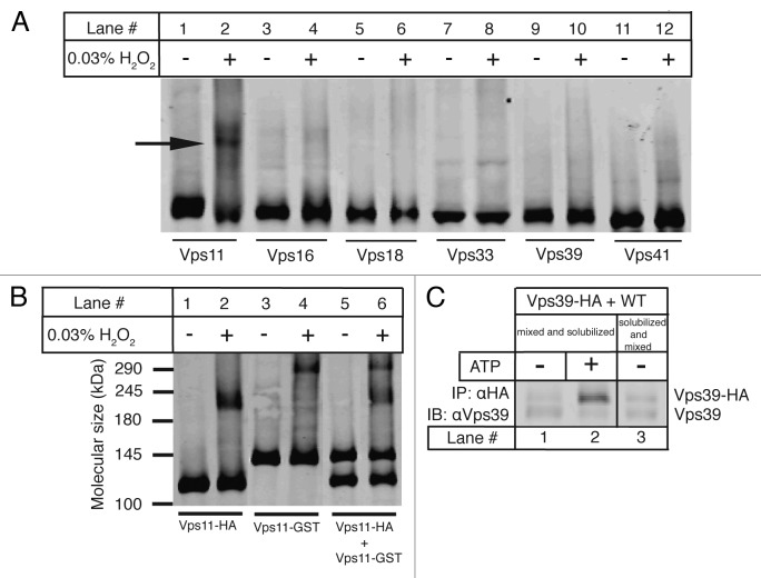
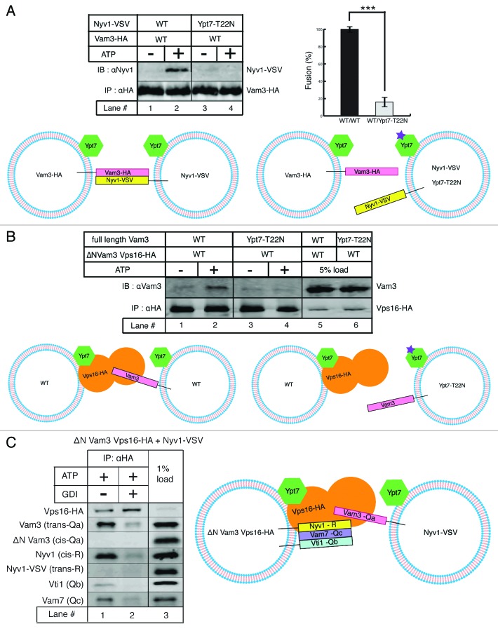
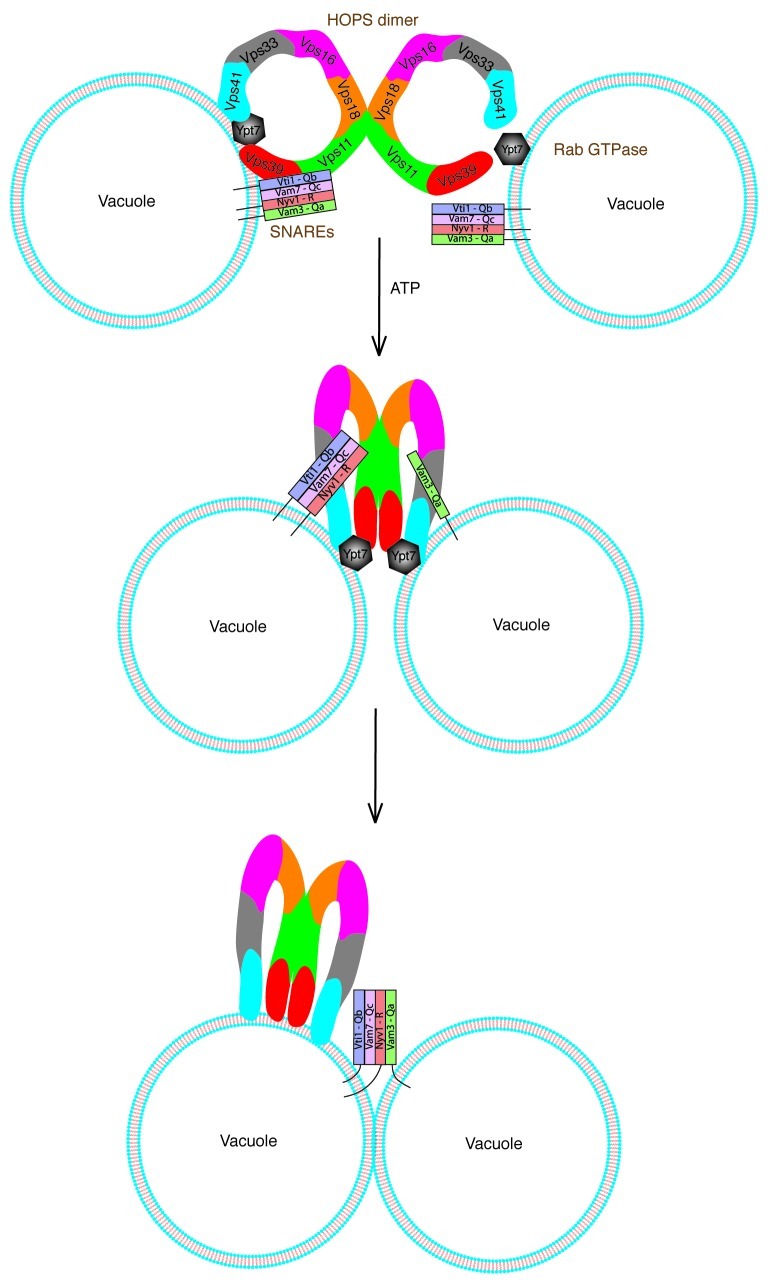
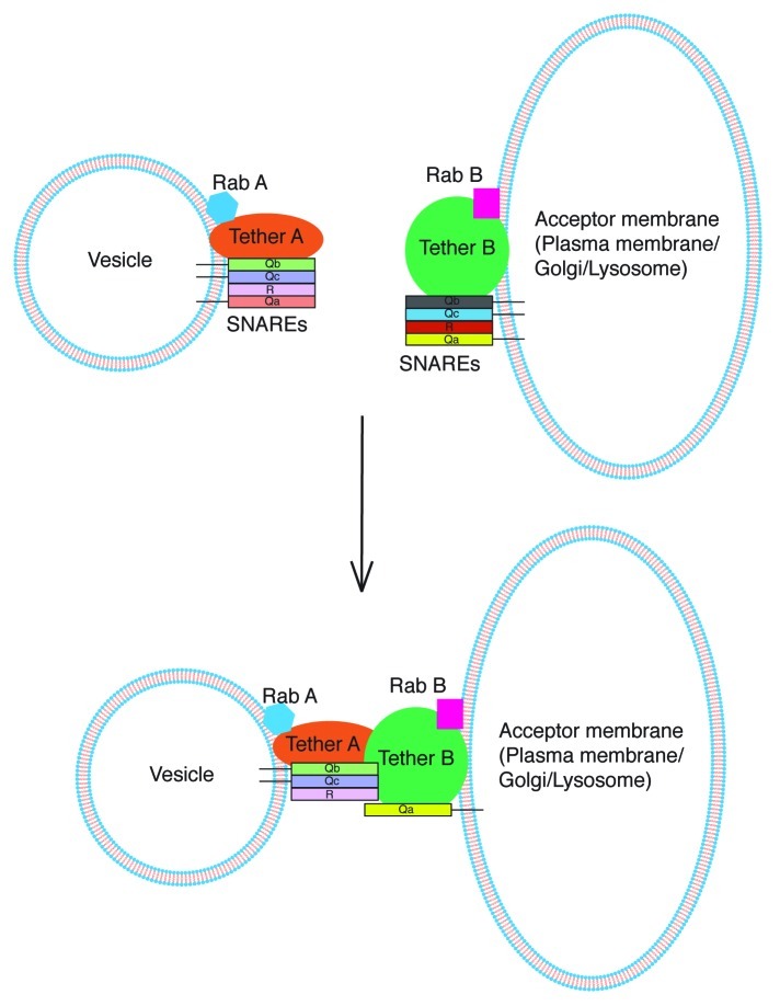
SNARE complexes mediate membrane fusion in the endomembrane system. They consist of coiled-coil bundles of four helices designated as Qa, Qb, Qc and R. A critical intermediate in the fusion pathway is the trans-SNARE complex generated by the assembly of SNAREs residing in opposing membranes. Mechanistic details of trans-SNARE complex formation and topology in a physiological system remain largely unresolved. Our studies on native yeast vacuoles revealed that SNAREs alone are insufficient to form trans-SNARE complexes and that additional factors, potentially tethering complexes and Rab GTPases, are required for the process. Here we report a novel finding that a HOPS tethering complex dimer catalyzes Rab GTPase-dependent formation of a topologically preferred QbQcR-Qa trans-SNARE complex.


Figures

None
Figure 1. HOPS is a dimeric complex on the surface of yeast vacuoles. (A) Gel filtration analysis of purified yeast vacuoles. Fractions 13–26 from Superose 6 gel filtration of solubilized vacuoles were inspected using SDS-PAGE followed by immunoblotting against Vps39. When vacuoles were processed at 150 mM KCl in the solubilization buffer, HOPS predominantly runs in fraction 16 (top lane). At 600 mM KCl, HOPS predominantly runs in fraction 21 (bottom lane). Arrows indicate molecular weights. (B) Cis-HOPS complexes assayed by differently tagged Vps41. Vacuoles from the Vps41-HA strain harboring a Vps41-TAP plasmid were processed as described in Materials and Methods. After IgG pull down of Vps41-TAP, co-precipitating proteins were analyzed by SDS-PAGE and western blotting. In the top panel, the left lane shows precipitated Vps41-TAP, co-precipitating Vps41-HA and Vps39 and the right lane displays corresponding protein inputs. The bottom panel depicts relative co-precipitation efficiencies of Vps41-HA and Vps39 quantified by Odyssey densitometry and normalized from three independent experiments. Vps39 is consistently found to co-precipitate at approximately twice the efficiency of that of Vps41-HA (p < 0.01).
None
Figure 2. HOPS dimerizes via the core subunit Vps11. (A) Crosslinking of HOPS subunits. Vacuoles harboring HA-tagged versions of all HOPS subunits were incubated under standard fusion conditions with or without 0.03% H2O2, centrifuged and analyzed on non-reducing SDS-PAGE followed by western blotting with antiHA antibody. Lanes 1–12 show the effect of peroxide treatment on each HOPS subunit labeled below. Only Vps11 shows a crosslinked product marked by the arrow (lane 2). (B) Crosslinking of Vps11-HA and Vps11-GST. Crosslinking of vacuoles harboring C-terminal 6HA or GST/3HA tags on Vps11 was done as described in 2A. Molecular weights in kDa are as marked. Vps11-HA (~120 kDa, lane 1) forms a higher molecular weight crosslinked product (~240 kDa, lane 2). Similarly Vps11-GST (~145 kDa, lane 3) forms a crosslinked product (~290 kDa, lane 4). A mixture of Vps11-HA and Vps11-GST (lane 5) shows two crosslinked products (~240 and 290 kDa, lane 6) identical to those observed in lanes 2 and 4, but no intermediate band. (C) Trans-HOPS complexes assayed by Vps39 variants. Vacuoles from Vps39-HA and wild type strains were processed as described in Materials and Methods. Vps39-HA was precipitated using antiHA antibody and co-precipitating Vps39 was analyzed using SDS-PAGE and western blotting. Interactions between Vps39-HA and non-tagged Vps39 from the two fusion partners include those observed in the absence of ATP (lane 1), in the presence of ATP (lane 2) and in the solubilization buffer (lane 3).
Figure 3.
Figure 3.
A HOPS dimer catalyzes Ypt7-dependent trans QbQcR-Qa SNARE complex formation. (A) trans-SNARE complexes assayed by tagged SNAREs. Vam3-HA vacuoles from the wild type strain were mixed with Nyv1-VSV vacuoles from either the wild type strain (lanes 1 and 2) or the Ypt7-T22N strain (lanes 3 and 4) and incubated with or without ATP under standard fusion conditions. Vam3-HA was precipitated using Protein-G/antiHA antibody and co-precipitating Nyv1-VSV was detected by SDS-PAGE and western blotting with antiNyv1 antibody. A trans-SNARE complex is observed only in the presence of ATP when both partners are wild type (lane 2) but not in a Rab mutant background for one of the partners (lane 4). The panel on the right side compares fusion efficiency between both wild type partners with that between the wild type and Ypt7-T22N mutant quantified and normalized from three independent experiments (p < 0.001). (B) trans-HOPS/Qa SNARE interactions. Vacuoles from the ΔN-terminal Vam3 strain harboring Vps16-HA were mixed with vacuoles from either the wild type strain (lanes 1 and 2) or the Ypt7-T22N strain (lanes 3 and 4) and incubated with or without ATP under standard fusion conditions. Vps16-HA was precipitated using Protein-G/antiHA antibody and co-precipitating Vam3 was detected by SDS-PAGE and western blotting with antiVam3 antibody. Vam3 (trans-Qa SNARE) co-precipitates with Vps16-HA only in presence of ATP when both partners are wild type (lane 2) but not in a Rab mutant background for one of the partners (lane 4). Lanes 5–8 show corresponding protein inputs. (C) Vacuoles from the ΔN-terminal Vam3 strain harboring Vps16-HA were mixed with vacuoles from the Nyv1-VSV strain and incubated with ATP alone (lane 1) or both ATP and GDI (lane 2) under standard fusion conditions. Vps16-HA was precipitated using Protein-G/antiHA antibody and co-precipitating SNAREs were analyzed by SDS-PAGE and western blotting against the indicated proteins. Full length Vam3 (trans Qa SNARE) and Nyv1 (cis R SNARE) co-precipitate with Vps16-HA in an ATP-dependent manner as. Lane 3 shows corresponding protein inputs.
None
Figure 4. Working model for trans-SNARE complex establishment in homotypic fusion. Each fusion partner (vacuole) is exactly identical and possesses similar fusion machinery including four vacuolar SNAREs, a HOPS dimer and Ypt7. In the presence of ATP, the HOPS dimer along with Ypt7 in cis coordinates a QbQcR acceptor subcomplex having displaced the Qa SNARE. A HOPS dimer on one fusion partner (left) recognizes activated Ypt7 on the opposing fusion partner (right) and incorporates the single Qa SNARE from the opposing partner. Ultimately a HOPS dimer-dependent QbQcR-Qa trans-SNARE complex is assembled. HOPS subunits are color coded throughout.
None
Figure 5. Proposed model for trans-SNARE complex establishment in heterotypic fusion. Each fusion partner possesses its own distinct set of fusion factors. Initial recognition between the non-identical fusion partners (vesicle and plasma membrane for example) is likely through occur through interaction between their cognate tethering complexes. This heterogeneous tether dimer catalyzes the formation of a QbQcR-Qa trans-SNARE complex in the presence of functional Rab GTPases on each partner.

Similar articles

Cited by

References

    1. Brunger AT. Structure and function of SNARE and SNARE-interacting proteins. Q Rev Biophys. 2005;38:1–47. doi: 10.1017/S0033583505004051. - DOI - PubMed
    1. Jahn R, Scheller RH. SNAREs--engines for membrane fusion. Nat Rev Mol Cell Biol. 2006;7:631–43. doi: 10.1038/nrm2002. - DOI - PubMed
    1. Alpadi K, Kulkarni A, Comte V, Reinhardt M, Schmidt A, Namjoshi S, et al. Sequential analysis of trans-SNARE formation in intracellular membrane fusion. PLoS Biol. 2012;10:e1001243. doi: 10.1371/journal.pbio.1001243. - DOI - PMC - PubMed
    1. Cai H, Reinisch K, Ferro-Novick S. Coats, tethers, Rabs, and SNAREs work together to mediate the intracellular destination of a transport vesicle. Dev Cell. 2007;12:671–82. doi: 10.1016/j.devcel.2007.04.005. - DOI - PubMed
    1. Ungermann C, von Mollard GF, Jensen ON, Margolis N, Stevens TH, Wickner W. Three v-SNAREs and two t-SNAREs, present in a pentameric cis-SNARE complex on isolated vacuoles, are essential for homotypic fusion. J Cell Biol. 1999;145:1435–42. doi: 10.1083/jcb.145.7.1435. - DOI - PMC - PubMed

LinkOut - more resources