De novo somatic mutations in components of the PI3K-AKT3-mTOR pathway cause hemimegalencephaly
- PMID: 22729223
- PMCID: PMC4417942
- DOI: 10.1038/ng.2329
De novo somatic mutations in components of the PI3K-AKT3-mTOR pathway cause hemimegalencephaly
Abstract
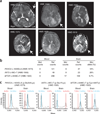
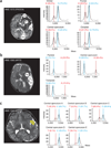
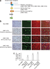
De novo somatic mutations in focal areas are well documented in diseases such as neoplasia but are rarely reported in malformation of the developing brain. Hemimegalencephaly (HME) is characterized by overgrowth of either one of the two cerebral hemispheres. The molecular etiology of HME remains a mystery. The intractable epilepsy that is associated with HME can be relieved by the surgical treatment hemispherectomy, allowing sampling of diseased tissue. Exome sequencing and mass spectrometry analysis in paired brain-blood samples from individuals with HME (n = 20 cases) identified de novo somatic mutations in 30% of affected individuals in the PIK3CA, AKT3 and MTOR genes. A recurrent PIK3CA c.1633G>A mutation was found in four separate cases. Identified mutations were present in 8-40% of sequenced alleles in various brain regions and were associated with increased neuronal S6 protein phosphorylation in the brains of affected individuals, indicating aberrant activation of mammalian target of rapamycin (mTOR) signaling. Thus HME is probably a genetically mosaic disease caused by gain of function in phosphatidylinositol 3-kinase (PI3K)-AKT3-mTOR signaling.
Figures


Similar articles
-
Profiling PI3K-AKT-MTOR variants in focal brain malformations reveals new insights for diagnostic care.Brain. 2022 Apr 29;145(3):925-938. doi: 10.1093/brain/awab376. Brain. 2022. PMID: 35355055 Free PMC article.
-
PI3K/AKT pathway mutations cause a spectrum of brain malformations from megalencephaly to focal cortical dysplasia.Brain. 2015 Jun;138(Pt 6):1613-28. doi: 10.1093/brain/awv045. Epub 2015 Feb 25. Brain. 2015. PMID: 25722288 Free PMC article.
-
Mammalian target of rapamycin pathway mutations cause hemimegalencephaly and focal cortical dysplasia.Ann Neurol. 2015 Apr;77(4):720-5. doi: 10.1002/ana.24357. Epub 2015 Feb 26. Ann Neurol. 2015. PMID: 25599672 Free PMC article.
-
Hemimegalencephaly, a paradigm for somatic postzygotic neurodevelopmental disorders.Curr Opin Neurol. 2013 Apr;26(2):122-7. doi: 10.1097/WCO.0b013e32835ef373. Curr Opin Neurol. 2013. PMID: 23449172 Review.
-
mTOR signaling in epilepsy: insights from malformations of cortical development.Cold Spring Harb Perspect Med. 2015 Apr 1;5(4):a022442. doi: 10.1101/cshperspect.a022442. Cold Spring Harb Perspect Med. 2015. PMID: 25833943 Free PMC article. Review.
Cited by
-
The genetics of the epilepsies.Curr Neurol Neurosci Rep. 2015 Jul;15(7):39. doi: 10.1007/s11910-015-0559-8. Curr Neurol Neurosci Rep. 2015. PMID: 26008807 Review.
-
The Neurodevelopmental Pathogenesis of Tuberous Sclerosis Complex (TSC).Front Neuroanat. 2020 Jul 14;14:39. doi: 10.3389/fnana.2020.00039. eCollection 2020. Front Neuroanat. 2020. PMID: 32765227 Free PMC article. Review.
-
Somatic mutations in GLI3 and OFD1 involved in sonic hedgehog signaling cause hypothalamic hamartoma.Ann Clin Transl Neurol. 2016 Mar 24;3(5):356-65. doi: 10.1002/acn3.300. eCollection 2016 May. Ann Clin Transl Neurol. 2016. PMID: 27231705 Free PMC article.
-
Cutaneous Squamous Cell Carcinoma in the Age of Immunotherapy.Cancers (Basel). 2021 Mar 8;13(5):1148. doi: 10.3390/cancers13051148. Cancers (Basel). 2021. PMID: 33800195 Free PMC article. Review.
-
Genetic Basis of Brain Malformations.Mol Syndromol. 2016 Sep;7(4):220-233. doi: 10.1159/000448639. Epub 2016 Aug 27. Mol Syndromol. 2016. PMID: 27781032 Free PMC article. Review.
References
-
- Salamon N, et al. Contralateral hemimicrencephaly and clinical-pathological correlations in children with hemimegalencephaly. Brain. 2006;129:352–365. - PubMed
-
- Di Rocco C, Battaglia D, Pietrini D, Piastra M, Massimi L. Hemimegalencephaly: clinical implications and surgical treatment. Childs Nerv. Syst. 2006;22:852–866. - PubMed
-
- Crino PB. mTOR: a pathogenic signaling pathway in developmental brain malformations. Trends Mol. Med. 2011;17:734–742. - PubMed
Publication types
MeSH terms
Substances
Grants and funding
LinkOut - more resources
Full Text Sources
Other Literature Sources
Medical
Molecular Biology Databases
Miscellaneous

