Review
. 2012;88(6):266-82.
doi: 10.2183/pjab.88.266.

Neuronal dysfunction with aging and its amelioration

Affiliations
Review

Neuronal dysfunction with aging and its amelioration

Susumu Ando. Proc Jpn Acad Ser B Phys Biol Sci. 2012.

Abstract

The author focused on the functional decline of synapses in the brain with aging to understand the underlying mechanisms and to ameliorate the deficits. The first attempt was to unravel the neuronal functions of gangliosides so that gangliosides could be used for enhancing synaptic activity. The second attempt was to elicit the neuronal plasticity in aged animals through enriched environmental stimulation and nutritional intervention. Environmental stimuli were revealed neurochemically and morphologically to develop synapses leading to enhanced cognitive function. Dietary restriction as a nutritional intervention restored the altered metabolism of neuronal membranes with aging, providing a possible explanation for the longevity effect of dietary restriction. These results obtained with aging and dementia models of animals would benefit aged people.


Figures

Figure 1.
Figure 1.
Synaptic mechanisms in relation to cholinergic neurotransmission.Acetylcholine is synthesized from choline and acetyl-CoA in synapses and stored in synaptic vesicles. Synaptic vesicles move and attach to the inside of synaptic membranes to be ready for the release of acetylcholine. Synaptic membranes are polarized by the action of ouabain-sensitive Na+,K+-ATPase to keep resting membrane potential negative inside. Electric impulses come down to nerve terminals or synapses to depolarize the synaptic membranes. The membrane depolarization very shortly open voltage-dependent calcium channels to allow calcium ions to enter into presynapses. The calcium ion influx triggers membrane fusion between synaptic vesicles and synaptic membranes by means of SNARE proteins to form pores, leading to the release of acetylcholine into synaptic cleft.22) Acetylcholine released binds to acetylcholine receptors on postsynaptic membranes to induce new action potentials. Most of acetylcholine molecules released are degraded, and free choline is taken up by presynapses through high-affinity choline transporters and reused for acetylcholine synthesis.
Figure 2.
Figure 2.
Age-related decrease of acetylcholine release from synaptosomes isolated from different age-groups of C57BL/6 mice.1) Synaptosomes were incubated with 50 mM KCL-containing Krebs-Ringer solution for 5 min at 37 ℃. Acetylcholine released was quantified and expressed as the means ± SD. *, p < 0.01 as compared to 3-month-old mice (two-tailed Student’s t-test).
Figure 3.
Figure 3.
Compositional changes in the lipids of synaptic membranes with aging (A) and correlation between Na+,K+-ATPase activity and phosphatidylcholine contents (B).2) PE, phosphatidylethanolamine; PC, phosphatidylcholine; PS, phosphatidylserine; and SM, sphingomyelin.
Figure 4.
Figure 4.
Effects of acetyl-l-carnitine administration on cholinergic parameters in rat cerebral cortical synaptosomes.4) High-affinity choline uptake (A), acetylcholine synthesis (B) and high-K+ (50 mM)-evoked acetylcholine release (C) are shown in comparison between the acetyl-l-carnitine group and control. *, p < 0.05 (vs. control, Student’s t-test).
Figure 6.
Figure 6.
Cognitive function is deteriorated with aging in rats reared in standard social condition (SC), and the age related cognitive dysfunction is largely ameliorated by an enriched environment (EC).12) To assess the cognitive function, Hebb-Williams maze problems were employed. A: The maze pattern of one problem out of Hebb-Williams maze problems is shown. Shadowed areas indicate error zones and broken lines indicate the borders for counting errors when rats cross the lines. S and G indicate start and goal boxes, respectively. B: Changes of the number of errors in the Hebb-Williams maze test with training (Trials 1–8) are drawn in logarithmic plot. Two fitted lines show the learning rates in the case of aged rats that were reared in SC or EC. Other plots for the cases of young and adult rats12) are not shown. C: Effects of different ages and environmental conditions on cognitive function are shown by the total number of errors in the Hebb-Williams maze test. Error bars indicate standard error of means.
Figure 5.
Figure 5.
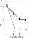
Effect of anti-Chol-1α antibody, GGR-41, on learning activity as assessed by Hebb-Williams maze #8.8) Learning rates were severely retarded in the rats given GGR-41 or mecamylamine, a nicotinic cholinergic receptor antagonist, compared with sham-operated rats. Open circles indicate controls that received only the vehicle; closed circles, rats given GGR-41 and closed squares, rats given mecamylamine.
Figure 7.
Figure 7.
Effects of different environmental conditions on synaptophysin contents.10) Two groups of rats (30 months) maintained in standard social condition (SC) or enriched environmental condition (EC) were examined for the synaptophysin contents in various brain regions. F, O, T and E are frontal, occipital, temporal and entorhinal cortices; H, S and C are hippocampus, striatum and cerebellum, respectively. The data represent mean values ± SEM. Statistically significant differences are observed for frontal and entorhinal cortices.
Figure 8.
Figure 8.
Electron micrographs of axospinous synapses in the fifth layer of rat cortices at 30 months of age.11) At, axon; and Sp, spine. Scale bars = 0.5 µm. A: An axospinous synapse in the rat reared in standard social condition. An asymmetric and nonperforated synapse contains spherical vesicles. B: An axospinous synapse in the rat reared in enriched environment. An asymmetric and perforated synapse is filled with more abundant vesicles.
Figure 9.
Figure 9.
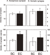
Effects of different environmental conditions on synaptic morphological parameters in the fifth layer of the neocortex of rats aged 30 months.11) A: Synaptic parameters of axospinous synapses. B: Synaptic parameters of somatic synapses. Top: Density of synapses (number per 100 µm2 for axospinous synapses, and number per 100 µm for somatic synapses). Bottom: Density of synaptic vesicles (number per µm2). SC and EC represent standard and enriched condition groups, respectively. Error bars show standard deviations. Asterisks indicate significant differences (P < 0.01).
Figure 10.
Figure 10.
Incorporation into and disappearance from myelin of deuterated cholesterol in mice given 30% D2O. The turnover rate of cholesterol as expressed by Fraction I (closed circles) was obtained by subtracting the contribution of recycling components, Fraction II (open triangles), from the total deuterated cholesterol (open circles).14)

Similar articles

Cited by

References

    1. Tanaka Y., Hasegawa A., Ando S. (1996) Impaired synaptic functions with aging as characterized by decreased calcium influx and acetylcholine release. J. Neurosci. Res. 43, 63–70 - PubMed
    1. Tanaka Y., Ando S. (1990) Synaptic aging as revealed by changes in membrane potential and decreased activity of Na+,K+-ATPase. Brain Res. 506, 46–52 - PubMed
    1. Ando S., Tanaka Y. (1990) Synaptic membrane aging in the central nervous system. Gerontology 36 (Supple. 1), 10–14 - PubMed
    1. Ando S., Tadenuma T., Tanaka Y., Fukui F., Kobayashi S., Ohashi Y., Kawabata T. (2001) Enhancement of learning activity and cholinergic synaptic function by carnitine in aging rats. J. Neurosci. Res. 66, 266–271 - PubMed
    1. Furuse H., Waki H., Kaneko K., Fujii S., Miura M., Sasaki H., Ito K.-I., Kato H., Ando S. (1998) Effect of the mono- and tetra-sialogangliosides, GM1 and GQ1b, on long-term potentiation in the CA1 hippocampal neurons of the guinea pig. Exp. Brain Res. 123, 307–314 - PubMed