. 2012 May 2;486(7403):420-8.
doi: 10.1038/nature10831.

Experimental adaptation of an influenza H5 HA confers respiratory droplet transmission to a reassortant H5 HA/H1N1 virus in ferrets

Affiliations

Experimental adaptation of an influenza H5 HA confers respiratory droplet transmission to a reassortant H5 HA/H1N1 virus in ferrets

Masaki Imai et al. Nature. .

Abstract

Highly pathogenic avian H5N1 influenza A viruses occasionally infect humans, but currently do not transmit efficiently among humans. The viral haemagglutinin (HA) protein is a known host-range determinant as it mediates virus binding to host-specific cellular receptors. Here we assess the molecular changes in HA that would allow a virus possessing subtype H5 HA to be transmissible among mammals. We identified a reassortant H5 HA/H1N1 virus-comprising H5 HA (from an H5N1 virus) with four mutations and the remaining seven gene segments from a 2009 pandemic H1N1 virus-that was capable of droplet transmission in a ferret model. The transmissible H5 reassortant virus preferentially recognized human-type receptors, replicated efficiently in ferrets, caused lung lesions and weight loss, but was not highly pathogenic and did not cause mortality. These results indicate that H5 HA can convert to an HA that supports efficient viral transmission in mammals; however, we do not know whether the four mutations in the H5 HA identified here would render a wholly avian H5N1 virus transmissible. The genetic origin of the remaining seven viral gene segments may also critically contribute to transmissibility in mammals. Nevertheless, as H5N1 viruses continue to evolve and infect humans, receptor-binding variants of H5N1 viruses with pandemic potential, including avian-human reassortant viruses as tested here, may emerge. Our findings emphasize the need to prepare for potential pandemics caused by influenza viruses possessing H5 HA, and will help individuals conducting surveillance in regions with circulating H5N1 viruses to recognize key residues that predict the pandemic potential of isolates, which will inform the development, production and distribution of effective countermeasures.


Conflict of interest statement

Competing financial interests

Y.K. has received speaker’s honoraria from Chugai Pharmaceuticals, Novartis, Daiichi-Sankyo, Toyama Chemical, Wyeth, and GlaxoSmithKline; grant support from Chugai Pharmaceuticals, Daiichi Sankyo Pharmaceutical, Toyama Chemical, Otsuka Pharmaceutical Co., Ltd.; is a consultant for Theraclone and Crucell, and is a founder of FluGen. G.N. is a consultant for Theraclone and a founder of FluGen.

Figures
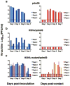
Figure 1
Figure 1
Respiratory droplet transmission of H5 avian-human reassortant viruses in ferrets. Groups of three or six ferrets were inoculated intranasally with 106 PFU of pdm09 (a), H5HA/pdm09 (b), or H5HA-mutant/pdm09 (c). One day post-infection, three or six naive ferrets were placed in each adjacent cage. Nasal washes were collected every other day from both inoculated (left panel) and contact (right panel) animals for virus titration. Virus titres in organs were determined by plaque assay in MDCK cells. The lower limit of detection is indicated by the horizontal dashed line.
Figure 2
Figure 2
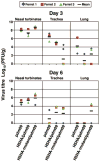
Virus replication in respiratory organs. Ferrets were infected intranasally with 106 PFU of virus. Three ferrets per group were euthanized on days 3 and 6 after infection for virus titration. Virus titres in nasal turbinates, trachea, and lung were determined by plaque assay in MDCK cells. Horizontal bars show the mean. Asterisks indicate virus titres significantly different from that of pdm09 (Dunnett’s test; P < 0.05).
Figure 3
Figure 3
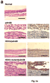
Pathological analyses of H5 avian-human reassortant viruses. a, Representative histological changes in nasal turbinates from influenza virus-infected ferrets. Three ferrets per group were infected intranasally with 106 PFU of virus, and tissues were collected on day 3 after infection for pathological examination. Uninfected ferret tissues served as negative controls (normal). Left panel, haematoxylin-eosin staining. Right panel, immunohistochemical staining for viral antigen detection (brown staining). Bars, 50 μm. b, Representative histological changes in lungs from influenza virus-infected ferrets. Three ferrets per group were infected intranasally with 106 PFU of virus, and tissues were collected on days 3 and 6 after infection for pathological examination. Left panel, the histological findings in the lungs are represented as schematic diagrams for one of the three animals per group. Cutting aspects of each lung lobe are illustrated with lesion distribution: blue, normal appearance; green, bronchopneumonia; red, alveolitis; and pink, interstitial thickening. Red circles indicate the areas shown in the histology sections. RU, right upper lobe; LU, left upper lobe; RM, right middle lobe; LL, left lower lobe; RL, right lower lobe; Ac, accessory lobe. Middle panel, haematoxylin-eosin staining. Right panel, immunohistochemical staining for viral antigen detection (brown staining). Bronchi are indicated by asterisks “*”. Inserts show a higher magnification of the antigen-positive cells. The lesions were diagnosed as bronchopneumonia, when the lung lesion was observed mainly around the inflammatory or viral antigen-positive bronchi/bronchioles. By contrast, the lesions were diagnosed as alveolitis when bronchi and bronchioles remained intact and inflammation was observed mainly in alveoli. Interstitial thickening was occasionally observed as stimulation by inoculum or secondary to the severe inflammation of adjacent tissue. Bars, 200 μm. c, Pathological severity scores in infected ferrets. To represent comprehensive histological changes, respiratory tissue slides were evaluated by scoring the pathological changes and viral antigen expression levels. The pathological scores were determined for each animal in each group (n = 3/group on days 3 and 6 pi) using the following scoring system: 0 = no pathological change/antigen negative; 1 = affected area (<30%) or only interstitial lesion/rare viral antigens; 2 = affected area (<80%, ≥30%)/moderate viral antigens; 3 = severe lesion ( ≥80%)/many viral antigens; Nasal: pathologic changes in the nasal mucosa, Nasal Ag: viral antigens in the nasal mucosa, Lung: pathologic changes in the lungs, Lung Ag: viral antigens in the lungs. Asterisks indicate virus pathological scores significantly different from that of pdm09 (Dunnett’s test; P < 0.05).
Figure 3
Figure 3
Pathological analyses of H5 avian-human reassortant viruses. a, Representative histological changes in nasal turbinates from influenza virus-infected ferrets. Three ferrets per group were infected intranasally with 106 PFU of virus, and tissues were collected on day 3 after infection for pathological examination. Uninfected ferret tissues served as negative controls (normal). Left panel, haematoxylin-eosin staining. Right panel, immunohistochemical staining for viral antigen detection (brown staining). Bars, 50 μm. b, Representative histological changes in lungs from influenza virus-infected ferrets. Three ferrets per group were infected intranasally with 106 PFU of virus, and tissues were collected on days 3 and 6 after infection for pathological examination. Left panel, the histological findings in the lungs are represented as schematic diagrams for one of the three animals per group. Cutting aspects of each lung lobe are illustrated with lesion distribution: blue, normal appearance; green, bronchopneumonia; red, alveolitis; and pink, interstitial thickening. Red circles indicate the areas shown in the histology sections. RU, right upper lobe; LU, left upper lobe; RM, right middle lobe; LL, left lower lobe; RL, right lower lobe; Ac, accessory lobe. Middle panel, haematoxylin-eosin staining. Right panel, immunohistochemical staining for viral antigen detection (brown staining). Bronchi are indicated by asterisks “*”. Inserts show a higher magnification of the antigen-positive cells. The lesions were diagnosed as bronchopneumonia, when the lung lesion was observed mainly around the inflammatory or viral antigen-positive bronchi/bronchioles. By contrast, the lesions were diagnosed as alveolitis when bronchi and bronchioles remained intact and inflammation was observed mainly in alveoli. Interstitial thickening was occasionally observed as stimulation by inoculum or secondary to the severe inflammation of adjacent tissue. Bars, 200 μm. c, Pathological severity scores in infected ferrets. To represent comprehensive histological changes, respiratory tissue slides were evaluated by scoring the pathological changes and viral antigen expression levels. The pathological scores were determined for each animal in each group (n = 3/group on days 3 and 6 pi) using the following scoring system: 0 = no pathological change/antigen negative; 1 = affected area (<30%) or only interstitial lesion/rare viral antigens; 2 = affected area (<80%, ≥30%)/moderate viral antigens; 3 = severe lesion ( ≥80%)/many viral antigens; Nasal: pathologic changes in the nasal mucosa, Nasal Ag: viral antigens in the nasal mucosa, Lung: pathologic changes in the lungs, Lung Ag: viral antigens in the lungs. Asterisks indicate virus pathological scores significantly different from that of pdm09 (Dunnett’s test; P < 0.05).
Figure 4
Figure 4
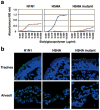
Characterization of the receptor-binding properties of an H5 HA mutant virus possessing four mutations in its HA protein. a, Binding of an H5 HA-mutant virus to sialylglycopolymers in solid-phase binding assays. A human seasonal H1N1 virus, a wild-type avian H5 HA virus, and a mutant H5 HA virus were compared for their ability to bind to sialylglycopolymers containing either α2,3-linked (blue) or α2,6-linked (red) sialic acids. b, Binding of an mutant H5 HA virus to human respiratory tissues. Human, wild-type H5, and mutant H5 HA viruses were incubated with human tissue sections and then stained with the appropriate antibody (anti-human virus antiserum to detect human influenza virus binding or anti-H5 HA antibodies to detect binding by wild-type and mutant H5 HA viruses). All sections were subsequently incubated with fluorescent-labeled secondary antibodies and Hoechst dye (blue). Green stain indicates virus binding.

Comment in

Similar articles

Cited by

References

    1. Glaser L, et al. A single amino acid substitution in 1918 influenza virus hemagglutinin changes receptor binding specificity. J Virol. 2005;79:11533–11536. - PMC - PubMed
    1. Matrosovich M, et al. Early alterations of the receptor-binding properties of H1, H2, and H3 avian influenza virus hemagglutinins after their introduction into mammals. J Virol. 2000;74:8502–8512. - PMC - PubMed
    1. Rogers GN, Paulson JC. Receptor determinants of human and animal influenza virus isolates: differences in receptor specificity of the H3 hemagglutinin based on species of origin. Virology. 1983;127:361–373. - PubMed
    1. Auewarakul P, et al. An avian influenza H5N1 virus that binds to a human-type receptor. J Virol. 2007;81:9950–9955. - PMC - PubMed
    1. Gambaryan A, et al. Evolution of the receptor binding phenotype of influenza A (H5) viruses. Virology. 2006;344:432–438. - PubMed

Publication types

MeSH terms

Substances