. 2012 Oct 25;432(2):306-15.
doi: 10.1016/j.virol.2012.05.031. Epub 2012 Jun 20.

The early UL31 gene of equine herpesvirus 1 encodes a single-stranded DNA-binding protein that has a nuclear localization signal sequence at the C-terminus

Affiliations

The early UL31 gene of equine herpesvirus 1 encodes a single-stranded DNA-binding protein that has a nuclear localization signal sequence at the C-terminus

Seongman Kim et al. Virology. .

Abstract

The amino acid sequence of the UL31 protein (UL31P) of equine herpesvirus 1 (EHV-1) has homology to that of the ICP8 of herpes simplex virus type 1 (HSV-1). Here we show that the UL31 gene is synergistically trans-activated by the IEP and the UL5P (EICP27). Detection of the UL31 RNA transcript and the UL31P in EHV-1-infected cells at 6h post-infection (hpi) as well as metabolic inhibition assays indicated that UL31 is an early gene. The UL31P preferentially bound to single-stranded DNA over double-stranded DNA in gel shift assays. Subcellular localization of the green fluorescent protein (GFP)-UL31 fusion proteins revealed that the C-terminal 32 amino acid residues of the UL31P are responsible for the nuclear localization. These findings may contribute to defining the role of the UL31P single-stranded DNA-binding protein in EHV-1 DNA replication.


Figures

Fig. 1
Fig. 1
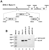
Location of UL31 gene in EHV-1 genome and detection of UL31 protein. (A) Location of UL31 gene in EHV-1 genome. Gene location was based on previously published Ab4 EHV-1 sequence (Telford et al., 1992). (B) Detection of UL31P from RK13 cells infected with EHV-1 and transfected with plasmid expressing UL31P or GFP-UL31P.
Fig. 2
Fig. 2
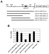
The UL31 promoter region cloned into luciferase reporter vector and UL31 promoter activity. (A) The UL31 promoter region cloned into luciferase reporter vector. The number indicates nucleotide distance from UL31 ORF first nucleotide (+ 1). The tentative TATA box sequence is located on UL31(-232/-227). Different regions of UL31 promoter were cloned into luciferase reporter vector as described in Materials and methods. (B) The UL31 promoter activities were examined without effector plasmid as described in Materials and methods. Promoter activity is expressed as relative luciferase units (RLU). Each experiment was carried out in triplicate. Error bars represent the mean ± standard deviation (SD) of three independent experiments.
Fig. 3
Fig. 3
The trans-activation of the UL31 promoter by a single or double effector vectors. Reporter plasmid [pUL31(-1033/-3)-Luc], and effector plasmids (pSVIE, pSVIR2, pCMV-IR4 (EICP22), pSVEICP0, pSVUL5 (EICP27), pCMV-ETIF, and pCMV-UL31) were used for promoter assays. Relative luciferase units (RLU). Error bars indicate the mean ± SD. (A) UL31 promoter activity by a single effector plasmid. (B) UL31 promoter activity by double effector plasmids.
Fig. 4
Fig. 4
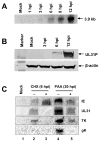
Detection of the UL31 transcript and the UL31P. Western blot and northern blot analysis were performed as described in Materials and methods. (A) Detection of the UL31 transcript from EHV-1-infected RK13 cells at indicated time points. (B) Detection of the UL31P from EHV-1-infected RK13 cells at different time points. Western blot was performed using an anti-UL31 monoclonal antibody YC3 (1:1,000) and an anti-β-actin monoclonal antibody (1:5,000) as an internal control. (C) Detection of EHV-1 representative gene and UL31 gene transcripts from EHV-1-infected RK13 cells in the presence of protein synthesis inhibitor CHX or viral DNA synthesis inhibitor PAA.
Fig. 5
Fig. 5
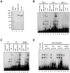
Interaction of the UL31P with DNA. Gel shift and supershift assays were performed by using radiolabeled single- and double-stranded DNA probes, GST-UL31P fusion protein, and anti-UL31P antibody YC3 as described in Materials and methods. (A) Western blot analysis of the purified GST-UL31 fusion protein with an anti-UL31 monoclonal antibody YC3. (B) Interaction of the UL31P with single-stranded DNA. The GST protein and purified GST-UL31P were mixed with 32P-labeled two single-stranded DNA probes 46-mer EHV-1 IE (+) oligonucleotide (positions −18 to −63 nt relative to the transcription start site, lanes 1–5) and its complementary oligonucleotide (lanes 6–10), and 46-bp double-stranded IE(+/−) DNA (lanes 11–15) in the presence or absence of an anti-UL31 antibody YC3 and EHV-1 IE region 2-specific monoclonal antibody E1.1. The complexes supershifted with YC3 monoclonal antibody are indicated by arrowheads. (C) Gel shift assay using three different sequence oligonucleotide probes. The GST-UL31P was mixed with radiolabeled 46-mer EHV-1 IE(+) oligonucleotide (lanes 3 and 4), 60-mer EHV-1 TK promoter sequence oligonucleotide (positions −290 to −349 nt relative to the translational start site of TK ORF, lanes 7 and 8), or 60-mer SV40 early promoter sequence oligonucleotide (positions −10 to −69 nt relative to the transcription start site, lanes 11 and 12). The complexes supershifted with YC3 monoclonal antibody are indicated by arrowheads. (D) Competition assay. The GST-UL31P was mixed with radiolabeled IE(+) DNA in the presence or absence of increasing amounts of 46-mer IE(+) oligonucleotide (lanes 3–5), 46-mer IE(−) oligonucleotide (lanes 6–8), 46-bp IE(+/−) double-stranded DNA (lanes 9–11), or 60-mer SV40E(+) oligonucleotide competitor (lanes 12–14).
Fig. 6
Fig. 6
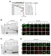
Mapping of the nuclear localization signal (NLS) of EHV-1 UL31P. Construction of GFP-UL31 fusion genes and fluorescence assays were performed as described in Materials and methods. The numbers within UL31 parentheses indicate amino acid sequences of UL31P. (A) The schematic illustrates the amino acid sequence regions of UL31P fused to the GFP and the subcellular localization of the GFP-UL31 fusion protein; N for nuclear, C for cytoplasmic, and C/N for both cytoplasmic and nuclear. (B and D) Expression of GFP-UL31 fusion proteins. The GFP fusion proteins containing truncations of UL31 ORF were detected from whole-cell lysates of transfected RK13 cells at 24 h post transfection by using an anti-rabbit GFP antibody. Arrows, bands corresponding to GFP and GFP-UL31 fusion proteins. (C and E) Localization of GFP-UL31 fusion proteins. Top, middle, and bottom panels indicate distribution of GFP-UL31 fusion protein, RK13 cell nucleus stained by DAPI, and merge of GFP and nucleus, respectively.
Fig. 7
Fig. 7
Alignment of the C-terminal 50 amino acids of EHV-1 UL31P with those of alphaherpesvirus homologues. The amino acid sequences of EHV-1 UL31 ORF and its homologs were aligned by use of CLUSTALW2 multiple sequence alignment program (http://www.ebi.ac.uk/Tools/es/cgi-bin/clustalw2). EHV-1 UL31 (Ab4p strain, accession number YP_053076; RacL11 strain, Kim et al., unpublished data); HSV-1 ICP8 (accession number BAE78520); PRV UL29 (accession number YP_068332); VZV ORF29 (accession number ABW06912). The numbers indicate the amino acid position relative to the N-terminus for each UL31P homologues. The NLS sequences of EHV-1 UL31P and HSV-1 ICP8 are underlined.

Similar articles

Cited by

References

    1. Ahn BC, Breitenbach JE, Kim SK, O’Callaghan DJ. The equine herpesvirus-1 IR3 gene that lies antisense to the sole immediate-early (IE) gene is trans-activated by the IE protein, and is poorly expressed to a protein. Virology. 2007;363:15–25. - PMC - PubMed
    1. Ahn BC, Kim S, Zhang Y, Charvat RA, O’Callaghan DJ. The early UL3 gene of equine herpesvirus-1 encodes a tegument protein not essential for replication or virulence in the mouse. Virology. 2011;420:20–31. - PMC - PubMed
    1. Ahn BC, Zhang Y, O’Callaghan DJ. The equine herpesvirus-1 (EHV-1) IR3 transcript downregulates expression of the IE gene and the absence of IR3 gene expression alters EHV-1 biological properties and virulence. Virology. 2010;402:327–337. - PMC - PubMed
    1. Allen GP, Bryans JT. Molecular epizootiology, pathogenesis, and prophylaxis of equine herpesvirus-1 infections. Prog Vet Microbiol Immunol. 1986;2:78–144. - PubMed
    1. Allen GP, O’Callaghan DJ, Randall CC. Purification and characterization of equine herpesvirus-induced DNA polymerase. Virology. 1977;76:395–408. - PubMed

Publication types

MeSH terms