Commensal bacteria calibrate the activation threshold of innate antiviral immunity
- PMID: 22705104
- PMCID: PMC3679670
- DOI: 10.1016/j.immuni.2012.04.011
Commensal bacteria calibrate the activation threshold of innate antiviral immunity
Abstract
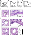
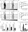
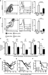
Signals from commensal bacteria can influence immune cell development and susceptibility to infectious or inflammatory diseases. However, the mechanisms by which commensal bacteria regulate protective immunity after exposure to systemic pathogens remain poorly understood. Here, we demonstrate that antibiotic-treated (ABX) mice exhibit impaired innate and adaptive antiviral immune responses and substantially delayed viral clearance after exposure to systemic LCMV or mucosal influenza virus. Furthermore, ABX mice exhibited severe bronchiole epithelial degeneration and increased host mortality after influenza virus infection. Genome-wide transcriptional profiling of macrophages isolated from ABX mice revealed decreased expression of genes associated with antiviral immunity. Moreover, macrophages from ABX mice exhibited defective responses to type I and type II IFNs and impaired capacity to limit viral replication. Collectively, these data indicate that commensal-derived signals provide tonic immune stimulation that establishes the activation threshold of the innate immune system required for optimal antiviral immunity.
Copyright © 2012 Elsevier Inc. All rights reserved.
Figures


Comment in
-
Maintaining poise: commensal microbiota calibrate interferon responses.Immunity. 2012 Jul 27;37(1):10-2. doi: 10.1016/j.immuni.2012.07.001. Immunity. 2012. PMID: 22840839
Similar articles
-
Deficiency of the B cell-activating factor receptor results in limited CD169+ macrophage function during viral infection.J Virol. 2015 May;89(9):4748-59. doi: 10.1128/JVI.02976-14. Epub 2015 Feb 11. J Virol. 2015. PMID: 25673724 Free PMC article.
-
The Intestinal Microbiome Primes Host Innate Immunity against Enteric Virus Systemic Infection through Type I Interferon.mBio. 2021 May 11;12(3):e00366-21. doi: 10.1128/mBio.00366-21. mBio. 2021. PMID: 33975932 Free PMC article.
-
Interleukin-27R Signaling Mediates Early Viral Containment and Impacts Innate and Adaptive Immunity after Chronic Lymphocytic Choriomeningitis Virus Infection.J Virol. 2018 May 29;92(12):e02196-17. doi: 10.1128/JVI.02196-17. Print 2018 Jun 15. J Virol. 2018. PMID: 29593047 Free PMC article.
-
Confounding roles for type I interferons during bacterial and viral pathogenesis.Int Immunol. 2013 Dec;25(12):663-9. doi: 10.1093/intimm/dxt050. Epub 2013 Oct 24. Int Immunol. 2013. PMID: 24158954 Free PMC article. Review.
-
Innate immunity to influenza virus infection.Nat Rev Immunol. 2014 May;14(5):315-28. doi: 10.1038/nri3665. Nat Rev Immunol. 2014. PMID: 24762827 Free PMC article. Review.
Cited by
-
Epidemiology of acute respiratory viral infections in children in Vientiane, Lao People's Democratic Republic.J Med Virol. 2021 Aug;93(8):4748-4755. doi: 10.1002/jmv.27004. Epub 2021 May 12. J Med Virol. 2021. PMID: 33830514 Free PMC article.
-
IKKβ in intestinal epithelial cells regulates allergen-specific IgA and allergic inflammation at distant mucosal sites.Mucosal Immunol. 2014 Mar;7(2):257-67. doi: 10.1038/mi.2013.43. Epub 2013 Jul 10. Mucosal Immunol. 2014. PMID: 23839064 Free PMC article.
-
Enteric immunity, the gut microbiome, and sepsis: Rethinking the germ theory of disease.Exp Biol Med (Maywood). 2017 Jan;242(2):127-139. doi: 10.1177/1535370216669610. Epub 2016 Oct 4. Exp Biol Med (Maywood). 2017. PMID: 27633573 Free PMC article. Review.
-
Macrophage Polarization in Virus-Host Interactions.J Clin Cell Immunol. 2015 Apr;6(2):311. doi: 10.4172/2155-9899.1000311. J Clin Cell Immunol. 2015. PMID: 26213635 Free PMC article.
-
Autologous Faecal Microbiota Transplantation to Improve Outcomes of Haematopoietic Stem Cell Transplantation: Results of a Single-Centre Feasibility Study.Biomedicines. 2023 Dec 11;11(12):3274. doi: 10.3390/biomedicines11123274. Biomedicines. 2023. PMID: 38137495 Free PMC article.
References
-
- Altman JD, Moss PA, Goulder PJ, Barouch DH, McHeyzer-Williams MG, Bell JI, McMichael AJ, Davis MM. Phenotypic analysis of antigen-specific T lymphocytes. Science. 1996;274:94–96. - PubMed
Publication types
MeSH terms
Substances
Grants and funding
- U19 AI083022/AI/NIAID NIH HHS/United States
- AI083022/AI/NIAID NIH HHS/United States
- R01 AI095466/AI/NIAID NIH HHS/United States
- T32 RR007063/RR/NCRR NIH HHS/United States
- AI091759/AI/NIAID NIH HHS/United States
- T32-AI007324/AI/NIAID NIH HHS/United States
- AI087990/AI/NIAID NIH HHS/United States
- P01 AI078897/AI/NIAID NIH HHS/United States
- 2-P30 CA016520/CA/NCI NIH HHS/United States
- R01 AI102942/AI/NIAID NIH HHS/United States
- R21 AI077098/AI/NIAID NIH HHS/United States
- K08 DK093784/DK/NIDDK NIH HHS/United States
- R21 AI083480/AI/NIAID NIH HHS/United States
- U01 AI095608/AI/NIAID NIH HHS/United States
- R01 AI061570/AI/NIAID NIH HHS/United States
- R01 AI074878/AI/NIAID NIH HHS/United States
- T32 AI007324/AI/NIAID NIH HHS/United States
- AI078897/AI/NIAID NIH HHS/United States
- AI061570/AI/NIAID NIH HHS/United States
- R01 AI091759/AI/NIAID NIH HHS/United States
- DK50306/DK/NIDDK NIH HHS/United States
- P30 DK050306/DK/NIDDK NIH HHS/United States
- T32 AI007532/AI/NIAID NIH HHS/United States
- R01 AI071309/AI/NIAID NIH HHS/United States
- K08-DK093784/DK/NIDDK NIH HHS/United States
- AI074878/AI/NIAID NIH HHS/United States
- T32-AI007532/AI/NIAID NIH HHS/United States
- T32-RR007063/RR/NCRR NIH HHS/United States
- R21 AI087990/AI/NIAID NIH HHS/United States
- AI095608/AI/NIAID NIH HHS/United States
- P30 CA016520/CA/NCI NIH HHS/United States
- T32-AI05528/AI/NIAID NIH HHS/United States
- AI095466/AI/NIAID NIH HHS/United States
- AI071309/AI/NIAID NIH HHS/United States
- HHSN266200500030C/AI/NIAID NIH HHS/United States
- AI077098/AI/NIAID NIH HHS/United States
LinkOut - more resources
Full Text Sources
Other Literature Sources
Molecular Biology Databases

