Nitric oxide releasing nanoparticles for treatment of Candida albicans burn infections
- PMID: 22701111
- PMCID: PMC3370663
- DOI: 10.3389/fmicb.2012.00193
Nitric oxide releasing nanoparticles for treatment of Candida albicans burn infections
Abstract
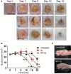
Candida albicans is a leading fungal cause of burn infections in hospital settings, and sepsis is one of the principle causes of death after a severe burn. The prevalence of invasive candidiasis in burn cases varies widely, but it accounts for 3-23% of severe infection with a mortality rate ranging from 14 to 70%. Therefore, it is imperative that we develop innovative therapeutics to which this fungus is unlikely to evolve resistance, thus curtailing the associated morbidity and mortality and ultimately improving our capacity to treat these infections. An inexpensive and stable nitric oxide (NO)-releasing nanoparticle (NO-np) platform has been recently developed. NO is known to have direct antifungal activity, modulate host immune responses and significantly regulate wound healing. In this study, we hypothesized that NO-np would be an effective therapy in the treatment of C. albicans burn infections. Using a murine burn model, NO-np demonstrated antifungal activity against C. albicans in vivo, most likely by arresting its growth and morphogenesis as demonstrated in vitro. NO-np demonstrated effective antimicrobial activity against yeast and filamentous forms of the fungus. Moreover, we showed that NO-np significantly accelerated the rate of wound healing in cutaneous burn infections when compared to controls. The histological evaluation of the affected tissue revealed that NO-np treatment modified leukocyte infiltration, minimized the fungal burden, and reduced collagen degradation, thus providing potential mechanisms for the therapeutics' biological activity. Together, these data suggest that NO-np have the potential to serve as a novel topical antifungal which can be used for the treatment of cutaneous burn infections and wounds.
Keywords: Candida albicans; burn healing; collagen; nanoparticles; nitric oxide.
Figures


Similar articles
-
Antimicrobial and healing efficacy of sustained release nitric oxide nanoparticles against Staphylococcus aureus skin infection.J Invest Dermatol. 2009 Oct;129(10):2463-9. doi: 10.1038/jid.2009.95. Epub 2009 Apr 23. J Invest Dermatol. 2009. PMID: 19387479
-
Sustained Nitric Oxide-Releasing Nanoparticles Induce Cell Death in Candida albicans Yeast and Hyphal Cells, Preventing Biofilm Formation In Vitro and in a Rodent Central Venous Catheter Model.Antimicrob Agents Chemother. 2016 Mar 25;60(4):2185-94. doi: 10.1128/AAC.02659-15. Print 2016 Apr. Antimicrob Agents Chemother. 2016. PMID: 26810653 Free PMC article.
-
Antifungal effects of indolicidin-conjugated gold nanoparticles against fluconazole-resistant strains of Candida albicans isolated from patients with burn infection.Int J Nanomedicine. 2019 Jul 17;14:5323-5338. doi: 10.2147/IJN.S207527. eCollection 2019. Int J Nanomedicine. 2019. PMID: 31409990 Free PMC article.
-
Candida and candidaemia. Susceptibility and epidemiology.Dan Med J. 2013 Nov;60(11):B4698. Dan Med J. 2013. PMID: 24192246 Review.
-
The in vivo anti-Candida albicans activity of flavonoids.J Oral Biosci. 2021 Jun;63(2):120-128. doi: 10.1016/j.job.2021.03.004. Epub 2021 Apr 9. J Oral Biosci. 2021. PMID: 33839266 Review.
Cited by
-
Inhibition of Classical and Alternative Modes of Respiration in Candida albicans Leads to Cell Wall Remodeling and Increased Macrophage Recognition.mBio. 2019 Jan 29;10(1):e02535-18. doi: 10.1128/mBio.02535-18. mBio. 2019. PMID: 30696734 Free PMC article.
-
Development of Novel Amphotericin B-Immobilized Nitric Oxide-Releasing Platform for the Prevention of Broad-Spectrum Infections and Thrombosis.ACS Appl Mater Interfaces. 2021 May 5;13(17):19613-19624. doi: 10.1021/acsami.1c01330. Epub 2021 Apr 27. ACS Appl Mater Interfaces. 2021. PMID: 33904311 Free PMC article.
-
Developments in the management of Chagas cardiomyopathy.Expert Rev Cardiovasc Ther. 2015 Dec;13(12):1393-409. doi: 10.1586/14779072.2015.1103648. Epub 2015 Oct 23. Expert Rev Cardiovasc Ther. 2015. PMID: 26496376 Free PMC article. Review.
-
Fidgetin-Like 2: A Microtubule-Based Regulator of Wound Healing.J Invest Dermatol. 2015 Sep;135(9):2309-2318. doi: 10.1038/jid.2015.94. Epub 2015 Mar 10. J Invest Dermatol. 2015. PMID: 25756798 Free PMC article.
-
Topical antimicrobials for burn infections - an update.Recent Pat Antiinfect Drug Discov. 2013 Dec;8(3):161-97. doi: 10.2174/1574891x08666131112143447. Recent Pat Antiinfect Drug Discov. 2013. PMID: 24215506 Free PMC article. Review.
References
-
- Alonso R., Llopis I., Flores C., Murgui A., Timoneda J. (2001). Different adhesins for type IV collagen on Candida albicans: identification of a lectin-like adhesin recognizing the 7S(IV) domain. Microbiology 147 (Pt 7), 1971–1981 - PubMed
-
- Baroli B., Ennas M. G., Loffredo F., Isola M., Pinna R., López-Quintela M. A. (2007). Penetration of metallic nanoparticles in human full-thickness skin. J. Invest. Dermatol. 127, 1701–1712 - PubMed
LinkOut - more resources
Full Text Sources
Other Literature Sources
Miscellaneous

