

Role of estrogen receptor signaling required for endometriosis-like lesion establishment in a mouse model
- PMID: 22700766
- PMCID: PMC3404357
- DOI: 10.1210/en.2012-1294
Role of estrogen receptor signaling required for endometriosis-like lesion establishment in a mouse model
Abstract
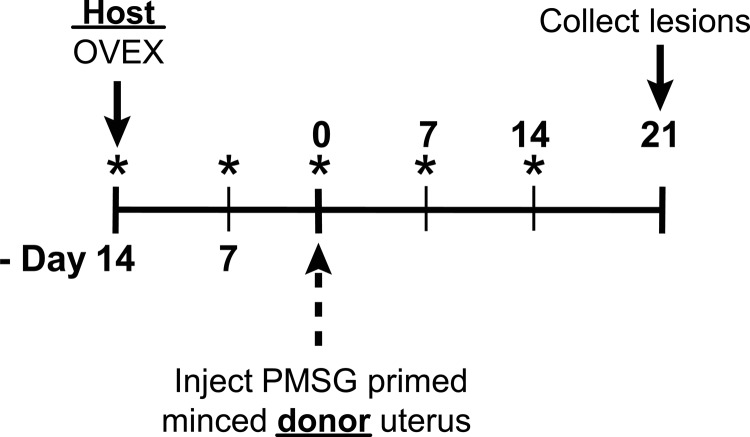
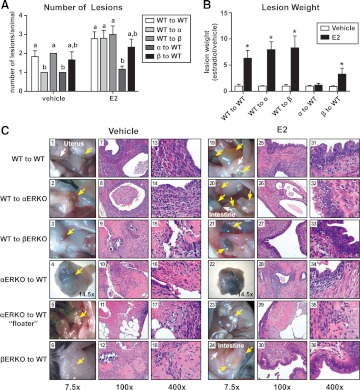
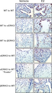
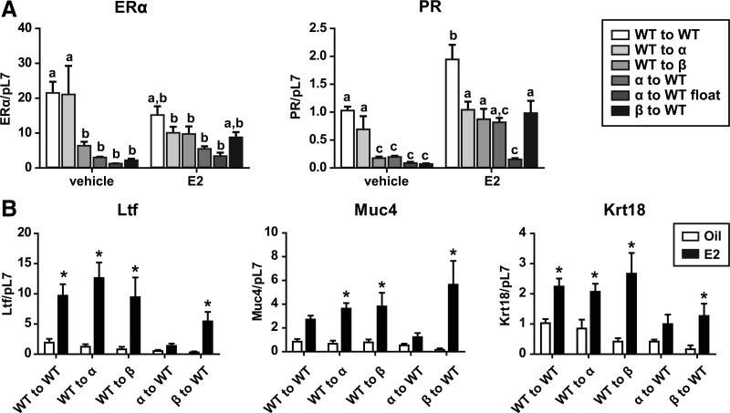
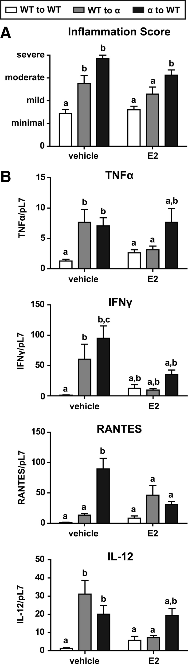
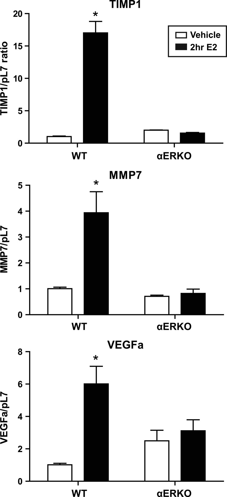
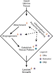
Endometriosis results from ectopic invasion of endometrial tissue within the peritoneal cavity. Aberrant levels of the estrogen receptor (ER), ERα and ERβ, and higher incidence of autoimmune disorders are observed in women with endometriosis. An immunocompetent mouse model of endometriosis was used in which minced uterine tissue from a donor was dispersed into the peritoneal cavity of a recipient. Wild-type (WT), ERα-knockout (αERKO), and βERKO mice were donors or recipients to investigate the roles of ERα, ERβ, and estradiol-mediated signaling on endometriosis-like disease. Mice were treated with vehicle or estradiol, and resulting location, number, and size of endometriosis-like lesions were assessed. In comparison with WT lesions in WT hosts, αERKO lesions in WT hosts were smaller and fewer in number. The effect of ER status and estradiol treatment on nuclear receptor status, proliferation, organization, and inflammation within lesions were examined. αERKO lesions in WT hosts did not form distal to the incision site, respond to estradiol, or proliferate but did have increased inflammation. WT lesions in αERKO hosts did respond to estradiol, proliferate, and show decreased inflammation with treatment, but surprisingly, progesterone receptor expression and localization remained unchanged. Only minor differences were observed between WT lesions in βERKO hosts and βERKO lesions in WT hosts, demonstrating the estradiol-mediated signaling responses are predominately through ERα. In sum, these results suggest ER in both endometriosis-like lesions and their environment influence lesion characteristics, and understanding these interactions may play a critical role in elucidating this enigmatic disease.
Figures

Similar articles
-
Krüppel-like factor 9 deficiency in uterine endometrial cells promotes ectopic lesion establishment associated with activated notch and hedgehog signaling in a mouse model of endometriosis.Endocrinology. 2014 Apr;155(4):1532-46. doi: 10.1210/en.2013-1947. Epub 2014 Jan 29. Endocrinology. 2014. PMID: 24476135 Free PMC article.
-
Krüppel-Like Factor 13 Deficiency in Uterine Endometrial Cells Contributes to Defective Steroid Hormone Receptor Signaling but Not Lesion Establishment in a Mouse Model of Endometriosis.Biol Reprod. 2015 Jun;92(6):140. doi: 10.1095/biolreprod.115.130260. Epub 2015 Apr 22. Biol Reprod. 2015. PMID: 25904015 Free PMC article.
-
Studies using the estrogen receptor alpha knockout uterus demonstrate that implantation but not decidualization-associated signaling is estrogen dependent.Biol Reprod. 2002 Oct;67(4):1268-77. doi: 10.1095/biolreprod67.4.1268. Biol Reprod. 2002. PMID: 12297545
-
Estrogenic effects on prostatic differentiation and carcinogenesis.Reprod Fertil Dev. 2001;13(4):285-96. doi: 10.1071/rd01010. Reprod Fertil Dev. 2001. PMID: 11800167 Review.
-
The role of estrogen and estrogen receptor-alpha in male adipose tissue.Mol Cell Endocrinol. 2001 Jun 10;178(1-2):147-54. doi: 10.1016/s0303-7207(01)00414-2. Mol Cell Endocrinol. 2001. PMID: 11403904 Review.
Cited by
-
Use of a Mouse Model of Experimentally Induced Endometriosis to Evaluate and Compare the Effects of Bisphenol A and Bisphenol AF Exposure.Environ Health Perspect. 2018 Dec;126(12):127004. doi: 10.1289/EHP3802. Environ Health Perspect. 2018. PMID: 30675821 Free PMC article.
-
A single gestational exposure to 2,3,7,8-tetrachlorodibenzo-p-dioxin disrupts the adult uterine response to estradiol in mice.Toxicol Sci. 2013 Dec;136(2):514-26. doi: 10.1093/toxsci/kft208. Epub 2013 Sep 19. Toxicol Sci. 2013. PMID: 24052564 Free PMC article.
-
Estrogen is essential but not sufficient to induce endometriosis.J Biosci. 2017 Jun;42(2):251-263. doi: 10.1007/s12038-017-9687-4. J Biosci. 2017. PMID: 28569249
-
Intricate Connections between the Microbiota and Endometriosis.Int J Mol Sci. 2021 May 26;22(11):5644. doi: 10.3390/ijms22115644. Int J Mol Sci. 2021. PMID: 34073257 Free PMC article. Review.
-
The Increase of Circulating PD-1- and PD-L1-Expressing Lymphocytes in Endometriosis: Correlation with Clinical and Laboratory Parameters.Mediators Inflamm. 2018 Nov 25;2018:7041342. doi: 10.1155/2018/7041342. eCollection 2018. Mediators Inflamm. 2018. PMID: 30595667 Free PMC article.
References
-
- Rawson JM. 1991. Prevalence of endometriosis in asymptomatic women. J Reprod Med 36:513–515 - PubMed
-
- Galle PC. 1989. Clinical presentation and diagnosis of endometriosis. Obstet Gynecol Clin North Am 16:29–42 - PubMed
-
- Bulun SE, Imir G, Utsunomiya H, Thung S, Gurates B, Tamura M, Lin Z. 2005. Aromatase in endometriosis and uterine leiomyomata. J Steroid Biochem Mol Biol 95:57–62 - PubMed
-
- Giudice LC, Kao LC. 2004. Endometriosis. Lancet 364:1789–1799 - PubMed
Publication types
MeSH terms
Substances
Grants and funding
LinkOut - more resources
Full Text Sources
Other Literature Sources
Medical
Molecular Biology Databases
Research Materials
