. 2012 Jun 12;22(6):1191-207.
doi: 10.1016/j.devcel.2012.04.018.

FGF9 and FGF20 maintain the stemness of nephron progenitors in mice and man

Affiliations

FGF9 and FGF20 maintain the stemness of nephron progenitors in mice and man

Hila Barak et al. Dev Cell. .

Abstract

The identity of niche signals necessary to maintain embryonic nephron progenitors is unclear. Here we provide evidence that Fgf20 and Fgf9, expressed in the niche, and Fgf9, secreted from the adjacent ureteric bud, are necessary and sufficient to maintain progenitor stemness. Reduction in the level of these redundant ligands in the mouse led to premature progenitor differentiation within the niche. Loss of FGF20 in humans, or of both ligands in mice, resulted in kidney agenesis. Sufficiency was shown in vitro where Fgf20 or Fgf9 (alone or together with Bmp7) maintained isolated metanephric mesenchyme or sorted nephron progenitors that remained competent to differentiate in response to Wnt signals after 5 or 2 days in culture, respectively. These findings identify a long-sought-after critical component of the nephron stem cell niche and hold promise for long-term culture and utilization of these progenitors in vitro.


Figures

Figure 1
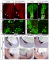
Figure 1. Fgf20 and Fgf9 are required for kidney development
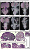
A-C) Normal urogenital system formed in control (A), Fgf20βGal/βGal (B) and Fgf9+/− Fgf20+/βGal (C) embryos. Note slightly smaller kidneys in (B) but not in (C). D) Fgf9+/−; Fgf20βGal/βGal embryos developed mild (D’) to severe (D”) reduction in kidney size. E) When both alleles were deleted the kidneys were absent (yellow arrowhead) and adrenal glands were smaller (white arrowhead). F,G) H&E staining of kidneys from control (F) and Fgf20βGal/βGal (G) mice shows bilateral reduction in kidney size of mutant compared to control. Nephrogenic zone thickness (white line) is indistinguishable between Fgf20βGal/βGal and control kidneys. High power image of boxed area in G shows normal intermediate steps in nephron development (SSB, arrow; early glomeruli, arrowhead in G’). H) Kidney section of Fgf9+/−; Fgf20βGal/βGal embryo showed regions where the nephrogenic zone was depleted (arrowheads). H’) High power image of boxed area in H. Glomeruli were counted from 4 control, 5 Fgf20+/βGal and 5 Fgf20βGal/βGal kidneys. Average count (in thousands with S.D) is shown in (I). Asterisk p= 8.1775×10⁻⁸. Kidney (K), Gonad (G) and Bladder (B). Scale bar: 500μm.
Figure 2
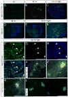
Figure 2. Loss of nephron progenitors in Fgf9/20 mutants
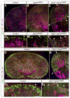
Sections of E18.5 kidneys stained with Ck8 (purple, UB), WT1 (Red; low, CMM; high, podocytes), LTL (blue, proximal tubules), Six2 (green, progenitors) or Pax2 (UB, nephron progenitors and nephron epithelia). (A, D). The nephrogenic zone in controls (green) is located distal to the UB and to glomeruli (red) attached to proximal tubules (blue). A’-D’) High power image of control nephrogenic zone showed progenitors (yellow arrowheads) packed in two compact cell layers distal UB tips. B) The nephrogenic zone near some tips in Fgf20βGal/βGal kidneys contains fewer Six2+ cells relative to controls but is otherwise indistinguishable (high power view in B’). C-E) Fgf9+/−; Fgf20βGal/βGal kidneys contained many cortical areas where the nephrogenic zone was replaced by mature structures (white arrows). C’-E’) High power image of nephrogenic zone from Fgf9+/−; Fgf20βGal/βGal kidneys. The cortex contained some UB tips surrounded by two cell layers of nephron progenitors (yellow arrowheads), other tips with fewer Six2+ cells (white arrowheads), and some lacking a nephrogenic zone altogether contained mature nephrons instead (white arrows). Scale bars: 100μm.
Figure 3
Figure 3. Reduced Fgf9/20 levels allow premature differentiation of nephron progenitors in the niche
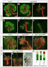
E11.5 metanephroi analyzed after 48hrs in organ culture (A-F). Fgf20βGal/βGal metanephroi (B,C) were smaller than controls (A). B-F) Some UB tips in Fgf20βGal/βGal metanephroi (B-C) or Fgf9+/−; Fgf20βGal/βGal metanephroi (D-F) are surrounded by fewer Six2+ cells (yellow arrow), while others lack Six2 expressing cells (blue arrows). G-K) Jag1 (epithelial RVs and SSBs), Ck8 (UB), and Six2 (nephron progenitors) demonstrate premature differentiation in the niche in metanephroi distal to some UB tips (white arrows). L) In situ hybridization of Wnt4 (blue) and antibody staining to Ck8 (brown) shows ectopic Wnt4 expression distal to UB tips in Fgf9+/−; Fgf20βGal/βGal metanephroi (white arrows). M) Quantification of tip subtypes (marked with yellow or blue arrows) in metanephroi of various genotypes (supplemental information). Normal progenitor numbers in dark green reduced progenitors numbers in light green; no progenitors in pink. Scale bars: 100μm.
Figure 4
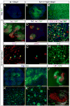
Figure 4. Fgf20 expressed in nephron progenitors and Fgf9 secreted from the UB collaborate to maintain the niche
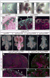
A-C) Whole-mount x-gal staining at E10.5-12.5 Fgf20/βGal embryos detects ßGal activity (blue) in the mesonephros (black arrowheads) and metanephric kidneys (red arrowheads). D) X-gal staining identified activity in the MM of E14.5 Fgf20/βGal kidneys (black dashed line-UB). E) Ck8, NCAM (epithelia) and ßGal (Fgf20 expression domain) staining of E16.5 Fgf20βGal/βGal kidneys. ßGal protein was detected in the CMM (white arrows). The weak signal in the nascent RV (yellow arrowheads) may be due to expression or perdurance. F) Six2 identified nephron progenitors that contained ßGal protein. G-J) Urogenital system in mice lacking Fgf9 in their UB. G) Control (HoxB7-Cre+/tg;Fgf20+/βGal; H) and HoxB7-Cre+/tg;Fgf9f/f;Fgf20+/βGal kidneys appeared normal. I) In contrast, HoxB7-Cre+/tg;Fgf9+/f;Fgf20βGal/βGal kidneys are smaller. J) Fully penetrant kidney agenesis (yellow arrowhead) seen in HoxB7-cre;Fgf9f/f; Fgf20βGal/βGal embryos. K-L) Ck8 (magenta), WT1 (red), LTL (blue) and Six2 (green) staining of HoxB7-Cretg; Fgf9f/f; Fgf20+/βGal (control, K) or HoxB7-Cretg; Fgf9+/f; Fgf20βGal/βGal (L) kidneys. Note large cortical areas where the nephrogenic zone was replaced by mature structures (white arrows, L). High power image of nephrogenic zone of control HoxB7-Cretg; Fgf9f/f; Fgf20+/βGal kidney (K’, yellow arrowheads) or HoxB7-Cretg; Fgf9+/f; Fgf20βGal/βGal kidney (L’). Note regions lacking Six2 with mature structures (podocytes: red; proximal tubules: blue) located instead at edge of kidney (white arrows). Kidney (K), Gonad (G), Bladder (B). Scale bars: 50μm (E-F), 500μm (G-J), 100μm (K-L’).
Figure 5
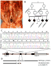
Figure 5. Identification of a homozygous frameshift mutation in FGF20 that segregates with bilateral renal agenesis
(A) Bilateral reno-ureteral agenesis in 18-week fetus; (a) adrenal glands with flat oval shape due to kidney agenesis. (B) Pedigree of the consanguineous family showing two first cousin couples and four fetuses presenting with anhydramnios / bilateral renal agenesis; segregation of the FGF20 c.337delG mutation is indicated. (C) Nucleotide sequence traces and deduced amino acid sequence within exon 2 shown for wild type (top) and mutated (bottom) alleles. Deleted guanine at position 337 of the coding sequence is marked with asterisk in the wild type sequence. The frameshift introduced missense amino acids from position 113 to 120 (in bold) and created a stop codon at position 121 (numbering as in NCBI sequence AB030648). (D) Wild type and deduced FGF20 proteins: Heparin-binding domains indicated by light grey boxes; position of mutation is marked with red asterisk; the 8 missense amino acids are in black.
Figure 6
Figure 6. Fgf9/20 deficient kidneys initiate UB outgrowth but branching fails due to high rates of apoptosis in the MM
E11.5 whole-mount Ck8 staining in controls (A) illustrated a branched UB (white arrowhead). In contrast, the UB in Fgf9−/−; Fgf20βGal/βGal E11.5 embryos (B) elongated (arrow) but failed to branch (yellow arrowhead). Sections of E11.5 kidneys stained with Pax2 (UB and MM) showed MM condensing around the UB tips (Ck8+) in controls (C) but not in Fgf9−/−; Fgf20βGal/βGal embryos (D). The mutant MM appeared diffuse and formed a smaller contact zone with the UB. MM in control is Pax2 positive and TUNEL negative (E); the dying MM cells in Fgf9−/−; Fgf20βGal/βGal embryos are TUNEL positive (white arrowheads in F). At E12.5, Ck8 and Pax2 outline a rudimentary MM remaining in Fgf9−/−; Fgf20βGal/βGal embryos (H) compared to controls (G) (yellow arrowheads in G-H).
Figure 7
Figure 7. Fgf9 promotes survival and maintains competence of nephron progenitors in vitro
Isolated E11.5 MM cultured in defined media (M; A), in Heparin-supplemented media (M+H; B), or in Heparin and Fgf9 media (M+H+Fgf9; C,E-K). Explants were fixed and stained as indicated. After 4 days in media, the majority of MM cells were TUNEL-positive (A). When supplemented with Heparin, cell survival improved, some entering mitosis (B). In the presence of Fgf9, survival and proliferation were further increased (C). Isolated E11.5 MM was grown in culture for 2 days with M+H (D) or M+H+Fgf9 (E-I). Six2+ cells were only detected when Fgf9 was present (E, E’). A few progenitors (left-pointing arrowheads) positive for Six2 (F), pHH3 (G) and DAPI (H) underwent mitosis (merged image; I). Nephron progenitors maintained in the presence of Fgf9 for 2 days are competent to respond to Wnt produced by dSC by forming epithelial aggregates expressing both distal (Cdh1+) and proximal (Jag1+) markers (J, J’, higher magnification; see text for detail). Note undifferentiated, Six2+ cells in (K). Colonies of Six2+ cells isolated by FACS were maintained for 2 days in culture in M+H+Fgf9 (L). Scale bars: 100μm.
Figure 8
Figure 8. Fgf9 synergized with Bmp7 to promote self-renewal of competent nephron progenitors in vitro
Isolated E11.5 MM was grown for 48hrs in media supplemented with Bmp7 (A) or Heparin with Fgf9 and Bmp7 (M+H+Fgf9+Bmp7) (B-J). Six2+ cells were not detected in explants grown with Bmp7 (A). In explants cultured in M+H+Fgf9+Bmp7, many Six2+ cells are detected (B-B’). Note that Six2+ cells sorted into a tight cluster with a clear boundary separating them from Six2− cells (B). Adding dSC to cells pretreated by M+H+Fgf9+Bmp7 induced robust differentiation (C). Epithelial aggregates (Cdh1+, Jag+) are surrounded by Six2+ cells (D). Isolated E11.5 MM cultured in M+H+Fgf9+Bmp7 media and EdU (5-ethynyl-2’-deoxyuridine) for 4-7hr, washed and cultured in M+H+Fgf9+Bmp7 for additional 44hrs. Explants were stained with EdU detection kit (E-F), pHH3 (E, G) and Six2 (E, H) Many Six2+ cells remained EDUbright (E-H, yellow arrows), suggesting that they did not divide again within 48hr. A few nephron progenitors Six2+, EDUDimm (cells that divided once after labeling), pHH3+ cells were able to divide at least once within two days of culture. Isolated E11.5 MM was grown for 5 days in M+H+Fgf9+Bmp7. Many Six2+ nephron progenitors survived (I, a close-up in I’). Adding dSC after 5 days induced robust epithelial differentiation of either distal (Cdh1+) or proximal (Cdh6+) character (J). Six2-GFP expressing cells were sorted, cultured with M+H+Fgf9+Bmp7 and stained for Six2 after 48hrs (K, close-up in K’). Adding dSC to these cells for an additional 4 day period induced differentiation of distal (Cdh1+) or proximal (Cdh6+) epithelia (L). Scale bars: 100μm.

Comment in

Similar articles

Cited by

References

    1. Barak H, Boyle SC. Organ culture and immunostaining of mouse embryonic kidneys. Cold Spring Harb Protoc 2011. 2011 pdb prot5558. - PubMed
    1. Barasch J, Qiao J, McWilliams G, Chen D, Oliver JA, Herzlinger D. Ureteric bud cells secrete multiple factors, including bFGF, which rescue renal progenitors from apoptosis. Am J Physiol. 1997;273:F757–767. - PubMed
    1. Bates CM. Role of fibroblast growth factor receptor signaling in kidney development. Pediatr Nephrol 2011 - PubMed
    1. Bertram JF, Douglas-Denton RN, Diouf B, Hughson MD, Hoy WE. Human nephron number: implications for health and disease. Pediatr Nephrol. 2011;26:1529–1533. - PubMed
    1. Blank U, Brown A, Adams DC, Karolak MJ, Oxburgh L. BMP7 promotes proliferation of nephron progenitor cells via a JNK-dependent mechanism. Development. 2009;136:3557–3566. - PMC - PubMed

Publication types

MeSH terms

Supplementary concepts