. 2012;7(6):e38416.
doi: 10.1371/journal.pone.0038416. Epub 2012 Jun 5.

Med5(Nut1) and Med17(Srb4) are direct targets of mediator histone H4 tail interactions

Affiliations

Med5(Nut1) and Med17(Srb4) are direct targets of mediator histone H4 tail interactions

Zhongle Liu et al. PLoS One. 2012.

Abstract

The Mediator complex transmits activation signals from DNA bound transcription factors to the core transcription machinery. In addition to its canonical role in transcriptional activation, recent studies have demonstrated that S. cerevisiae Mediator can interact directly with nucleosomes, and their histone tails. Mutations in Mediator subunits have shown that Mediator and certain chromatin structures mutually impact each other structurally and functionally in vivo. We have taken a UV photo cross-linking approach to further delineate the molecular basis of Mediator chromatin interactions and help determine whether the impact of certain Mediator mutants on chromatin is direct. Specifically, by using histone tail peptides substituted with an amino acid analog that is a UV activatible crosslinker, we have identified specific subunits within Mediator that participate in histone tail interactions. Using Mediator purified from mutant yeast strains we have evaluated the impact of these subunits on histone tail binding. This analysis has identified the Med5 subunit of Mediator as a target for histone tail interactions and suggests that the previously observed effect of med5 mutations on telomeric heterochromatin and silencing is direct.


Conflict of interest statement

Competing Interests: The authors have declared that no competing interests exist.

Figures

Figure 1
Figure 1. H4 10 Bpa and H4 22 Bpa retain wild type levels of Mediator binding.
(A) Sequence alignment of WT H4, H4 10 Bpa and H4 22 Bpa peptide used in binding and cross-linking experiments. (B) Western blot analysis of histone tail binding experiment comparing Mediator binding affinity of WT H4, H4 10 Bpa and H4 22 Bpa. WT Mediator complex (∼3 nM) was mixed with each biotinylated peptide (2 µM or 1 µM) (Input). After incubation with streptavidin beads, Mediator not associated with peptide was in the supernatant and saved as the flow-through fraction (F.T.). Peptide-bound Mediator complex was eluted by boiling the beads in SDS-PAGE loading dye (Elution). The indicated percent of each sample was analyzed by a Western blot in which the specific amount of Mediator was quantified by the specified antibodies against subunits from different structural modules.
Figure 2
Figure 2. H4 10 Bpa has four specific cross-linking targets within Mediator.
(A) SDS-PAGE blot probed with streptavidin poly-HRP to detect biotinylated peptide cross-linked to Mediator subunits. WT H4, H4 10 Bpa and H4 22 Bpa (4 µM) were incubated in the presence or absence of the WT Mediator complex (∼7.5 nM) and exposed to UV for 15 min when indicated. Cross-linking products were resolved on 6% SDS-polyacrylamide gel, transferred to to PVDF, detected by streptavidin poly-HRP, and referred to as BCTs (H4 10 Bpa Cross-linking Targets). A weak band with relatively poor reproducibility was asterisked. (B) SDS-PAGE blot probed with streptavidin poly-HRP to detect biotinylated H4 10 Bpa peptide cross-linked to Mediator subunits after different UV exposure times. Identical mixtures, which contain 7.5 nM Mediator complex and 4 µM H4 10 Bpa, were exposed to UV for 0, 5, 10 or 15 min. (C) SDS-PAGE blot probed with streptavidin poly-HRP to detect biotinylated H4 10 Bpa peptide cross-linked to Mediator subunits in reactions with varient H4 10 Bpa concentration. WT Mediator complex (∼7.5 nM) was incubated with 1 µM, 2 µM or 4 µM H4 10 Bpa peptide and exposed to UV irradiation for 15 min. (D) Western blot analysis of histone tail peptide binding experiment comparing Mediator binding affinity for H4 10 Bpa and H2B tail peptide under the identical concentrations to the cross-linking reactions. WT Mediator complex (∼3 nM) was mixed with H4 10 Bpa (4 µM) or synthetic biotinylated histone H2B N’-tail peptide (4 µM or 8 µM) as the inputs. The basic steps and layout of the analysis were as described earlier (Fig. 1). (E) and (F) SDS-PAGE blot probed with streptavidin poly-HRP to detect biotinylated H4 10 Bpa peptide cross-linked to Mediator subunits, after H4 22 Bpa (E) or H2B tail peptide (F) was added at the indicated concentration.
Figure 3
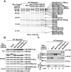
Figure 3. Med5(Nut1)p, Med14(Rgr1)p, Med17(Srb4)p and Med1p are H4 10 Bpa cross-linking targets.
Silver staining (A) and immunoblotting analysis (B) comparing the composition of affinity-purified MYC-tagged and non-MYC-tagged WT Mediator complexes after 10% SDS-PAGE. (C) A comparison of the cross-linking patterns of MYC-tagged Mediator complexes with the WT pattern using an SDS-PAGE blot probed with streptavidin poly-HRP to detect biotinylated H4 10 Bpa peptide cross-linked to Mediator subunits. Each indicated Mediator species (∼7.5 nM) was incubated with H4 10 Bpa (4 µM). A ‘Long Exposure’ of the 79 kD region on the SDS-PAGE blot probed with streptavidin poly-HRP is shown for a better view of the weak BCT4 signal in each sample.
Figure 4
Figure 4. Med5(Nut1)p is important for Mediator-H4 interaction, while Med1p is not.
Silver staining (A) and immunoblotting analysis (B) comparing the composition of affinity-purified Δmed5(nut1), Δmed1, and Δmed5(nut1)/Δmed1 and WT Mediator complexes after 10% SDS-PAGE. The amount of each complex in the individual lanes was normalized by adjusting the load such that an equal signal from the α-flag and α-Med7 antibodies was present. (C) Western blot analysis of histone tail binding experiment comparing the H4 tail binding affinity of WT and Δmed5(nut1) Mediator complexes. An equal concentration (∼3 nM) of either WT or Δmed5(nut1) Mediator complex was mixed with WT H4 peptide (1 µM) and with H2B peptide (1 µM) as the inputs. The basic steps and layout of the analysis were as described earlier (Fig. 1). (D) Western blot analysis of histone tail binding experiment comparing the H4 tail binding affinity between WT and Δmed1 Mediator complexes. An equal concentration (∼3 nM) of WT or Δmed1 Mediator complex was mixed with WT H4 peptide (1 µM) and with H2B peptide (1 µM) as the inputs. (E) Western blot analysis of histone tail binding experiment comparing the H4 tail binding affinity of WT, Δmed5(nut1) and Δmed5(nut1)/Δmed1 Mediator complexes. An equal concentration (∼6 nM) of WT, Δmed5(nut1) or Δmed5(nut1)/Δmed1 Mediator complex was mixed with WT H4 peptide (1.4 µM) as the inputs. (F) Western blot analysis of GST-CTD pull down experiment comparing the GST-CTD binding affinity for WT and Δmed5(nut1) Mediator complexes. An equal concentration (∼15 nM) of WT or Δmed5(nut1) Mediator complex (Input) was incubated with glutathione beads, which were pre-loaded with equal amounts of GST-CTD or GST. After incubation, the supernatant was saved as Flow-though (F.T.). Bound protein was eluted from the beads by boiling them in SDS-PAGE loading dye (Elution). An indicated percent of each fraction was analyzed by immunoblotting using the specified antibodies against Mediator subunits from different structural modules.
Figure 5
Figure 5. The C-terminus of Med14(Rgr1)p does not directly contribute to H4-Mediator interaction.
(A) Immunoblotting analysis comparing the composition of affinity-purified rgr1(Med14)-Δ2 and WT Mediator complexes after 10% SDS-PAGE. (B) Western blot analysis of histone tail binding experiment comparing the H4 tail binding affinity of WT, Δmed5(nut1) and rgr1(Med14)-Δ2 Mediator complexes. An equal concentration (∼6 nM) of WT, Δmed5(nut1) or rgr1(Med14)-Δ2 Mediator complex was mixed with WT H4 peptide (1.4 µM) as the inputs. The basic steps and layout of the analysis were as described earlier (Fig. 1).
Figure 6
Figure 6. Med16(Sin4)p and Med9(Cse2)p do not directly influence H4 tail binding.
Silver staining (A) and immunoblotting analysis (B) comparing the composition of affinity-purified Δmed16(sin4), Δmed9(cse2) and WT Mediator complexes after 10% SDS-PAGE. (C) Western blot analysis of histone tail binding experiment comparing the H4 tail binding affinity of WT, Δmed5(nut1), and Δmed16(sin4) Mediator complexes. An equal concentration (∼4.7 nM) of WT, Δmed5(nut1), or Δmed16(sin4) Mediator complex was mixed with WT H4 peptide (1.4 µM) as the inputs. The basic steps and layout of the analysis were as described earlier (Fig. 1). (D) Western analysis of histone tail binding experiment comparing the H4 tail binding affinity of WT and Δmed9(cse2) Mediator complexes. An equal concentration (∼3 nM) of WT or Δmed9(cse2) Mediator complex was mixed with WT H4 peptide (1 µM) as the inputs.
Figure 7
Figure 7. H4 10 Bpa cross-linking patterns of the mutant Mediator complexes.
SDS-PAGE blot probed with streptavidin poly-HRP to detect and compare the pattern of biotinylated peptide cross-linked to Mediator subunits in WT Mediator and the mutant Mediator complexes. Equimolar amounts (∼7.5 nM) of each indicated Mediator complex were incubated with H4 10 Bpa (4 µM) and exposed to UV for 15 min. Asterisk refers to the same weak signal as in Fig. 2-A.

Similar articles

Cited by

References

    1. Conaway RC, Conaway JW. Function and regulation of the Mediator complex. Curr Opin Genet Dev. 2011;21:225–230. - PMC - PubMed
    1. Myers LC, Kornberg RD. Mediator of transcriptional regulation. Annu Rev Biochem. 2000;69:729–749. - PubMed
    1. Guglielmi B, van Berkum NL, Klapholz B, Bijma T, Boube M, et al. A high resolution protein interaction map of the yeast Mediator complex. Nucleic Acids Res. 2004;32:5379–5391. - PMC - PubMed
    1. Linder T, Gustafsson CM. The Soh1/MED31 protein is an ancient component of Schizosaccharomyces pombe and Saccharomyces cerevisiae Mediator. J Biol Chem. 2004;279:49455–49459. - PubMed
    1. Bjorklund S, Gustafsson CM. The yeast Mediator complex and its regulation. Trends Biochem Sci. 2005;30:240–244. - PubMed

Publication types

MeSH terms

LinkOut - more resources