. 2012 Aug 1;31(15):3239-51.
doi: 10.1038/emboj.2012.161. Epub 2012 Jun 12.

Impairment of GABAB receptor dimer by endogenous 14-3-3ζ in chronic pain conditions

Affiliations

Impairment of GABAB receptor dimer by endogenous 14-3-3ζ in chronic pain conditions

Sophie Laffray et al. EMBO J. .

Abstract

In the central nervous system, the inhibitory GABAB receptor is the archetype of heterodimeric G protein-coupled receptors (GPCRs). However, the regulation of GABAB dimerization, and more generally of GPCR oligomerization, remains largely unknown. We propose a novel mechanism for inhibition of GPCR activity through de-dimerization in pathological conditions. We show here that 14-3-3ζ, a GABAB1-binding protein, dissociates the GABAB heterodimer, resulting in the impairment of GABAB signalling in spinal neurons. In the dorsal spinal cord of neuropathic rats, 14-3-3ζ is overexpressed and weakens GABAB inhibition. Using anti-14-3-3ζ siRNA or competing peptides disrupts 14-3-3ζ/GABAB1 interaction and restores functional GABAB heterodimers in the dorsal horn. Importantly, both strategies greatly enhance the anti-nociceptive effect of intrathecal Baclofen in neuropathic rats. Taken together, our data provide the first example of endogenous regulation of a GPCR oligomeric state and demonstrate its functional impact on the pathophysiological process of neuropathic pain sensitization.


Conflict of interest statement

The authors declare that they have no conflict of interest.

Figures
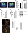
Figure 1
Figure 1
Increased expression of 14-3-3ζ in the spinal cord of neuropathic rats. (A1) In-situ hybridization to 14-3-3ζ mRNA illustrated with autoradiographic films showing an increase in ipsilateral dorsal horn of SNL rats (double arrowhead in (b)). Bar: 200 μm. (A2) Quantification of in-situ hybridization by densitometry (n=5 in each group). (B) qRT–PCR measurement of 14-3-3ζ mRNA (n=5 in each group). (C1) Mean intensity of 14-3-3ζ immunoblot. (C2) Example of 14-3-3ζ immunoblot. (B, C1) %±s.e.m. of the reference value (sham) set to 100%, n=5 in each condition. (D1) Double immunohistochemistry for B1 (arrowheads in (a)) and 14-3-3ζ (arrow in (a)) in spinal cord sections of sham (a) and SNL (b) rats. B1 subunit and 14-3-3ζ colocalization is enhanced after nerve injury (b, double arrow). (D2). Quantification of colocalization between GABAB1 and 14-3-3ζ (n=4 rats in each group). ***P<0.001; **P<0.01; NS=non-significant; Contra=Contralateral; Ipsi=Ipsilateral to the nerve ligation.
Figure 2
Figure 2
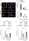
14-3-3ζ dissociation of the GABAB heterodimer in cultured spinal neurons. (A) (a–c) Double transfected cells (B1b/B2) showing myc–B1b (a, red) and HA–B2 (b, blue) colocalization at the plasma membrane (double arrowheads). Bar: 20 μm. (d–g) Triple transfected cells (B1b/B2/14-3-3ζ) showing that B1b (d, red) and 14-3-3ζ (f, green) colocalize in domains lacking B2 (double arrows) while B1b and B2 (e, blue) remain colocalized in the absence of 14-3-3ζ (double arrowhead). Bar: 10 μm. (B) Colocalization in double (B1b/B2; n=18) and triple (B1b/B2/14-3-3ζ; n=16) transfected neurons. *P<0.05; **P<0.01. (C) Correlation of labelling intensity in double (B1b/B2; n=18) and triple (B1b/B2/14-3-3ζ; n=16) transfected neurons. (D) In double transfected COS-7 cells, HA–B2 co-precipitates with myc–B1b (left panel). In triple transfected cells, the interaction between B1 and B2 subunits is abolished (right panel) (n=3 independent experiments). (E) In double and triple transfected cells, HA–B2 co-precipitates with B1ASA-GFP that does not interact with 14-3-3ζ (see Supplementary Figure S3B and C) (n=3 independent experiments). IP lanes: co-IP; S lanes: supernatant. (F, G) FRET efficiency (mean %±s.e.m., n=31 or 21 cells) between N-ter GFP (B1b), and t-dimer DsRed (B2) in primary cultures transfected with GFP-B1b and t-dimer DsRed-B2 alone (B1b/B2), or together with Flag–14-3-3ζ (B1b/B2/14-3-3ζ). Cultures are further incubated with iFect (veh), anti-14-3-3 siRNA (+siRNA), or mismatch RNA (+mmRNA) (F) or competing peptides (+pVec-B1 919–927 or 922–930) (G). *P<0.05. NS=non-significant.
Figure 3
Figure 3
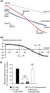
Dissociation of the GABAB heterodimer at the plasma membrane. (A1) FRET analysis in regions of interest identified with a mask (delineated with a purple line) drawn over areas positive for anti-calreticulin (a, ER marker), anti-mannosidase II (b, Golgi marker) or DiD (c, lipophilic carbocyanine used as a marker for the plasma membrane). (A2) FRET efficiency between GFP–B1b and t-dimer DsRed-B2 is decreased at the plasma membrane (right). No changes are found in the Golgi, and an increase in FRET efficiency is even detected in the ER (n=15 cells in each conditions). (B) COS-7 cells were transfected and processed for double immunoprecipitation. The surface fraction of the GABAB complexes was then isolated and probed on western blot with an anti-B2 antibody. The association between B2 and B1 at the plasma membrane was reduced after 14-3-3ζ overexpression (right panel). (C) In control COS-7 cells (myc–B1b/HA–B2/GFP), B1/B2 co-IP is independent of the Brefeldin A treatment (left panel). In Flag–14-3-3ζ overexpressing cells, the interaction between B1 and B2 subunits is abolished in the absence of Brefeldin A, but remains intact upon treatment with Brefeldin A (right panel). As a control, B1/B2 interaction decreases when Brefeldin A is washed out, thus allowing proteins to be properly retargeted to the plasma membrane (third band in right panel) (n=3 independent experiments).
Figure 4
Figure 4
In-vitro impairment of GABAB signalling. (A1) Voltage ramp-evoked currents in control ACSF (blue), 25 μM Baclofen (red) and Baclofen+CGP 55845A (50 μM) (grey). (A2) Baclofen current resulting from the subtraction of blue plot (1) from red plot (2) as shown in Figure 3A1. (B) Baclofen-induced current during a voltage ramp in control (black bar, n=13), in the presence of 50 μM CGP 55845 (light grey bar, P<0.01, n=6), in neurons overexpressing 14-3-3ζ (dark grey bar, P<0.01, n=9), and in neurons overexpressing the DsRed vector alone (white bar, n=8). Results are expressed as a percentage of increase above the basal level measured in the absence of Baclofen. NT cells=non-transfected cells.
Figure 5
Figure 5
Decrease of endogenous co-expression of B1 and B2 in neuropathic rats. (A) Immunohistochemistry for endogenous B1 and B2 in lamina II of the dorsal horn of three groups of rat (n=4 in each group). The colocalization is prominent in sham rats (arrowhead, a). In neuropathic rats with control RNA (mmRNA) injection, B1 (green) and B2 (red) labelling were often found alone at the plasma membrane of spinal neurons (arrow, b). After siRNA injection in neuropathic rats, the colocalization is restored (arrowhead, c). Bar=5 μm. (B) Quantification of the percentage of B1 colocalized with B2 labelling in lamina I–II of Sham (black bars) and SNL (grey bars) rats (n=20 cells in each conditions). *P<0.05 versus Sham. (C1) Co-immunoprecipitation in vivo showing a decrease of B1/B2 interaction in neuropathic conditions that is abolished after siRNA or competing peptide injection. No changes are detected in Sham rats upon siRNA or peptide injections. **P<0.01 versus Sham; a, b: P<0.05, P<0.01 versus SNL with mmRNA injection, respectively. (C2) Increase of B1/14-3-3ζ interaction in neuropathic conditions that is abolished after competing peptide injection. n=5 in each conditions. **P<0.01.
Figure 6
Figure 6
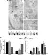
Changes in B1/B2 colocalization in spinal neurons at the ultrastructural level. (A) Triple immunogold in dorsal horn neurons of naive (a, d) and SNL (b, c, e) rats. (a, d) Isolated B2 (arrowheads) or B2 colocalization with B1b (double arrowheads). Rare association between 14-3-3ζ and B1b at the plasma membrane (arrow). (b, c and e) Prominent association between 14-3-3ζ and B1b at the plasma membrane or in the cytoplasm (arrows). Colocalization between B2 and B1b, mainly in the cytoplasm (double arrowhead). Triple colocalization showing 14-3-3ζ between B2 and B1b (double arrows). Bar: 100 nm. (B1) Quantification of the percentage of B1 colocalized with B2 or 14-3-3ζ (distance <20 nm). (B2) Quantification of distances (nm) between gold particles. Mean distances±s.e.m. *P<0.05; NS=non-significant (n=15 cells in each condition).
Figure 7
Figure 7
In-vivo alteration of GABAB signalling and alleviation of mechanical allodynia in SNL rats. (A) Autoradiographic images of [35S]GTPγS in sham and SNL rat spinal cord (n=5 in each condition) showing the basal binding (left), 1 μM Baclofen-stimulated binding (right), and 1 μM Baclofen-stimulated binding after anti-14-3-3ζ siRNA injections (bottom right). Arrowheads: side ipsilateral to nerve ligation. Bar: 1.5 mm. (B) The quantification of GTP binding shows a decrease of GABAB activation in the ipsilateral dorsal horn of SNL rats. (C–F) Threshold to mechanical stimulation measured in SNL (C, E) and sham (D, F) rats before and after intrathecal injections (n=5 in each condition). The threshold before surgery (day −1) is set to 100%. Baclofen produces an anti-nociceptive effect in SNL rats (C, Baclo, 12d; E, Baclo, 14d). Anti-14-3-3ζ siRNA has no effect per se in SNL rats (C, grey bars; RNA, 15d). In contrast, pVEC-B1 (922–930) has an effect similar to that of Baclofen (E, grey bars; peptide, 15d). The effects of Baclofen are significantly enhanced after injection of siRNA (C, grey bars; RNA+Baclo, 15d) or competing peptide conjugate (E, grey bars; peptide+Baclo, 15d). Mismatch RNA (C, black bars) or pVEC only (E, black bars) have no effect when injected alone or in conjugation with Baclofen. No changes of the mechanical threshold are seen in sham rats after intrathecal injections of Baclofen (D, F), siRNA (D, grey bars), or pVEC-B1 (922–930) (F, grey bars). Data are mean values±s.e.m. **P<0.01; ***P<0.001; a: P<0.01 versus iFect (12d) (C); b: P<0.01 versus SNL/siRNA, Baclo (12d) (C); c: P<0.001 versus Saline (14d) (E); d: P<0.05 versus SNL/pVEC-B1 (922–930), Baclo (14d) (E).

Comment in

Similar articles

Cited by

References

    1. Attal N, Cruccu G, Haanpaa M, Hansson P, Jensen TS, Nurmikko T, Sampaio C, Sindrup S, Wiffen P (2006) EFNS guidelines on pharmacological treatment of neuropathic pain. Eur J Neurol 13: 1153–1169 - PubMed
    1. Bardoni R, Ghirri A, Salio C, Prandini M, Merighi A (2007) BDNF-mediated modulation of GABA and glycine release in dorsal horn lamina II from postnatal rats. Dev Neurobiol 67: 960–975 - PubMed
    1. Benke D, Honer M, Michel C, Bettler B, Mohler H (1999) gamma-aminobutyric acid type B receptor splice variant proteins GBR1a and GBR1b are both associated with GBR2 in situ and display differential regional and subcellular distribution. J Biol Chem 274: 27323–27330 - PubMed
    1. Berg D, Holzmann C, Riess O (2003) 14-3-3 proteins in the nervous system. Nat Rev Neurosci 4: 752–762 - PubMed
    1. Bouvier M (2001) Oligomerization of G-protein-coupled transmitter receptors. Nat Rev Neurosci 2: 274–286 - PubMed

Publication types

MeSH terms

LinkOut - more resources