. 2012 Jan 1;2(1):36-49.
doi: 10.4161/bact.19775.

The C-terminal cysteine annulus participates in auto-chaperone function for Salmonella phage P22 tailspike folding and assembly

Affiliations

The C-terminal cysteine annulus participates in auto-chaperone function for Salmonella phage P22 tailspike folding and assembly

Takumi Takata et al. Bacteriophage. .

Abstract

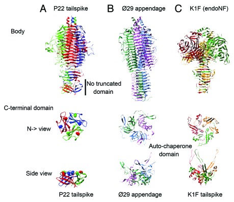
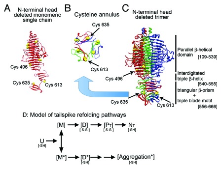
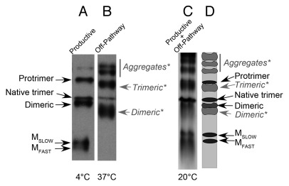
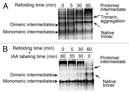
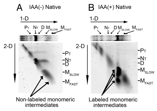
Elongated trimeric adhesins are a distinct class of proteins employed by phages and viruses to recognize and bind to their host cells, and by bacteria to bind to their target cells and tissues. The tailspikes of E. coli phage K1F and Bacillus phage Ø29 exhibit auto-chaperone activity in their trimeric C-terminal domains. The P22 tailspike is structurally homologous to those adhesins. Though there are no disulfide bonds or reactive cysteines in the native P22 tailspikes, a set of C-terminal cysteines are very reactive in partially folded intermediates, implying an unusual local conformation in the domain. This is likely to be involved in the auto-chaperone function. We examined the unusual reactivity of C-terminal tailspike cysteines during folding and assembly as a potential reporter of auto-chaperone function. Reaction with IAA blocked productive refolding in vitro, but not off-pathway aggregation. Two-dimensional PAGE revealed that the predominant intermediate exhibiting reactive cysteine side chains was a partially folded monomer. Treatment with reducing reagent promoted native trimer formation from these species, consistent with transient disulfide bonds in the auto-chaperone domain. Limited enzymatic digestion and mass spectrometry of folding and assembly intermediates indicated that the C-terminal domain was compact in the protrimer species. These results indicate that the C-terminal domain of the P22 tailspike folds itself and associates prior to formation of the protrimer intermediate, and not after, as previously proposed. The C-terminal cysteines and triple β-helix domains apparently provide the staging for the correct auto-chaperone domain formation, needed for alignment of P22 tailspike native trimer.


Figures

None
Figure 1. Structures of the C-terminal and auto-chaperone domains in Salmonella phage P22 tailspike and homologous proteins. (A) The structure of Salmonela phage P22 tailspike (PDB 1TYU). C-terminal domain remains in mature protein. (B) The structure of Bacillus phage Ø29 gp12 appendage (PDB 3GQ7). C-terminal domain is cleaved off after folding completion. (C) The structure of E.coli phage K1F tailspike (endoNF) (PDB 1V0E). C-terminal trimeric domain with long tentacles assists folding, and is cleaved off in the mature protein (3GUD, 3GW6).
None
Figure 2. Structure of P22 tailspike with cysteine locations, and schematic of the folding and assembly pathway. (A) P22 tailspike single chain crystal structure with the N-terminal head domain deleted. Yellow spheres indicate cysteine residues. (B) A cross-section of the native trimer cysteine annulus bottom to top view. The three individual subunits are colored red, blue and green. Two cysteines from each subunit (C613 and C635) are located in this region. (C) The N-terminal head binding domain deleted native trimer showing the eight cysteines (PDB 1TSP). The P22 tailspike major structural domains are parallel β-helical domain (109–539), interdigitated triple β-helix domain (540–555), triangular β-prism and triple blade motif structure (556–666). All eight cysteines are reduced in the naive structure.36 (D) Schematic diagram of intermediates in the in vitro refolding, assembly, and aggregation of tailspike chain. U: unfolded polypeptides. NT: native trimer. The folding pathway depicts intermediates and disulfide bonds distributions: The state of the reactive cysteines in the [M] and [M*] species are described in the Results section. Dimeric intermediate: [D] and protrimer intermediate: [PT] have been reported to contain inter-disulfide bonds: [-S-S-]. All disulfide bonds are reduced: [-SH] in native trimer. The aggregation prone intermediates are marked as [*]. No disulfide bonds have been reported in aggregation species.
None
Figure 3. During the refolding of P22 tailspike, productive folding intermediates and non-productive multimeric aggregation, can be resolved with Native-PAGE. (A) At 4°C productive intermediates predominate as visualized by western blot analysis of a Native-PAGE, probed by a mixture of monoclonal antibodies recognizing non-native epitopes. (B) At 37°C non-productive folding intermediates and multimeric aggregation are detected by the same western blot analysis in (A). (C) At 20°C both productive and non-productive species are observed as seen in this silver stain of a Native-PAGE. (D) The graphic depicts the bands, the productive folding intermediates and the non-productive species from the gel in (C). Black labeled bands are productive species, gray and italic labeled bands are non-productive species. (A and B) were adapted from Betts and King 1998.63
None
Figure 4. In vitro refolding of P22 tailspike with or without iodoacetic acid (IAA). The monomeric species were labeled and stalled by alkylation treatment. P22 tailspike was denatured and refolded as described in Materials and Methods. At indicated times, IAA was added to stall folding reaction and analyzed by non-reducing 1-D native-PAGE. (A) In vitro refolding at 24°C shows the native trimer formation as a function of the refolding time. (B) Samples were made up to 30 mM IAA at indicated times and incubated for total 60 min at 24°C. IAA reacted monomeric species that aggregated, remained at the top of gel.
None
Figure 5. IAA treatment altered monomeric tailspike intermediates. (A) Non-reducing 2-D native-PAGE of refolded P22 tailspike without IAA labeling. P22 tailspike was denatured, refolded for 2 h at 24°C, and then additionally incubated for 1 h at 24°C without IAA. (B) Non-reducing 2-D native-PAGE of refolded P22 tailspike with IAA labeling. P22 tailspike was denatured, refolded for 2 h at 4°C, and then additionally incubated for 1 h at 4°C with 30 mM IAA. NT, PT, and D indicate native trimer, protrimer, and dimeric intermediates. MSLOW and MFAST refer to electrophoretically slower and faster migrating monomeric intermediates.
None
Figure 6. P22 tailspike monomeric intermediates were heat labile and aggregated in native gel. (A) Refolding tailspike was electrophoresed in the first dimension, then excised gel lane was incubated in 1 X native-PAGE running buffer on wet ice for 8 h before non-reducing 2-D native-PAGE. (B) The excised 1-D gel lane was incubated at 24°C for 1 h, then stored on wet ice until loaded on non-reducing 2-D native-PAGE. MSLOW was decreased with increased MFAST, indicated intra-disulfide bond formation, or folding collapse. (C) The 1-D excised gel lane was incubated at 24°C for 8 h before non-reducing 2-D native-PAGE. All monomeric species aggregated to complexes too large to enter gel.
None
Figure 7. The effects of DTT reduction on the various tailspike folding and assembly intermediates as monitorized by 2-D native-gel electrophoresis. (A) The excised gel lane from 1-D native-PAGE was incubated in native running buffer in the absence of DTT at 24°C for 1 h before 2-D native-PAGE. The productive folding and assembly intermediates migrated on the diagonal in the second dimension. (B) The excised gel lane from 1-D native-PAGE was incubated in native running buffer in the presence of 0.1 M DTT at 24°C for 1 h before 2-D native-PAGE. Native trimer formed from slower migrating monomeric MSLOW intermediates during incubation with DTT can be seen (larger circle). Reducing induced native trimer formation from protrimer PT intermediate (smaller circle). Aggregation intermediates were formed from faster migrating monomeric MFAST intermediate (triangles at the right of gel).
None
Figure 8. Comparison of tryptic peptide recovery between P22 tailspike monomeric intermediates and protrimer intermediates. Yellow region represents peptide identified in both species. Red region represents only identified in monomer species. White region peptide could not be identified in either. (A) The difference in trypsin digested peptides between WT monomeric intermediate and WT protrimer. (B) The difference in trypsin digested peptide between WT monomeric intermediate and ΔN N547Y protrimer. C-terminal domain was protected in both protrimer intermediates.
None
Figure 9. The folding and aggregation pathway of P22 tailspike adhesin. This schematic diagram depicts the C-terminal auto-chaperone directing productive folding and assembly. In the pathway each intermediates identified on 2-D gel are represented. The productive slower migrating monomeric (MSLOW) intermediate has reactive cysteine [S]§; the non-productive faster migrating monomeric (MFAST) intermediate may have intra-disulfide bond, or be more collapse; the productive dimeric and protrimer intermediates contain inter-disulfide bond [-S-S-] formed from reactive cysteines in MSLOW intermediates. The aggregation prone off-pathway species (*) do not contain productive intramolecular disulfide bonds.

Similar articles

Cited by

References

    1. Philipson L, Lonberg-Holm K, Pettersson U. Virus-receptor interaction in an adenovirus system. J Virol. 1968;2:1064–75. - PMC - PubMed
    1. Kells SS, Haselkorn R. Bacteriophage T4 short tail fibers are the product of gene 12. J Mol Biol. 1974;83:473–85. doi: 10.1016/0022-2836(74)90508-7. - DOI - PubMed
    1. Riede I. Receptor specificity of the short tail fibres (gp12) of T-even type Escherichia coli phages. Mol Gen Genet. 1987;206:110–5. doi: 10.1007/BF00326544. - DOI - PubMed
    1. Brennan MJ, David JL, Kenimer JG, Manclark CR. Lectin-like binding of pertussis toxin to a 165-kilodalton Chinese hamster ovary cell glycoprotein. J Biol Chem. 1988;263:4895–9. - PubMed
    1. Thomassen E, Gielen G, Schütz M, Schoehn G, Abrahams JP, Miller S, et al. The structure of the receptor-binding domain of the bacteriophage T4 short tail fibre reveals a knitted trimeric metal-binding fold. J Mol Biol. 2003;331:361–73. doi: 10.1016/S0022-2836(03)00755-1. - DOI - PubMed

LinkOut - more resources