Phage display on the base of filamentous bacteriophages: application for recombinant antibodies selection
- PMID: 22649612
- PMCID: PMC3347532
Phage display on the base of filamentous bacteriophages: application for recombinant antibodies selection
Abstract
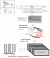
The display of peptides and proteins on the surface of filamentous bacteriophage is a powerful methodology for selection of peptides and protein domains, including antibodies. An advantage of this methodology is the direct physical link between the phenotype and the genotype, as an analyzed polypeptide and its encoding DNA fragment exist in one phage particle. Development of phage display antibody libraries provides repertoires of phage particles exposing antibody fragments of great diversity. The biopanning procedure facilitates selection of antibodies with high affinity and specificity for almost any target. This review is an introduction to phage display methodology. It presents recombinant antibodies display in more details:, construction of phage libraries of antibody fragments and different strategies for the biopanning procedure.
Figures


Similar articles
-
Phage display technology: clinical applications and recent innovations.Clin Biochem. 2002 Sep;35(6):425-45. doi: 10.1016/s0009-9120(02)00343-0. Clin Biochem. 2002. PMID: 12413604 Review.
-
Isolation of monoclonal antibody fragments from phage display libraries.Methods Mol Biol. 2009;502:341-64. doi: 10.1007/978-1-60327-565-1_20. Methods Mol Biol. 2009. PMID: 19082566
-
Phage display as a tool for identifying HIV-1 broadly neutralizing antibodies.Vavilovskii Zhurnal Genet Selektsii. 2021 Sep;25(5):562-572. doi: 10.18699/VJ21.063. Vavilovskii Zhurnal Genet Selektsii. 2021. PMID: 34595378 Free PMC article.
-
Considerations on antibody-phage display methodology.Comb Chem High Throughput Screen. 2005 Mar;8(2):117-26. doi: 10.2174/1386207053258532. Comb Chem High Throughput Screen. 2005. PMID: 15777175 Review.
-
Selection of full-length IgGs by tandem display on filamentous phage particles and Escherichia coli fluorescence-activated cell sorting screening.FEBS J. 2010 May;277(10):2291-303. doi: 10.1111/j.1742-4658.2010.07645.x. FEBS J. 2010. PMID: 20423457
Cited by
-
An Engineered M13 Filamentous Nanoparticle as an Antigen Carrier for a Malignant Melanoma Immunotherapeutic Strategy.Viruses. 2024 Feb 1;16(2):232. doi: 10.3390/v16020232. Viruses. 2024. PMID: 38400008 Free PMC article.
-
Naïve Human Antibody Libraries for Infectious Diseases.Adv Exp Med Biol. 2017;1053:35-59. doi: 10.1007/978-3-319-72077-7_3. Adv Exp Med Biol. 2017. PMID: 29549634 Free PMC article. Review.
-
Architectural insight into inovirus-associated vectors (IAVs) and development of IAV-based vaccines inducing humoral and cellular responses: implications in HIV-1 vaccines.Viruses. 2014 Dec 17;6(12):5047-76. doi: 10.3390/v6125047. Viruses. 2014. PMID: 25525909 Free PMC article. Review.
-
In Vitro Characterization of Neutralizing Hen Antibodies to Coxsackievirus A16.Int J Mol Sci. 2021 Apr 16;22(8):4146. doi: 10.3390/ijms22084146. Int J Mol Sci. 2021. PMID: 33923724 Free PMC article.
-
Hybridoma technology: is it still useful?Curr Res Immunol. 2021 Mar 22;2:32-40. doi: 10.1016/j.crimmu.2021.03.002. eCollection 2021. Curr Res Immunol. 2021. PMID: 35492397 Free PMC article. Review.
References
-
- Smith G.. Science. 1985;228:1315–1317. - PubMed
-
- Parmley S., Smith G.. Gene. 1988;73:305–318. - PubMed
-
- Ilyichev A., Minenkova O., Tat'kov S. Doklady. Vol. 307. AN USSR: 1989. pp. 481–483. - PubMed
-
- Minenkova O., Ilyichev A., Kishchenko G.. Gene. 1993;128:85–88. - PubMed
-
- MacCafferty J., Griffiths A., Winter G.. Nature. 1990;348:552–554. - PubMed
LinkOut - more resources
Full Text Sources
Other Literature Sources
