Review
. 2012 Jul 24;24(28):3724-46.
doi: 10.1002/adma.201200446. Epub 2012 May 29.

Engineering nano- and microparticles to tune immunity

Affiliations
Review

Engineering nano- and microparticles to tune immunity

James J Moon et al. Adv Mater. .

Abstract

The immune system can be a cure or cause of disease, fulfilling a protective role in attacking cancer or pathogenic microbes but also causing tissue destruction in autoimmune disorders. Thus, therapies aimed to amplify or suppress immune reactions are of great interest. However, the complex regulation of the immune system, coupled with the potential systemic side effects associated with traditional systemic drug therapies, has presented a major hurdle for the development of successful immunotherapies. Recent progress in the design of synthetic micro- and nano-particles that can target drugs, deliver imaging agents, or stimulate immune cells directly through their physical and chemical properties is leading to new approaches to deliver vaccines, promote immune responses against tumors, and suppress autoimmunity. In addition, novel strategies, such as the use of particle-laden immune cells as living targeting agents for drugs, are providing exciting new approaches for immunotherapy. This progress report describes recent advances in the design of micro- and nano-particles for immunotherapies and diagnostics.


Figures

Figure 1
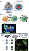
Figure 1. Engineered particles as synthetic antigen presenting cells
(A) Schematic view of key receptor-ligand interactions at the immunological synapse formed between an antigen presenting cell (APC, such as a dendritic cell) and a T-cell during T-cell activation. (B) Upper panel, schematic view of microparticles engineered as artificial APCs (aAPCs), which display ligands and release cytokines to stimulate T-cells. Lower panel, confocal microscopy view of immunological synapse formed between aAPC microparticle (red) and several T-cells (nuclei, blue; actin, green). (C) Fabrication of anisotropic “patchy” protein-coated microparticles by (i) forming colloidal crystals of microparticles, (ii) applying polydimethylsiloxane as a masking agent, (iii) PDMS masking at particle contact points, (iv) separation of particles from the scaffold, and finally (iv) two-step protein coating. Lower right, confocal micrograph illustrates dual protein patterning on patchy microspheres. Panel (B) reproduced with permission from [16]. Copyright 2008, Nature Publishing Group. Panel (C) reproduced with from [38]. Copyright 2011, Wiley.
Figure 2
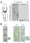
Figure 2. Modulation of particle interaction with phagocytes
(A) The effect of the contact angle between particles and cell membranes on the rate of particle internalization, demonstrating poor phagocytosis of highly anisotropic particles. (B) A flow chamber assay demonstrating rapid uptake of small, isotropic micelles by macrophages, but minimal uptake for long, flexible filomicelles. Scale bars, 5 μm. Panel (A) reproduced with permission from [47]. Panel (B) reproduced with permission from [69]. Copyright 2007, Nature Publishing Group.
Figure 3
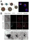
Figure 3. Active targeting of lymphoid organs with particle-carrying leukocytes
(A) Left panel, schematic of layer-by-layer capsule assembly by (i, ii) incubating a colloidal template with antigen, (iii, iv) alternate deposition of interacting polymers to form (v) a multilayered structure, followed by (vi) dissolution of core template. Right panel, confocal image of capsules (green) internalized into dendritic cells (membrane in red and nuclei in blue). (B) Upper panel, confocal images of untreated DCs, and DCs loaded with αAl2O3 NPs (green) and stained with antibody against the autophagosome marker, LC3 (red). Lower panel, TEM images showing that internalized αAl2O3 NPs are located inside endosomes/phagosomes, autophagosomes, and autolysosomes of DCs. Panel (A) reproduced with permission from [81]. Copyright 2008, Wiley. Panel (B) reproduced with permission from [61]. Copyright 2011, Nature Publishing Group.
Figure 4
Figure 4. Active targeting of lymphoid organs with particle-carrying leukocytes
(A) Schematic illustration of Fe3O4-ZnO core-shell nanoparticles coated with tumor antigens fused to ZnO-binding peptides. (B) In vivo MRI image showing accumulation of dendritic cells labeled with nanoparticles in draining lymph nodes. (C) Enhanced suppression of tumor growth after injection of dendritic cells carrying tumor antigen-loaded iron oxide NPs (open red squares) compared to administration with antigen only (open blue triangles), DCs only (filled black circles), or NPs (open black circles). Reproduced with permission from [13]. Copyright 2011, Nature Publishing Group.
Figure 5
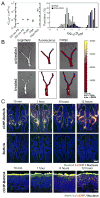
Figure 5. Delivery of vaccine particles to lymphoid organs
(A) Direct draining of sub-50 nm nanoparticles to lymph nodes from s.c. injection sites. Upper panel, histological sections of draining lymph nodes 1 day after administration of poly(propylene sulfide) NPs (red) with mean diameters of 100 nm or 25 nm. Scale bar, 200 μm. Lower panel, percentage of dendritic cells that internalized PPS NPs in the lymph node. (B) Histological section of draining lymph nodes over time after s.c. injection of either soluble antigen (ovalbumin, shown in red) or antigen encapsulated in180 nm diam. multilamellar lipid nanoparticles (blue). Germinal centers in the LN detected by staining with GL-7 antibody on day 14 are shown in green. Scale bar, 10 μm. (C) Intranodal administration of fluorescent PLGA microparticles detected by whole-animal fluorescence imaging (upper panels) or imaging of excised intact lymph nodes (lower panels). (D) Histological section of lymph node following direct intranodal injection of fluorescent PLGA particles (green) with staining for markers of B-cells (B220, blue) and T-cells (CD3, red). Panel (A) reproduced with permission from [9]. Copyright 2007, Nature Publishing Group. Panel (B) reproduced with permission from [121]. Panel (C) reproduced with permission from [127]. Panel (D), courtesy of C.M. Jewell, S.C. Bustamante López, and D.J. Irvine.
Figure 6
Figure 6. Particles designed to penetrate mucosal barriers
(A) Improved mucus-penetrating ability of poly(sebacic acid) (PSA) NPs after PEGylation as evidenced by increase in effective diffusivity and fraction of particles penetrating human cervicovaginal mucus. (B) Fluorescence image of reproductive tract on day 1 after intravaginal administration of siRNA-loaded PLGA NPs. (C) Mucus-binding particles for intranasal vaccine delivery. Upper panel, wide distribution and attachment of antigen-loaded nanogels on nasal epithelium after intransal administration. Scale bar, 500 um. Lower panel, release and transport of antigen (green) from nanogels (red) into the epithelial layer over time. Scale bar, 20 um. Panel (A) reproduced with permission from [142]. Panel (B) reproduced with permission from [151]. Copyright 2009, Nature Publishing Group. Panel (C) reproduced with permission from [152]. Copyright 2010, Nature Publishing Group.
Figure 7
Figure 7. Nanoparitcle delivery of immunomodulatory drugs in tumors
(A, B) Encapsulation of STAT3-sirNA/PEI polyplexes in PLGA nanoparticles to reduce cytotoxicity while maintaining gene silencing activity. (A) Schematic view of polyplex encapsulation. (B) Kockdown of STAT3 in dendritic cells by encapsulated polyplexes, compared to controls with scrambled siRNA (sc) or naked siRNA. (C–F) Blockade of systemic side effects by anchoring immunostimulatory ligands to lipid vesicles for intratumoral injection. (C) PEGylated liposomes displaying aCD40 and CpG were synthesized by surface-conjugation of anti-CD40, followed by post-insertion of CpG-lipid conjugates into the outer leaflet of the vesicle bilayer. Suppression of tumor growth (D) without weight loss (E), liver damage (not shown), or systemic cytokine release (F) after intratumoral injection of liposomes displaying anti-CD40 and CpG, compared to equal doses of soluble ligands. Panels (A, B) reproduced with permission from [164]. Copyright 2010, American Chemical Society. Panel (C–E) reproduced with permission from [175]. Copyright 2011, Elsevier.
Figure 8
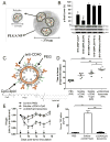
Figure 8. Leukocyte-mediated delivery of nanoparticles to tumors
(A, B) Gold nanoshells transported into tumors by macrophages for photothermal therapy. (A) TEM micrographs of a gold nanoshell-laden macrophage (upper panel) and monocytes (lower panel). Higher magnification views at right show aggregates of nanoshells inside the cells. (B) Histological tissue section of T47D tumor spheroid showing infiltrating nanoshell-laden macrophages (black) within the viable tumor as well as near areas of necrosis (pink staining; white arrow). (C) T-cells can carry internalized gold nanoparticles into tumors. Resected subcutaneous LCL xenograft tumors were analyzed by bright field imaging (top row) and immunohistochemistry for human CD3 expression and dark field imaging (bottom row) to indicate the presence of gold NPs. Red arrows indicate the colocalization of CD3+ T cells and AuNPs within the tumor. (D–F) T-cells carry surface-bound nanoparticles into tumors in vivo. (D) Lipid nanoparticles were stably conjugated to the surfaces of T-cells via maleimide-thiol reaction. (E) Nanoparticles remained on the surfaces of T-cells after 4 days of stimulation in vitro. (F) TRAMP mice bearing spontaneous prostate tumors were injected with fluorescent lipid NPs alone, Luciferase-expressing tumor-targeting T-cells alone, or luc-expressing T-cells carrying surface-bound NPs. Upper panels, whole-animal bioluminescence imaging of T-cell trafficking to the prostate tumors (dashed circles). Lower panels, fluorescence imaging of dissected intact prostates showing that free NPs achieve no entry into tumor site, while T-cells carry substantial quantities of particles into the tumor. Panels (A,B) reproduced with permission from [177]. Copyright 2007, American Chemical Society. Panel (C) reproduced with permission from [179]. Panel (D,E) reproduced with permission from [17]. Copyright 2010, Nature Publishing group. Panel (F), courtesy of M. Stephan, E. Higham, K.D. Wittrup, and J. Chen.
Figure 9
Figure 9. Crossing blood-brain-barrier with particle-carrying leukocytes
Bone marrow macrophages (BMM) loaded with drug-carrying nanoparticles have been used to treat HIV-1 infection in the brain. (Left panels) Histological images demonstrating migration of macrophages loaded with iron oxide NPs (blue staining) into neuroinflammatory HIV-1-infected brain sites (upper panels), but not control brain sections (lower panels). Right panels, dramatic reduction in HIV-1 infected brain sites (detected by staining for HIV p24 protein, blue) after treatment with macrophages carrying NPs loaded with anti-retroviral drugs. Reproduced with permission from [18]. Copyright 2009, American Association of Immunologists.

Similar articles

Cited by

References

    1. Pulendran B, Ahmed R. Nat Immunol. 2011;12:509. - PMC - PubMed
    1. Belyakov IM, Derby MA, Ahlers JD, Kelsall BL, Earl P, Moss B, Strober W, Berzofsky JA. Proc Natl Acad Sci U S A. 1998;95:1709. - PMC - PubMed
    1. Holmgren J, Czerkinsky C. Nat Med. 2005;11:S45. - PubMed
    1. Sallusto F, Lanzavecchia A, Araki K, Ahmed R. Immunity. 2010;33:451. - PMC - PubMed
    1. Hodi FS, O’Day SJ, McDermott DF, Weber RW, Sosman JA, Haanen JB, Gonzalez R, Robert C, Schadendorf D, Hassel JC, Akerley W, van den Eertwegh AJ, Lutzky J, Lorigan P, Vaubel JM, Linette GP, Hogg D, Ottensmeier CH, Lebbe C, Peschel C, Quirt I, Clark JI, Wolchok JD, Weber JS, Tian J, Yellin MJ, Nichol GM, Hoos A, Urba WJ. N Engl J Med. 2010;363:711. - PMC - PubMed

Publication types

LinkOut - more resources