. 2012 Aug;82(2):333-43.
doi: 10.1124/mol.112.078162. Epub 2012 May 17.

Differential modulation of drug-induced structural and functional plasticity of dendritic spines

Affiliations

Differential modulation of drug-induced structural and functional plasticity of dendritic spines

Eric C Miller et al. Mol Pharmacol. 2012 Aug.

Abstract

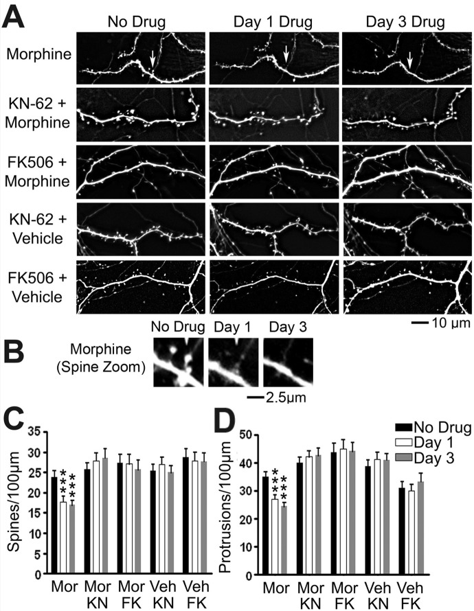
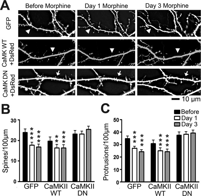
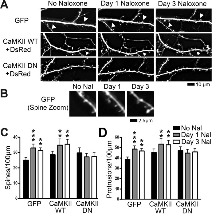
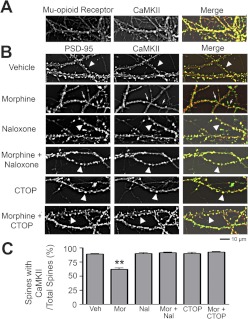
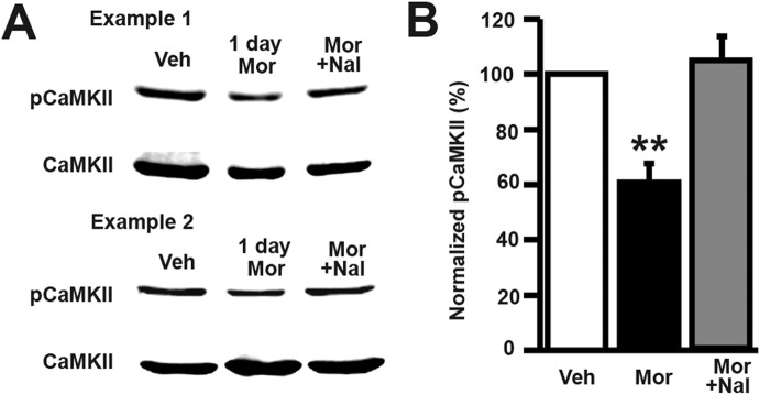
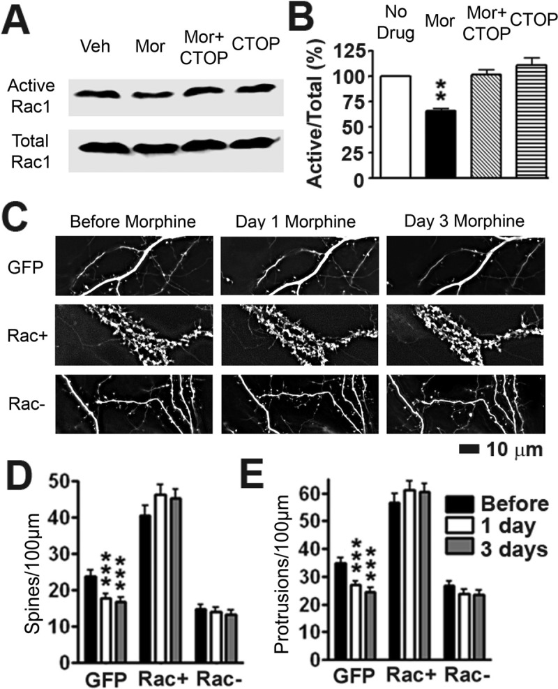
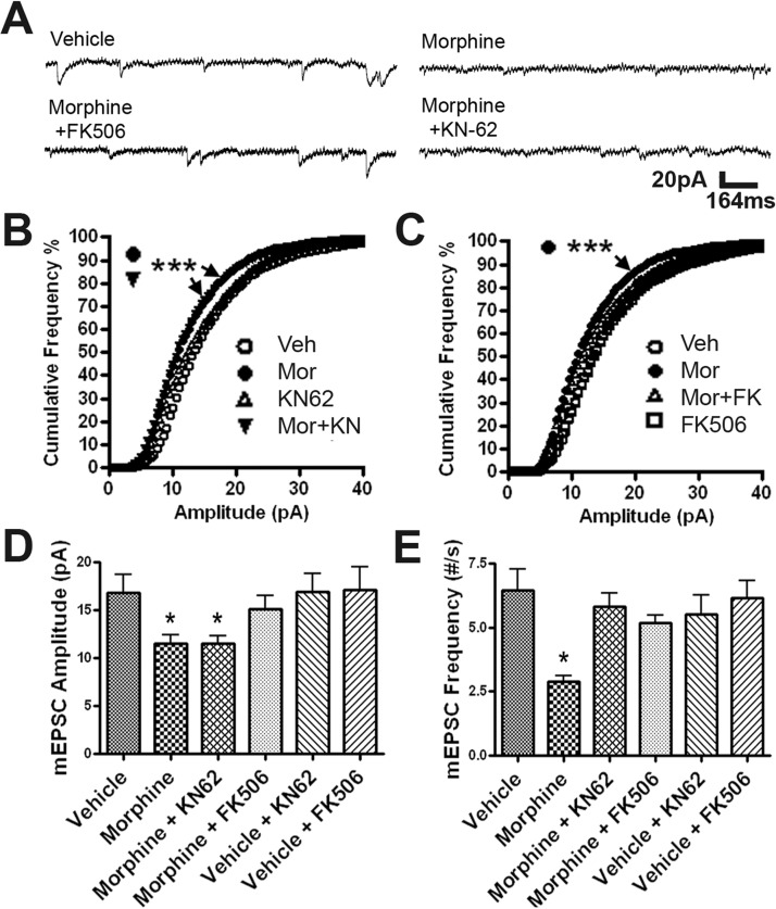
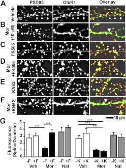
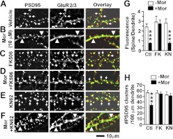
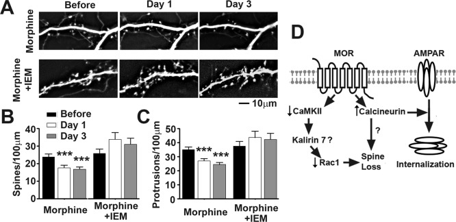
Drug-induced plasticity of excitatory synapses has been proposed to be the cellular mechanism underlying the aberrant learning associated with addiction. Exposure to various drugs of abuse causes both morphological plasticity of dendritic spines and functional plasticity of excitatory synaptic transmission. Chronic activation of μ-opioid receptors (MOR) in cultured hippocampal neurons causes two forms of synaptic plasticity: loss of dendritic spines and loss of synaptic α-amino-3-hydroxy-5-methyl-4-isoxazolepropionic acid (AMPA) receptors. With use of live imaging, patch-clamp electrophysiology, and immunocytochemistry, the present study reveals that these two forms of synaptic plasticity are mediated by separate, but interactive, intracellular signaling cascades. The inhibition of Ca(2+)/calmodulin-dependent protein kinase II with 1-[N,O-bis(5-isoquinolinesulfonyl)-N-methyl-l-tyrosyl]-4-phenylpiperazine (KN-62) blocks MOR-mediated structural plasticity of dendritic spines, but not MOR-mediated cellular redistribution of GluR1 and GluR2 AMPA receptor subunits. In contrast, the inhibition of calcineurin with tacrolimus (FK506) blocks both cellular processes. These findings support the idea that drug-induced structural and functional plasticity of dendritic spines is mediated by divergent, but interactive, signaling pathways.


Conflict of interest statement

The authors have no conflicts of interest to disclose.

Figures

Fig. 1.
Fig. 1.
Inhibition of both CaMKII and calcineurin prevents morphine-induced spine collapse. A, representative live images of GFP-labeled cultured hippocampal neurons first taken at 21 DIV and then imaged again 1 and 3 days after drug treatment. Scale bar, 10 μm. B, zoomed image of a collapsing spine from the morphine-treated lane of A. Scale bar, 2.5 μm. C, quantification of spine density per 100 μm of dendrite length in neurons described in A. Morphine (Mor) treatment causes a significant decrease in spine density that is blocked by cotreatment with KN-62 (KN) or FK506 (FK). D, quantification of protrusion density of neurons described in A. Morphine treatment causes a significant decrease in protrusion density that is blocked by cotreatment with KN-62 or FK506. Repeated-measures two-way ANOVA; Bonferroni post-test: ***, p < 0.001; n = 8 to 10 neurons/group. Veh, vehicle.
Fig. 2.
Fig. 2.
Expression of dominant-negative (DN) mutant CaMKII prevents morphine-induced spine collapse. A, representative live images of cultured hippocampal neurons first taken at 21 DIV and then imaged again 1 and 3 days after drug treatment. Neurons were transfected with plasmids encoding either GFP alone, DsRed and GFP-tagged CaMKII WT, or DsRed and CaMKII DN tagged with GFP at 7 DIV. Row 1 shows GFP; rows 2 and 3 show DsRed. Arrows denote spine collapse. Scale bar, 10 μm. B, quantification of spine density in A. Morphine treatment causes a significant decrease in spine density. The effect persists when neurons are transfected with CaMKII WT but is blocked when neurons are transfected with CaMKII DN. C, quantification of protrusion density in A. Morphine treatment causes a significant decrease in protrusion density. The effect persists when neurons are transfected with CaMKII WT but is blocked when neurons are transfected with CaMKII DN. Repeated-measures two-way ANOVA; Bonferroni post-test: **, p < 0.01; ***, p < 0.001; n = 8 to 10 neurons/group.
Fig. 3.
Fig. 3.
Expression of dominant-negative (DN) mutant CaMKII prevents naloxone-induced increases in spine density. A, representative live images of cultured hippocampal neurons first taken at 21 DIV and then imaged again 1 and 3 days after drug treatment. Neurons were transfected with plasmids encoding either GFP alone, DsRed and GFP-tagged CaMKII WT, or DsRed and CaMKII DN tagged with GFP at 7 DIV. Row 1 shows GFP; rows 2 and 3 show DsRed. Arrowheads indicate growth of spines. Scale bar, 10 μm. B, zoomed image of a new spine from the GFP-transfected, naloxone (Nal)-treated lane of A. Scale bar, 2.5 μm. C, quantification of spine density in A. Naloxone treatment causes a significant increase in spine density. The effect persists when neurons are transfected with CaMKII WT but is blocked when neurons are transfected with CaMKII DN. D, quantification of protrusion density in A. Naloxone treatment causes a significant increase in protrusion density. The effect persists when neurons are transfected with CaMKII WT but is blocked when neurons are transfected with CaMKII DN. Repeated-measures two-way ANOVA; Bonferroni post-test: **, p < 0.01; ***, p < 0.001; n = 8 to 10 neurons/group.
Fig. 4.
Fig. 4.
Morphine causes CaMKII translocalization from dendritic spines. A, neurons stained with anti-MOR (green in the overlay) and anti-CaMKII (red in the overlay) antibodies. MOR and CaMKII colocalize in >90% of dendritic spines. B, representative images of neurons stained with anti-PSD-95 (green in overlay) and anti-CaMKII (red in overlay) antibodies. Neurons were treated with drug at 21 DIV and then fixed at 24 DIV. Arrowheads indicate colocalization of PSD-95 and CaMKII; arrows indicate dendritic spines where CaMKII is not found. Scale bar, 10 μm. C, quantification of proportion of dendritic spines (as indicated by PSD-95) that contain CaMKII. Morphine causes the translocation of CaMKII from dendritic spines. This effect is blocked by MOR antagonists. One-way ANOVA; Bonferroni post-test: **, p < 0.01; n = 9 neurons/group. Veh, vehicle; Mor, morphine, Nal, naloxone.
Fig. 5.
Fig. 5.
Morphine exposure in vitro causes a decrease in phosphorylated CaMKII after 1 day of treatment. A, in the two representative samples, the top rows are Western blots of total cell lysates probed with a phospho (p)-CaMKII antibody (Thr286); the bottom rows are the same lysates that have been probed with a total CaMKII antibody. B, quantification of the blots shown in A. Relative optical density of phosphorylated CaMKII over total CaMKII was normalized by the nontreated control. Morphine decreases phospho-CaMKII, and this effect is blocked by naloxone. One-way ANOVA, Bonferroni post-test, **, p < 0.01; n = 4 sets of lysates. Veh, vehicle; Mor, morphine, Nal, naloxone.
Fig. 6.
Fig. 6.
Rac1 is necessary for morphine-induced spine loss. A, the top row shows active Rac1 (see Materials and Methods). The bottom row shows input Rac1 proteins in total cell lysates. B, quantification of the blots shown in A. Relative optical density of active Rac1 over total Rac1 was normalized by the nontreated control. Morphine decreases Rac1 activity, which is blocked by CTOP (one-way ANOVA, Bonferroni post-test, p < 0.01; n = 4 sets of lysates). C, representative live images of cultured hippocampal neurons first taken at 21 DIV and then imaged again 1 and 3 days after drug treatment. Neurons were transfected with either GFP, DsRed and GFP-tagged Rac+ or DsRed and GFP-tagged Rac- at 7 DIV. GFP images are displayed. For Rac+ and Rac− groups, DsRed images were used for quantification. Arrows indicate collapse of spines. Scale bar, 10 μm. D, quantification of spine density in C. Morphine treatment causes a significant decrease in spine density that is blocked when neurons are transfected with Rac1− or Rac+. E, quantification of protrusion density in C. Morphine treatment causes a significant decrease in protrusion density that is blocked when neurons are transfected with Rac1− or Rac+. Repeated-measures two-way ANOVA, main effect of transfection, p < 0.0001, interaction p < 0.0001; Bonferroni post-test: **, p < 0.01; ***, p < 0.001; n = 8 to 10 neurons/group. Veh, vehicle; Mor, morphine.
Fig. 7.
Fig. 7.
Calcineurin inhibition, but not CaMKII inhibition, blocks the morphine-induced decrease in the amplitude of AMPA receptor-mediated mEPSC responses. A, representative traces of mEPSC recordings in neurons with four different treatments. Scale bar, x-axis, 164 ms; y-axis, 20 pA. B, cumulative frequency graph of mEPSC amplitude of neurons treated with vehicle (Veh), morphine (Mor), KN-62, and morphine plus KN-62 (Mor+KN). Neurons treated with morphine and morphine plus KN-62 have significantly more small amplitude mEPSCs than neurons treated with vehicle. Kolmogorov-Smirnov test between vehicle and morphine, between vehicle and morphine plus KN-62, p < 0.0001. C, cumulative frequency graph of mEPSC amplitude of neurons treated with vehicle, morphine, morphine plus FK506, and vehicle plus FK506. Neurons treated with morphine were found to be significantly different from vehicle. Kolmogorov-Smirnov test between vehicle and morphine, p < 0.0001. D, average mEPSC amplitudes of each group. The mEPSC amplitude of neurons treated with morphine or morphine plus KN-62 was significantly decreased compared with that for vehicle. E, Average mEPSC frequencies of each group. The mEPSC frequency of neurons treated with morphine was significantly decreased compared with that for vehicle. One-way ANOVA, Bonferroni post-test: *, p < 0.05; ***, p < 0.001; n = 10 neurons/group.
Fig. 8.
Fig. 8.
Calcineurin inhibition, but not CaMKII inhibition, protects against morphine (Mor)-induced removal of GluR1 AMPA receptor subunits from the synapse. A–F, representative images of neurons stained with anti-PSD-95 (red in overlay) and anti-GluR1 (green in overlay) antibodies. Arrows indicate colocalization of PSD-95 and GluR1; arrowheads indicate dendrites where GluR1 is found in the dendritic shaft. Scale bar, 10 μm. G, quantification of the GluR1 clustering ratio (spine/dendrite). Morphine causes the cellular redistribution of GluR1; this effect is blocked by FK506 (F) but not by KN-62 (K). One-way ANOVA, Bonferroni post-test: ***, p < 0.001; n = 8 neurons/group. Veh, vehicle; Mor, morphine, Nal, naloxone.
Fig. 9.
Fig. 9.
Calcineurin inhibition, but not CaMKII inhibition, protects against morphine (Mor)-induced removal of GluR2 AMPA receptor subunits from the synapse. A–F, representative images of neurons stained with anti-PSD-95 (red in overlay) and anti-GluR2 (green in overlay) antibodies. Arrows indicate colocalization of PSD-95 and GluR2; arrowheads indicate dendrites where GluR2 is found in the dendritic shaft. Scale bar, 10 μm. G, quantification of the GluR2 florescence ratio (spine/dendrite). Morphine causes the cellular redistribution of GluR2; this effect is blocked by FK506 (FK) but not by KN-62 (KN). One-way ANOVA, Bonferroni post-test. H, quantification of spine density by counting the number of PSD-95 clusters per 100 μm of dendrite length. Both FK506 and KN-62 prevent morphine-induced spine loss. One-way ANOVA, Bonferroni post-test. ***, p < 0.001; n = 8 neurons/group. Ctl, control.
Fig. 10.
Fig. 10.
CP-AMPA receptor antagonist IEM-1460 (IEM) prevents morphine-induced spine collapse. A, representative live images of GFP-labeled cultured hippocampal neurons first taken at 21 DIV and then imaged again 1 and 3 days after drug treatment. Scale bar, 10 μm. B, quantification of spine density per 100 μm of dendritic length in neurons described in A. Morphine treatment causes a significant decrease in spine density that is blocked by cotreatment with IEM-1460, an inhibitor of CP-AMPA receptors. C, quantification of protrusion density of neurons described in A. Morphine treatment causes a significant decrease in protrusion density that is blocked by cotreatment with IEM-1460. Repeated-measures two-way ANOVA, Bonferroni post-test: ***, p < 0.001; n = 10 neurons/group. D, diagram illustrating the roles of CaMKII and calcineurin in morphine-induced plasticity of dendritic spines.

Similar articles

Cited by

References

    1. Arvidsson U, Riedl M, Chakrabarti S, Lee JH, Nakano AH, Dado RJ, Loh HH, Law PY, Wessendorf MW, Elde R. (1995) Distribution and targeting of a mu-opioid receptor (MOR1) in brain and spinal cord. J Neurosci 15:3328–3341 - PMC - PubMed
    1. Biala G, Betancur C, Mansuy IM, Giros B. (2005) The reinforcing effects of chronic d-amphetamine and morphine are impaired in a line of memory-deficient mice overexpressing calcineurin. Eur J Neurosci 21:3089–3096 - PMC - PubMed
    1. Billa SK, Liu J, Bjorklund NL, Sinha N, Fu Y, Shinnick-Gallagher P, Morón JA. (2010a) Increased insertion of glutamate receptor 2-lacking α-amino-3-hydroxy-5-methyl-4-isoxazole propionic acid (AMPA) receptors at hippocampal synapses upon repeated morphine administration. Mol Pharmacol 77:874–883 - PMC - PubMed
    1. Billa SK, Xia Y, Morón JA. (2010b) Disruption of morphine-conditioned place preference by a δ2-opioid receptor antagonist: study of μ-opioid and δ-opioid receptor expression at the synapse. Eur J Neurosci 32:625–631 - PMC - PubMed
    1. Bowers MS, Chen BT, Bonci A. (2010) AMPA receptor synaptic plasticity induced by psychostimulants: the past, present, and therapeutic future. Neuron 67:11–24 - PMC - PubMed

Publication types

MeSH terms