. 2012 Jul;23(13):2516-26.
doi: 10.1091/mbc.E11-12-1030. Epub 2012 May 16.

The Msb3/Gyp3 GAP controls the activity of the Rab GTPases Vps21 and Ypt7 at endosomes and vacuoles

Affiliations

The Msb3/Gyp3 GAP controls the activity of the Rab GTPases Vps21 and Ypt7 at endosomes and vacuoles

Jens Lachmann et al. Mol Biol Cell. 2012 Jul.

Abstract

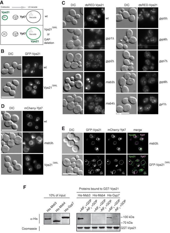
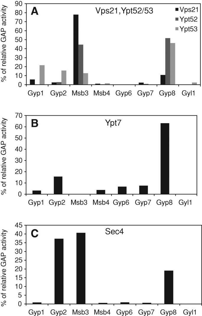
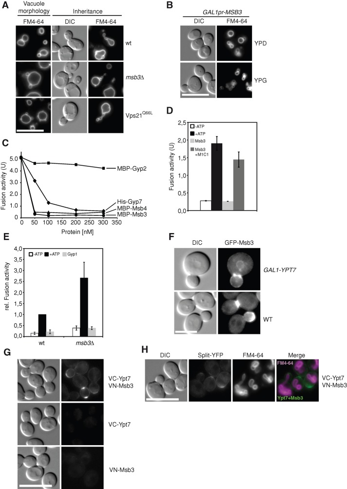
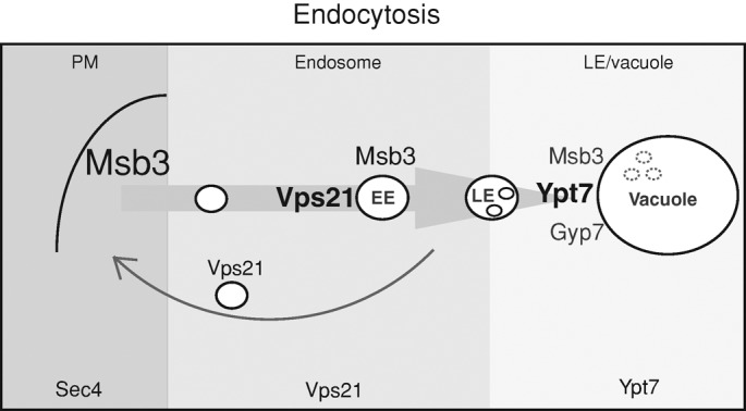
Fusion of organelles in the endomembrane system depends on Rab GTPases that interact with tethering factors before lipid bilayer mixing. In yeast, the Rab5 GTPase Vps21 controls fusion and membrane dynamics between early and late endosomes. Here we identify Msb3/Gyp3 as a specific Vps21 GTPase-activating protein (GAP). Loss of Msb3 results in an accumulation of Vps21 and one of its effectors Vps8, a subunit of the CORVET complex, at the vacuole membrane in vivo. In agreement, Msb3 forms a specific transition complex with Vps21, has the highest activity of all recombinant GAPs for Vps21 in vitro, and is found at vacuoles despite its predominant localization to bud tips and bud necks at the plasma membrane. Surprisingly, Msb3 also inhibits vacuole fusion, which can be rescued by the Ypt7 GDP-GTP exchange factor (GEF), the Mon1-Ccz1 complex. Consistently, msb3 vacuoles fuse more efficiently than wild-type vacuoles in vitro, suggesting that GAP can also act on Ypt7. Our data indicate that GAPs such as Msb3 can act on multiple substrates in vivo at both ends of a trafficking pathway. This ensures specificity of the subsequent GEF-mediated activation of the Rab that initiates the next transport event.


Figures

FIGURE 1:
FIGURE 1:
Identification of Msb3 as a Vps21-specific GAP. (A) A screen to identify a Vps21-specific GAP in yeast. In wild type, Vps21 is found in endosomes, whereas it is expected to shift to the vacuole if maintained in the GTP form. See text for details. (B) Localization of GTP-locked Vps21 to the vacuole. GFP-tagged Vps21 and its Q66L variant were introduced into a vps21∆ strain by integration of a linearized plasmid in which Vps21 is under the control of the NOP1 promotor. Yeast cells were monitored by fluorescence microscopy. DIC, differential interference contrast. (C) Localization of Vps21 in all GAP deletion strains. The indicated strains were transformed with a single-copy (CEN) plasmid encoding an additional copy of N-terminal dsRED-tagged Vps21 and analyzed as in A. (D) Ypt7 localization. mCherry-tagged Ypt7 was expressed as an additional copy in wild type, msb3∆, and Vps21Q66L strains and analyzed as in A. (E) Colocalization of GFP-Vps21 and mCherry-Ypt7 in the indicated strains by fluorescence microscopy. The grayscale pictures were two-dimensionally deconvolved with AutoQuant X software to reduce background fluorescence and were combined to an overlay with the GFP signal in the green channel and the mCherry signal in the red and blue channel. Merged signals can be observed in white. (F) In vitro interaction of Vps21 with GAPs. Purified His-Msb3, His-Msb4, or His-Gyp7 was incubated in the presence or absence of AlF3 with GST-Vps21, which was preloaded with GDP and coupled to GSH agarose. Bound proteins were eluted by boiling, resolved on SDS–PAGE, and detected with antibodies to the histidine tag (see Materials and Methods for details). A Coomassie-stained loading control of GST-Vps21 is shown.
FIGURE 2:
FIGURE 2:
Effect of msb3 deletion and mutants on the localization of Vps21 and effectors. (A–E) Wild-type, msb3∆, and Vps21 Q66L strains were transformed with a single-copy CEN plasmid expressing Vps8-GFP under the control of a NOP1 promotor (A), dsRED-Pep12 under the control of a PHO5 promotor (D), and Vps3-GFP under the control of a NOP1 promotor (E). Mon1 (B) and Vac1 (C) were genomically tagged with GFP or tomato, respectively. All strains were analyzed by fluorescence microscopy (see Materials and Methods). (F) Localization of GFP-tagged Msb3 and dsRED-tagged Vps21 was analyzed by fluorescence microscopy. (G) Localization of Vps21 in Msb3 active-site mutants. Variations of MSB3 were generated by QuikChange mutagenesis and stably integrated in the yeast genome of msb3∆ strains via a pRS shuttle vector. The proteins were expressed as GFP fusions under the control of the NOP1 promotor. dsRED-Vps21 was expressed from a single-copy CEN plasmid under the control of the PHO5 promotor. Cells were analyzed by fluorescence microscopy as before. DIC, differential interference contrast.
FIGURE 3:
FIGURE 3:
Activity of Msb3 and its interaction with Vps21. (A–C) Relative GTP-hydrolysis activity of the analyzed Rab–GAP pairs. All GAPs and Rabs were purified as full-length proteins. Rabs were precharged with [γ-32P]GTP, and release of radioactive 32P was analyzed over time as described in Materials and Methods. The entire GTP-hydrolysis of the respective Rab in all reactions with GAPs was set to 100%, and all Rab–GAP values are shown as a fraction of this hydrolysis rate. Each value was determined in duplicate and in three repetitions. All other GAP activities for each Rab are shown in Table 1.
FIGURE 4:
FIGURE 4:
Colocalization of Msb3 and Vps21 in vivo. (A) Colocalization of GAPs with endosomal membranes. Cells expressing the indicated GFP-tagged GAPs were labeled with FM4-64 and washed, and the dye was monitored after 10 and 60 min by fluorescence microscopy. (B) Localization of GAPs in the vps4 deletion. FM4-64 labeling and microscopy of the indicated strains was done as before. (C) Subcellular fractionation of cells expressing GFP-tagged Gyp2, Msb3, and Msb4. Cells of the indicated strains were lysed as described in Materials and Methods, and proteins were separated into a low-speed (P13) and a high-speed (P100) pellet. The pellets and the supernatants of the high-speed pellets (S100) were subjected to TCA precipitation and analyzed by Western blotting against the GFP tag. The vacuolar membrane protein Vac8 and the plasma membrane marker Sso2 served as control. We assume that the additional bands of Msb4 and Msb3 represent degradation products that appear due to the preparation, although we cannot exclude possible posttranslational modifications. (D, E) Analysis of Msb3 and Msb4 in isolated vacuole fractions. Vacuoles were purified from BY strains carrying GFP-tagged Msb3 and Msb4 (see Materials and Methods), and 40 μg of the isolated vacuoles was analyzed by SDS–PAGE and Western blotting against the GFP tag (D). In addition, vacuoles were directly analyzed by fluorescence microscopy (E). (F) Split-YFP analysis of Vps21 and Msb3. Vps21 and Msb3 were N-terminally tagged with the C-terminal (VC) or N-terminal (VN) Venus fragment, respectively (see Materials and Methods). Individually expressed proteins, as well as both in the same strain, were monitored by fluorescence microscopy. N-Terminally tagged VC-Vps39, together with VN-Msb3, was included as a control for unspecific binding. (G) Colocalization of the Vps21-Msb3 signal with genomically tomato-tagged Vac1 at its C-terminus by fluorescence microscopy.
FIGURE 5:
FIGURE 5:
Protein sorting in msb3∆ or overproduction strains. (A) Analysis of biosynthetic cargo, AP-3, and retrograde cargo to the vacuole. Wild-type, msb3∆, and Vps21 Q66L strains were transformed with a Cen-plasmid encoding an additional copy of N-terminal GFP-tagged Cps1, the GNS construct (GFP-Nyv1-Snc1; Reggiori et al., 2000), or the C-terminal GFP-tagged Vps10. As a positive control, we analyzed marker constructs in deletion strains. Vps8 is a class D subunit of the CORVET tethering complex, and its deletion causes Cps1 missorting to the vacuole membrane. Apl5 is a subunit of the AP-3 protein coat, causing missorting of the GNS construct to the plasma membrane upon deletion (Reggiori et al., 2000), and Vps10-GFP is missorted if retrograde transport is interrupted as in the vps26 mutant, a subunit of the retromer. (B) Effect of GAP deletions on macroautophagy. GAP deletions were transformed with a CEN plasmid harboring GFP-Atg8, and the cells were grown to mid exponential phase in a synthetic full medium lacking uracil. The cells were shifted to synthetic starvation medium and harvested at indicated time points, and protein extract was analyzed on a Western blot using anti-GFP antibodies. (C) Sorting of endocytic cargo to the vacuole lumen. The methionine transporter Mup1 was GFP tagged in wt cells, cells expressing inactive Vps21 S22N, or those carrying MSB3 under the control of the strong GPD promoter (Janke et al., 2004). Cells were grown in the absence of methionine (0 min). Methionine was added to a final concentration of 20 μg/ml, and Mup1-GFP was monitored immediately and 30 or 60 min thereafter.
FIGURE 6:
FIGURE 6:
Influence of Msb3 on vacuole morphology and fusion. (A) Typical vacuole morphology and morphological structures during inheritance. Vacuoles of wild-type, msb3∆, and Vps21 Q66L strains from a BY background (vacuole morphology) and a BJ background (inheritance) were labeled with FM4-64 as before and analyzed by fluorescence microscopy. BJ vacuoles have consistently large vacuoles and thus facilitate the scoring of possible inheritance defects. (B) Vacuole morphology upon Msb3 overexpression. Cells without (YPD) or with overexpressed (YPG) Msb3 were stained with FM4-64 and monitored by fluorescence microscopy. (C) Titration of purified GAPs into the vacuole fusion reaction. Vacuoles from the two tester strains were isolated as described in Materials and Methods and incubated in the presence of ATP and the indicated amounts of recombinant Gyp2 or Msb3. Fusion was determined after 90 min at 26°C (see Materials and Methods). (D) Recovery of fusion activity by Mon1–Ccz1. Fusion of wild-type vacuoles was done as in C. Where indicated, 0.5 μM Msb3 was added, and Mon1–Ccz1 (300 nM; Nordmann et al., 2010) was included in the presence of the same amounts of Msb3. (E) Fusion activity of msb3∆ vacuoles. Fusion of wild-type tester vacuoles was performed in parallel to tester vacuoles without Msb3. Gyp1-46 (0.5 μM) was added where indicated. (F) Localization of Msb3 in the absence and presence of overproduced Ypt7. Ypt7 was overproduced from the GAL1-promoter in cells expressing GFP-tagged Msb3. Cells were monitored by fluorescence microscopy. (G) Split-YFP analysis of Ypt7 and Msb3. Ypt7 and Msb3 were N-terminally tagged with the C-terminal (VC) or N-terminal (VN) Venus fragment, respectively (see Materials and Methods). Individually expressed proteins, as well as both in the same strain, were monitored by fluorescence microscopy. (H) Colocalization of the Ypt7-Msb3 signal with vacuoles. Vacuoles of a strain expressing both VC-Ypt7 andVN-Msb3 were labeled with FM4-64 as before and analyzed by fluorescence microscopy.
FIGURE 7:
FIGURE 7:
Working model. The role of Msb3 in the endocytic pathway. Relative amounts of Msb3, Vps21, and Ypt7 along the endocytic pathway are indicated by the font size. The different shades of gray indicate the predominant zones of each Rab.

Similar articles

Cited by

References

    1. Abenza JF, Galindo A, Pantazopoulou A, Gil C, de Los Ríos V, Peñalva MA. Aspergillus RabB Rab5 integrates acquisition of degradative identity with the long distance movement of early endosomes. Mol Biol Cell. 2010;21:2756–2769. - PMC - PubMed
    1. Albert S, Gallwitz D. Two new members of a family of Ypt/Rab GTPase activating proteins. Promiscuity of substrate recognition. J Biol Chem. 1999;274:33186–33189. - PubMed
    1. Albert S, Gallwitz D. Msb4p, a protein involved in Cdc42p-dependent organization of the actin cytoskeleton, is a Ypt/Rab-specific GAP. Biol Chem. 2000;381:453–456. - PubMed
    1. Albert S, Will E, Gallwitz D. Identification of the catalytic domains and their functionally critical arginine residues of two yeast GTPase-activating proteins specific for Ypt/Rab transport GTPases. EMBO J. 1999;18:5216–5225. - PMC - PubMed
    1. Barr F, Lambright DG. Rab GEFs and GAPs. Curr Opin Cell Biol. 2010;22:461–470. - PMC - PubMed

Publication types

MeSH terms

Substances

LinkOut - more resources