. 2012 May 16;32(20):6785-94.
doi: 10.1523/JNEUROSCI.0634-12.2012.

Calcium-dependent but action potential-independent BCM-like metaplasticity in the hippocampus

Affiliations

Calcium-dependent but action potential-independent BCM-like metaplasticity in the hippocampus

Sarah R Hulme et al. J Neurosci. .

Abstract

The Bienenstock, Cooper and Munro (BCM) computational model, which incorporates a metaplastic sliding threshold for LTP induction, accounts well for experience-dependent changes in synaptic plasticity in the visual cortex. BCM-like metaplasticity over a shorter timescale has also been observed in the hippocampus, thus providing a tractable experimental preparation for testing specific predictions of the model. Here, using extracellular and intracellular electrophysiological recordings from acute rat hippocampal slices, we tested the critical BCM predictions (1) that high levels of synaptic activation will induce a metaplastic state that spreads across dendritic compartments, and (2) that postsynaptic cell-firing is the critical trigger for inducing that state. In support of the first premise, high-frequency priming stimulation inhibited subsequent long-term potentiation and facilitated subsequent long-term depression at synapses quiescent during priming, including those located in a dendritic compartment different to that of the primed pathway. These effects were not dependent on changes in synaptic inhibition or NMDA/metabotropic glutamate receptor function. However, in contrast to the BCM prediction, somatic action potentials during priming were neither necessary nor sufficient to induce the metaplasticity effect. Instead, in broad agreement with derivatives of the BCM model, calcium as released from intracellular stores and triggered by M1 muscarinic acetylcholine receptor activation was critical for altering subsequent synaptic plasticity. These results indicate that synaptic plasticity in stratum radiatum of CA1 can be homeostatically regulated by the cell-wide history of synaptic activity through a calcium-dependent but action potential-independent mechanism.


Figures
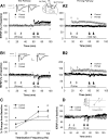
Figure 1.
Figure 1.
BCM-like heterosynaptic priming of LTP and LTD in stratum radiatum. Diagram of a hippocampal slice with electrode configurations shown. S1, Pathway on which priming stimulation was delivered; S2: pathway on which conditioning was delivered; R1, recording electrode. A1, When high-frequency priming stimulation was delivered on one radiatum pathway (light arrows, data for the primed pathway shown in A2), the level of LTP induced by subsequent 100 Hz stimulation (dark arrows) on the test pathway was significantly reduced (control, 27 ± 5%, n = 8; primed, 12 ± 5%, n = 6; p = 0.046). B1, Under control conditions 10 Hz conditioning induced stable LTD (−9 ± 3%; n = 6). In primed slices (data for the primed pathway shown in B2) significantly greater LTD of −18 ± 2% was induced (n = 7; p = 0.025). Insets, Representative waveforms are averages of 5 synaptic responses before LTP induction (1) and at the conclusion of the experiment (2). Calibration: 0.5 mV, 10 ms. C, Summary plot of mean fEPSP slope in the last 5 min of recording expressed as percentage change from baseline for control (filled circles) and primed (unfilled circles) groups across the range of stimulation frequencies from 10 to 100 Hz. Asterisks indicate significant difference between groups. D, Conditioning stimulation at 10 Hz with stronger stimulation (1.5 mV) induces a small potentiation which can be converted to depression when priming has been previously given (control, 6 ± 3%, n = 9; primed, −5 ± 4%, n = 6; p = 0.044). Inset, Summary plot showing the shift in LTD/LTP crossover point observed when stronger stimulation is used to induce synaptic plasticity. All data in this and subsequent figures are expressed as mean ± SEM.
Figure 2.
Figure 2.
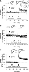
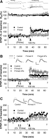
The metaplastic state can be cell-wide or compartmentally restricted. Diagram of a hippocampal slice with electrode configurations shown for A and B. S1, Stratum oriens pathway on which priming stimulation was delivered; S2, stratum radiatum pathway on which conditioning was delivered; R1, recording electrode in stratum oriens; R2, recording electrode in stratum radiatum. A, When high-frequency priming stimulation was delivered to stratum oriens (light arrows), the level of LTP induced by subsequent 100 Hz stimulation (dark arrow) on the test pathway in stratum radiatum was significantly reduced (control, n = 6; primed, n = 6). B, Priming in stratum oriens also facilitated LTD induction in stratum radiatum (control, 1 ± 1%, n = 6; primed, −12 ± 4%, n = 6). C, Priming stimulation in stratum radiatum did not inhibit LTP subsequently induced in stratum oriens (control, n = 8; primed, n = 7). Insets, Representative waveforms are averages of 5 synaptic responses before LTP induction (1) and at the conclusion of the experiment (2). Calibration: 0.5 mV, 10 ms.
Figure 3.
Figure 3.
Expression of the metaplastic effect is not through altered NMDAR or GABAergic function. A, Priming stimulation in stratum oriens reduced the NMDAR EPSC evoked on the homosynaptic pathway (control, 103 ± 7%, n = 5; primed, 73 ± 8%, n = 5). B, There was no change in the NMDAR EPSC evoked on the heterosynaptic (stratum radiatum) pathway after oriens priming (control, 106 ± 5%, n = 5; primed, 104 ± 7%, n = 6). C, Summated NMDAR EPSC area expressed as a ratio of the average single EPSC recorded in the last 5 min of recording before 100 Hz tetanization. There was no difference in summated area between primed and control slices in either the response to the first or second HFS (First HFS: control, 81 ± 16%, n = 5; primed 100 ± 33%, n = 5; p = 0.62; Second HFS: control, 91 ± 15%, primed, 114 ± 36%; p = 0.57). D, Summated NMDAR EPSC amplitude expressed as a ratio of the average single EPSC recorded in the last 5 min of recording before 100 Hz tetanization. There was no difference in summated amplitude between primed and control slices in either the response to the first or second HFS (First HFS: control, 12 ± 1%, n = 5; primed, 10 ± 1%, n = 5; p = 0.35; Second HFS: control, 12 ± 1%, primed, 11 ± 1%; p = 0.42). E, Blocking GABAergic transmission for the duration of the experiment did not prevent the inhibition of LTP following priming (control, 38 ± 5%, n = 7; primed, 15 ± 6%, n = 5). Insets, Representative waveforms are average of 5 synaptic responses before LTP induction (1) and at the conclusion of the experiment (2). Calibration: 0.5 mV, 10 ms.
Figure 4.
Figure 4.
Cell-firing is neither necessary nor sufficient to inhibit LTP. A, Priming significantly inhibited LTP observed with intracellular recording (control, 59 ± 9%, n = 6; primed, 14 ± 17%, n = 6; p = 0.037). Above graphs, Neither priming nor conditioning stimulation produced a change in input resistance (expressed as a percentage of pre-priming baseline); in the last 5 min of recording there was no significant difference in input resistance changes between primed (9 ± 6%) and control groups (0 ± 2%, p = 0.187). Top panel waveforms are representative single traces from one cell showing the first two trains of each bout of priming, and from the two trains of conditioning stimulation. Note the relatively low level of depolarization in all cases, and lack of substantial postsynaptic spiking. Calibration: 10 mV, 100 ms. B, Under control conditions (n = 10), which included a period of hyperpolarization before HFS, 100 Hz stimulation induced stable LTP. Hyperpolarization to completely prevent action potentials during priming stimulation in stratum radiatum (Primed H, n = 9) did not prevent the subsequent inhibition of LTP. The delivery of action potentials, patterned according to the standard priming stimulation (Primed APs, n = 6), facilitated subsequent LTP. C, Hyperpolarization to completely prevent action potentials during priming stimulation in stratum oriens (Primed H, n = 8) did not prevent the subsequent inhibition of LTP induced in stratum radiatum. Insets, Representative waveforms are the average of 10 synaptic responses before LTP induction (1) and at the conclusion of the experiment (2). Calibration: 5 mV, 10 ms.
Figure 5.
Figure 5.
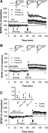
The inhibition of LTP is not dependent on NMDAR activation during priming. A, Priming in the presence of d-APV (50 μm, gray bar) resulted in most slices showing subsequently inhibited LTP [Primed (I), n = 6], while a subset of slices showed dramatically facilitated LTP [Primed (F) n = 3; control level of LTP (drug-treated as per primed slices) is shown by the black/gray dashed line, n = 7]. B, A similar result was observed when priming was given in the presence of d-APV (50 μm) and nimodipine (10 μm, gray bar) with some slices showing subsequently inhibited LTP [Primed (I), n = 6) and the same proportion as in A, showing facilitated LTP [Primed (F) n = 3; Control LTP, black/gray dashed line, n = 6]. C, When priming was given in the presence of d-APV (50 μm) and LY341495 (100 μm), only inhibited LTP was observed (control, n = 5; primed, n = 5). Insets (A, B), Representative waveforms are average of 5 synaptic responses before LTP induction (1) and at the conclusion of the experiment (2). Calibration: 0.5 mV, 10 ms. Inset (C), Distribution of fEPSP potentiation expressed as a percentage of baseline, measured in the last 5 min of recording, for the d-APV, d-APV + Nimodipine, and d-APV + LY341495 conditions.
Figure 6.
Figure 6.
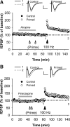
Dependence of priming on release of calcium from intracellular stores. A, When priming stimulation was delivered in stratum radiatum in the presence of 50 μm CPA (gray bar), there was no effect of priming on the level of subsequent LTP (control, n = 5; primed, n = 5). B, The inhibition of LTP by priming stimulation in stratum oriens was also prevented by CPA (control, n = 8; primed, n = 7). C, The facilitation of LTD by oriens priming was also blocked by CPA (control, 1 ± 1%, n = 6; primed, −1 ± 1%, n = 6; p = 0.11). D, When priming stimulation was delivered in stratum radiatum in the presence of 3 μm xestospongin C (Xesto, gray bar), there was no effect of priming on the level of subsequent LTP (control, n = 4; primed, n = 4). Insets, Representative waveforms are average of 5 synaptic responses before LTP induction (1) and at the conclusion of the experiment (2). Calibration: 0.5 mV, 10 ms.
Figure 7.
Figure 7.
M1-AChR activation is required for priming. A, When priming stimulation was delivered to stratum oriens in the presence of atropine (10 μm, gray bar), there was no effect of priming on the level of LTP (control, n = 6; primed, n = 6). B, The effect of priming stimulation on LTP was also prevented by the specific M1-AChR antagonist pirenzepine (20 μm; control, n = 5; primed, n = 5). Insets, Representative waveforms are averages of 5 synaptic responses before LTP induction (1) and at the conclusion of the experiment (2). Calibration: 0.5 mV, 10 ms.

Similar articles

Cited by

References

    1. Abraham WC. Metaplasticity: tuning synapses and networks for plasticity. Nat Rev Neurosci. 2008;9:387. - PubMed
    1. Abraham WC, Bear MF. Metaplasticity: the plasticity of synaptic plasticity. Trends Neurosci. 1996;19:126–130. - PubMed
    1. Abraham WC, Mason-Parker SE, Bear MF, Webb S, Tate WP. Heterosynaptic metaplasticity in the hippocampus in vivo: a BCM-like modifiable threshold for LTP. Proc Natl Acad Sci U S A. 2001;98:10924–10929. - PMC - PubMed
    1. Araque A, Martín ED, Perea G, Arellano JI, Buño W. Synaptically released acetylcholine evokes Ca2+ elevations in astrocytes in hippocampal slices. J Neurosci. 2002;22:2443–2450. - PMC - PubMed
    1. Artola A, Singer W. Long-term depression of excitatory synaptic transmission and its relationship to long-term potentiation. Trends Neurosci. 1993;16:480–487. - PubMed

Publication types

MeSH terms