. 2012 Jun 29;287(27):23216-26.
doi: 10.1074/jbc.M111.335927. Epub 2012 May 14.

Protein inhibitor of activated STAT3 (PIAS3) protein promotes SUMOylation and nuclear sequestration of the intracellular domain of ErbB4 protein

Affiliations

Protein inhibitor of activated STAT3 (PIAS3) protein promotes SUMOylation and nuclear sequestration of the intracellular domain of ErbB4 protein

Maria Sundvall et al. J Biol Chem. .

Abstract

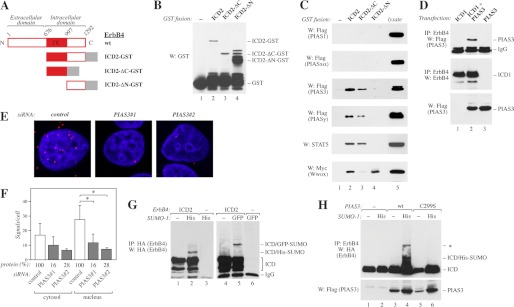
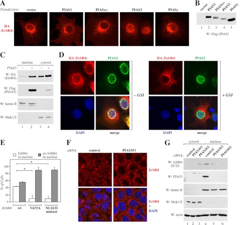
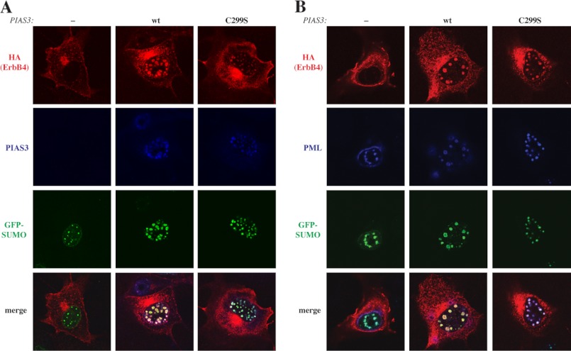
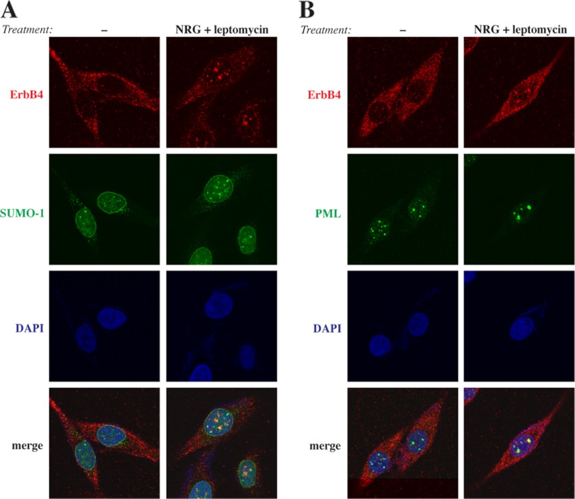
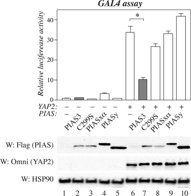
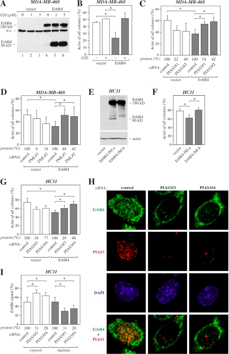
ErbB4 is a receptor tyrosine kinase implicated in the development and homeostasis of the heart, central nervous system, and mammary gland. Cleavable isoforms of ErbB4 release a soluble intracellular domain (ICD) that can translocate to the nucleus and function as a transcriptional coregulator. In search of regulatory mechanisms of ErbB4 ICD function, we identified PIAS3 as a novel interaction partner of ErbB4 ICD. In keeping with the small ubiquitin-like modifier (SUMO) E3 ligase function of protein inhibitor of activated STAT (PIAS) proteins, we showed that the ErbB4 ICD is modified by SUMO, and that PIAS3 stimulates the SUMOylation. Upon overexpression of PIAS3, the ErbB4 ICD generated from the full-length receptor accumulated into the nucleus in a manner that was dependent on the functional nuclear localization signal of ErbB4. In the nucleus, ErbB4 colocalized with PIAS3 and SUMO-1 in promyelocytic leukemia nuclear bodies, nuclear domains involved in regulation of transcription. Accordingly, PIAS3 overexpression had an effect on the transcriptional coregulatory activity of ErbB4, repressing its ability to coactivate transcription with Yes-associated protein. Finally, knockdown of PIAS3 with siRNA partially rescued the inhibitory effect of the ErbB4 ICD on differentiation of MDA-MB-468 breast cancer and HC11 mammary epithelial cells. Our findings illustrate that PIAS3 is a novel regulator of ErbB4 receptor tyrosine kinase, controlling its nuclear sequestration and function.


Figures

FIGURE 1.
FIGURE 1.
Interaction of ErbB4 with PIAS3. A, schematic structure of full-length ErbB4 JM-a CYT-2, and GST fusion proteins containing the whole ErbB4 intracellular domain of CYT-2-type (ICD2), ICD2 with C- (ΔC), or N-terminal (ΔN) deletion. TK, tyrosine kinase. B, GST fusion proteins (A) were pulled down with glutathione-Sepharose and analyzed by Western blotting (W) with anti-GST. C, GST-fusion proteins (A) were incubated with lysates of COS-7 cells transiently expressing FLAG-tagged PIAS proteins, STAT5A, or Myc-tagged Wwox. Pull-downs were analyzed by Western blotting with indicated antibodies. D, COS-7 cells were transfected with indicated constructs. Lysates were immunoprecipitated (IP) with anti-ErbB4 (HFR-1) followed by Western blotting with anti-FLAG to detect PIAS3. The membrane was reblotted with anti-ErbB4 (sc-283). Expression of PIAS3 was controlled by Western blotting with anti-FLAG. E, MCF-7 cells were transfected with the indicated control siRNA (Ambion) or siRNAs targeting PIAS3, stimulated for 15 min with 50 ng/ml NRG-1, and fixed. Complexes of ErbB4 and PIAS3 were visualized with anti-ErbB4 (HFR-1) and anti-PIAS3 (ab22856) antibodies using in situ PLA. Each red dot represents a single interaction. The nuclei were stained with DAPI (blue). F, quantification of PLA signals per cell. Signals were classified as cytosolic or nuclear on the basis of their colocalization with DAPI. Data of two independent experiments are shown (mean ± S.D.). *, p < 0.05. Densitometric data of PIAS3 protein levels relative to control knockdown as detected by Western blotting are indicated (protein (%)). G, COS-7 cells transfected with the indicated HA-tagged ErbB4 ICD2 and either His- or GFP-tagged SUMO-1 constructs were lysed in the presence of 20 mm N-ethylmaleimide. Lysates were analyzed by immunoprecipitation and Western blotting with anti-HA. H, COS-7 cells were transfected with HA-tagged ErbB4 ICD2 and the indicated FLAG-tagged PIAS3 and His-tagged SUMO-1 constructs. Lysates prepared as in G were immunoprecipitated with anti-ErbB4 (HFR-1) followed by Western blotting with anti-HA. Expression of PIAS3 was controlled by Western blotting with anti-FLAG. The band marked with an asterisk presumably represents another SUMO modification site.
FIGURE 2.
FIGURE 2.
Regulation of ErbB4 nuclear localization by PIAS3. A, COS-7 cells expressing HA-tagged ErbB4 JM-a CYT-2 with or without FLAG-tagged PIAS proteins were stained with anti-HA and photographed under a fluorescence microscope with a ×40 objective. B, expression of FLAG-tagged PIAS proteins in COS-7 cells was controlled by Western blotting with anti-FLAG. C, COS-7 cells expressing HA-tagged ErbB4 ICD2 and FLAG-tagged PIAS3 were subjected to subcellular fractionation. Nuclear and cytosolic fractions were analyzed by Western blotting (W) with indicated antibodies. Lamin B, nuclear marker; Mek1/2, cytosolic marker. D, COS-7 cells expressing HA-tagged ErbB4 JM-a CYT-2 and FLAG-tagged PIAS3 were treated for 4 h with 0 or 5 μm GSI IX and stained with anti-HA (red) and anti-PIAS3 (sc-46682, green). The nuclei were stained with DAPI (blue). The cells were visualized by confocal microscopy with a ×63 objective. E, COS-7 cells were transfected with constructs encoding HA-tagged ErbB4 JM-a CYT-2, ErbB4 JM-a CYT-2-V675A, or ErbB4 JM-a CYT-2-NLSI/II mutant and FLAG-tagged PIAS3 and stained with anti-HA. Cells were visualized by fluorescence microscopy and scored for cytosolic or nuclear staining. Columns (mean ± S.D.) show representative data from one of three independent experiments (*, p < 0.05). F, MCF-7 cells transfected with non-targeting (Qiagen) or PIAS3 siRNA were treated for 3 h with 25 ng/ml leptomycin B and for 45 min with 50 ng/ml NRG-1 and stained with anti-ErbB4 (HFR-1, red). The nuclei were stained with DAPI (blue). The cells were visualized by confocal microscopy with a ×40 objective. G, MCF-7 cells transfected with the indicated control siRNA (Ambion) or siRNAs targeting PIAS3 were subjected to subcellular fractionation. Nuclear and cytosolic fractions were analyzed by Western blotting with anti-ErbB4 (E200), anti-PIAS3 (sc-46682), anti-lamin B, and anti-Mek1/2. Loading was controlled by reblotting with anti-actin.
FIGURE 3.
FIGURE 3.
Colocalization of ErbB4 with SUMO-1, PIAS3, and PML. A, COS-7 cells expressing HA-tagged ErbB4 JM-a CYT-2, GFP-SUMO-1, and PML-3 in the presence or absence of PIAS3 or PIAS3C299S were stained with anti-HA (red) and anti-PIAS3 (sc-46682, blue). B, COS-7 cells expressing HA-tagged ErbB4 JM-a CYT-2, GFP-SUMO-1, and PML-3 in the presence or absence of PIAS3 or PIAS3C299S were stained with anti-HA (red) and anti-PML (sc-966, blue) antibodies. The cells were visualized by confocal microscopy with a ×63 objective.
FIGURE 4.
FIGURE 4.
Colocalization of endogenous ErbB4 with SUMO-1 and PML. WM-266-4 cells were treated for 3 h with 0 or 25 ng/ml leptomycin B and for 45 min with 0 or 50 ng/ml NRG-1. Cells were stained with anti-ErbB4 (HFR-1, red) and anti-SUMO-1 (green) (A) or anti-ErbB4 (HFR-1, red) and anti-PML (sc-5621, green) (B). The nuclei were stained with DAPI (blue). The cells were visualized by confocal microscopy with a ×63 objective.
FIGURE 5.
FIGURE 5.
PIAS proteins in ErbB4-regulated transcription. HEK 293 cells were transfected with a construct encoding ErbB4 ICD2 fused to the GAL4 DNA-binding domain and pFR-luc together with constructs encoding Omni-YAP2 and FLAG-tagged PIAS proteins as indicated. Columns represent relative luciferase activity normalized to signal from the cotransfected Renilla luciferase construct (mean ± S.D.). *, p < 0.05. Expression of PIAS proteins and YAP2 were controlled with anti-FLAG and anti-Omni, respectively. Loading was controlled by reblotting with anti-HSP90.W, Western blot.
FIGURE 6.
FIGURE 6.
PIAS3 in ErbB4-regulated differentiation. A, MDA-MB-468 cells expressing ErbB4 JM-a CYT-2 and the vector control cells were cultured for 6 h in the presence of 0, 1, or 5 μm GSI IX. Cell lysates were analyzed by Western blotting with anti-ErbB4 (sc-283). n.s., non-specific. B, MDA-MB-468 cells expressing ErbB4 JM-a CYT-2 and the vector control cells were suspended into Matrigel and cultured in the presence of 50 ng/ml NRG-1 and in the presence or absence of 5 μm GSI IX. On day 6 the colonies were counted. The columns represent the percentage of acini of all colonies in two independent experiments (mean ± S.D.). *, p < 0.05. C, MDA-MB-468 cells expressing ErbB4 JM-a CYT-2 and the vector control cells were treated with non-targeting (Qiagen) or the indicated PIAS3-specific siRNAs for 24 h, suspended into Matrigel, and cultured for 6 days. The columns represent the percentage of acini of all colonies in three independent experiments (mean ± S.D.). *, p < 0.05. Densitometric data of PIAS3 protein levels relative to control knockdown as detected by Western blotting are indicated (protein (%)). D, MDA-MB-468 cells expressing ErbB4 JM-a CYT-2 and the vector control cells were treated with non-targeting (Qiagen) or the indicated PML-specific siRNAs for 24 h, suspended into Matrigel, and cultured for 6 days. The columns represent the percentage of acini of all colonies in four independent experiments (mean ± S.D.). *, p < 0.05. Densitometric data of PML protein levels relative to control knockdown as detected by Western blotting are indicated (protein (%)). E, HC11 cells expressing ErbB4 JM-a CYT-2 or JM-b CYT-2 and the vector control cells were treated for 15 min with 50 ng/ml NRG-1 and analyzed by Western blotting with anti-ErbB4 (E200). The membrane was reprobed with anti-actin to control loading. F, HC11 cells expressing the indicated constructs were suspended into growth factor-reduced Matrigel and cultured for 14 days in the presence of 50 ng/ml NRG-1. The columns represent the percentage of acini of all colonies in three independent experiments (mean ± S.D.). *, p < 0.05. G, HC11 cells expressing ErbB4 JM-a CYT-2 and the vector control cells were treated with non-targeting (Ambion) or the indicated PIAS3-specific siRNAs for 24 h, suspended into Matrigel, and cultured for 15 days. The columns represent the percentage of acini of all colonies in two independent experiments (mean ± S.D.). *, p < 0.05. Densitometric data of PIAS3 protein levels relative to control knockdown as detected by Western blotting are indicated (protein (%)). H, representative confocal images (visualized with a ×63 objective) of HC11 cells expressing ErbB4 JM-a CYT-2 treated with non-targeting (Ambion) or PIAS3-specific siRNAs as in G. Cells were stained with anti-ErbB4 (HFR-1, green) and anti-PIAS3 (ab22856, red). The nuclei were stained with DAPI (blue). I, Quantification of cytosolic and nuclear ErbB4 immunoreactivity (mean ± S.D.). *, p < 0.05. Densitometric data of PIAS3 protein levels relative to control knockdown as detected by Western blotting are indicated (protein (%)).

Similar articles

Cited by

References

    1. Hynes N. E., Lane H. A. (2005) ERBB receptors and cancer. The complexity of targeted inhibitors. Nat. Rev. Cancer 5, 341–354 - PubMed
    1. Elenius K., Corfas G., Paul S., Choi C. J., Rio C., Plowman G. D., Klagsbrun M. (1997) A novel juxtamembrane domain isoform of HER4/ErbB4. Isoform-specific tissue distribution and differential processing in response to phorbol ester. J. Biol. Chem. 272, 26761–26768 - PubMed
    1. Elenius K., Choi C. J., Paul S., Santiestevan E., Nishi E., Klagsbrun M. (1999) Characterization of a naturally occurring ErbB4 isoform that does not bind or activate phosphatidyl inositol 3-kinase. Oncogene 18, 2607–2615 - PubMed
    1. Rio C., Buxbaum J. D., Peschon J. J., Corfas G. (2000) Tumor necrosis factor α-converting enzyme is required for cleavage of erbB4/HER4. J. Biol. Chem. 275, 10379–10387 - PubMed
    1. Ni C. Y., Murphy M. P., Golde T. E., Carpenter G. (2001) γ-Secretase cleavage and nuclear localization of ErbB-4 receptor tyrosine kinase. Science 294, 2179–2181 - PubMed

Publication types

MeSH terms

LinkOut - more resources