. 2012 May 9;20(5):899-910.
doi: 10.1016/j.str.2012.02.023.

Interaction of the mediator head module with RNA polymerase II

Affiliations

Interaction of the mediator head module with RNA polymerase II

Gang Cai et al. Structure. .

Abstract

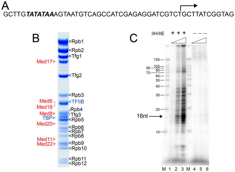
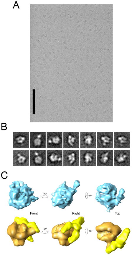
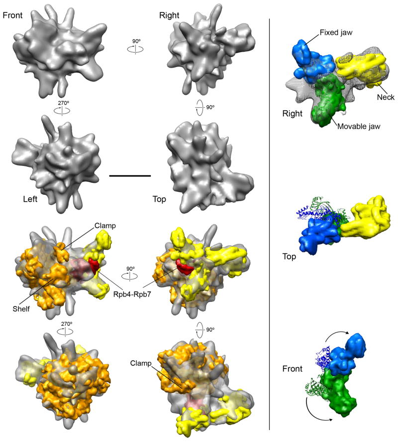
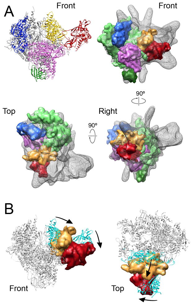
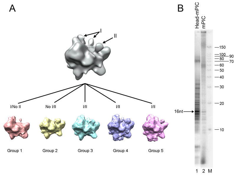
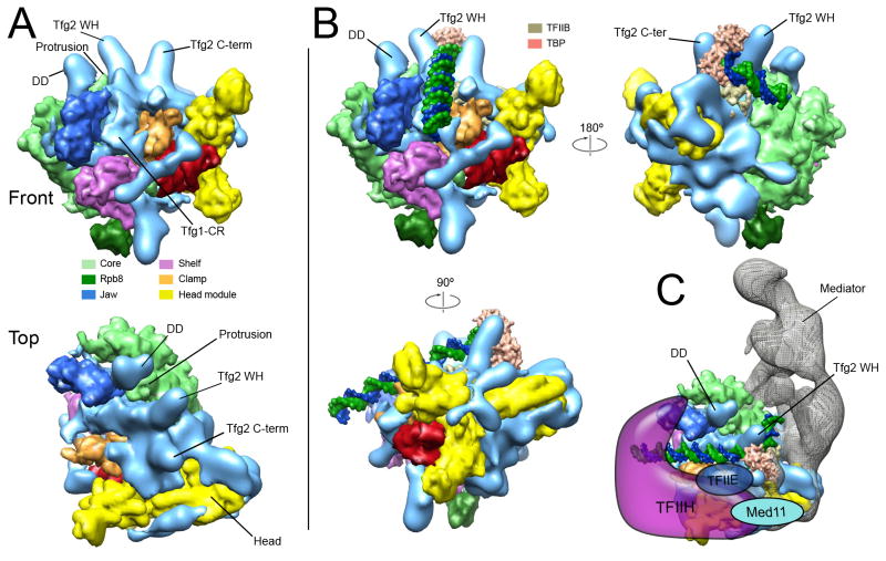
Mediator, a large (21 polypeptides, MW ∼1 MDa) complex conserved throughout eukaryotes, plays an essential role in control of gene expression by conveying regulatory signals that influence the activity of the preinitiation complex. However, the precise mode of interaction between Mediator and RNA polymerase II (RNAPII), and the mechanism of regulation by Mediator remain elusive. We used cryo-electron microscopy and reconstituted in vitro transcription assays to characterize a transcriptionally-active complex including the Mediator Head module and components of a minimum preinitiation complex (RNAPII, TFIIF, TFIIB, TBP, and promoter DNA). Our results reveal how the Head interacts with RNAPII, affecting its conformation and function.


Figures

Figure 1
Figure 1. Assembly and functional characterization of the Head-mPIC complex
(A) Sequence of the 53 base pair promoter DNA used for assembly of the Head-mPIC complex. The TATA box sequence (in italic bold) located about 30 bp upstream of the initiation start site (bent arrow) was fused to the DNA template used previously for RNAPII elongation complex assembly (Westover et al., 2004). (B) SDS-PAGE analysis of the Head-mPIC complex. The subunits of RNAPII (Rpb1-12), TFIIF (Tfg1-3), and the Mediator Head module (Med17, Med6, Med20, Med8, Med18, Med11, and Med22) were identified as indicated. (C) Transcriptional activity of the Head-mPIC complex was tested in a reconstituted transcription assays performed in the presence (lanes 1–3; 0.37, 0.75, and 1.5 pmol Head-mPIC, respectively) or absence (lanes 4–6; same Head-mPIC concentrations as 1–3) of TFIIE and TFIIH. Transcripts were separated in a 15% denaturing gel and imaged with a fluorescent image analyzer. The predominant transcript (16 nucleotides long) is indicated. M: labeled RNA Decade marker. See also Figure S1.
Figure 2
Figure 2. Initial cryo-EM analysis of a Head-mPIC complex
(A) A raw micrograph showing images of Head-mPIC particles preserved in amorphous ice. The scale bar corresponds to 100 nm. (B) Some initial class averages including clear non-RNAPII features obtained after reference-free alignment and clustering of Head-mPIC cryo-EM images. (C) Initial 3D structure of the Head-mPIC complex (top) obtained by back projection after determining the relative orientations of Head-mPIC class averages, and a distortion-free model of the Head-mPIC structure (bottom) constructed by fitting a ~35 Å resolution molecular map of RNAPII derived from its X-ray structure (Bushnell and Kornberg, 2003) (in orange) and a ~35 Å EM map of the open conformation of the Head module (Cai et al., 2010) (in bright yellow) into the initial Head-mPIC structure. See also Figures S2 and S3.
Figure 3
Figure 3. Cryo-EM structure of the Head-mPIC complex and docking of the RNAPII and Head module structures
(A) Different views of a ~16 Å resolution cryo-EM map of the Head-mPIC complex illustrate how the main portion of the structure is strongly reminiscent of RNAPII, and show that a large segment of additional density matches the EM structure of the open conformation of the Head module (Cai et al., 2010) (shown in red in the Front, Right, and Top views). The scale bar corresponds to 10 nm. (B) Molecular models (at 10 Å resolution) of RNAPII (in orange) and the Head module (in bright yellow), derived from their X-ray structures (Cramer et al., 2001; Imasaki et al., 2011) were fitted into the Head-mPIC cryo-EM map. RNAPII could be docked in a single orientation. The Head structure was segmented into its known structural modules (neck, fixed and movable jaws) and these were re-positioned to match the distribution of Head density in the Head-mPIC map. RNAPII subunits Rpb4-Rpb7 are positioned between the Head module jaws. (C) The conformation of the Head module in the Head-mPIC complex (top panel) can be compared to that revealed by X-ray crystallography analysis by matching the position of the neck and considering changes in the positions of the fixed and movable jaws. Upon interaction with RNAPII, both the fixed and movable jaws undergo large rotations (original positions in the X-ray structure are shown as ribbons in the middle and bottom panels; positions in the cryo-EM map of the Head-mPIC complex as solid surfaces), resulting in a conformation that allows the jaws to interact closely with RNAPII. See also Figures S4, S5, S6 and S7.
Figure 4
Figure 4. Conformation of RNAPII in the Head-mPIC complex
(A) Molecular maps (at 18 Å resolution) derived from RNAPII structural modules (top left; core in light green, Rpb8 in dark green, jaw-lobe in blue, shelf in magenta, clamp in yellow, and Rpb4-Rpb7 in red) defined by X-ray crystallography studies of RNAPII (Cramer et al., 2001) docked into the Head-mPIC EM map (shown as a semi-transparent gray mesh). All modules were docked independently and computed docking positions were minimally adjusted to preserve inter-module crystal contacts. (B) Comparing the positions of the clamp and the Rpb4-Rpb7 subunits in the Head-mPIC cryo-EM map (solid surfaces; clamp in gold and Rpb4-Rpb7 in red) with those in the X-ray structure of RNAPII (the RNAPII crystal structure is shown as a gray ribbon model with the clamp and Rpb4-Rpb7 colored in teal) reveals that Head interaction results in a large rearrangement of the clamp, likely facilitated by repositioning of Rpb4-Rpb7 (movement of the clamp and Rpb4-Rpb7 also indicated by the arrows). See also Figures S8 and S9.
Figure 5
Figure 5. Conformational variability and transcriptional activity of the Head-mPIC complex
(A) Five Head-mPIC maps generated through Codimensional Principal Component Analysis (CD-PCA) provide a detailed description of variability in the conformation of the Head-mPIC complex. Domains at the upstream (I) and downstream (II) ends of the active site cleft are variably present/ordered in the different conformations of the Head-mPIC complex revealed by CD-PCA. (B) Comparison between transcriptional activity of the Head-mPIC and mPIC complexes in the presence of TFIIH and TFIIE. Assays were performed with the Head–mPIC (1.5 pmol, lane 1), and mPIC (1.5 pmol, lane 2) and results were analyzed as before. The amounts of 16-nucleotide long transcript generated were 2.2 fmol for the Head-mPIC, and 0.12 fmol for mPIC alone. M: labeled RNA Decade marker. See also Figure S10.
Figure 6
Figure 6. Implications for PIC organization and RNAPII-promoter interaction
(A) Distribution of additional (non-RNAPII and non-Head) density (depicted as a light blue surface) along the RNAPII surface. Individual features are positioned adjacent to the locations of specific TFIIF domains determined through biochemical studies (see Figure S6) (Chen et al., 2007; Chen et al., 2010; Eichner et al., 2010). Densities that likely correspond to the Tfg1-Tfg2 dimerization domain (DD) and the Tfg2 wing-helix domain (Tfg2 WH) flank the RNAPII protrusion. Density matching the expected location of the Tfg1 charged region (Tfg1-CR) and Tfg2 C-terminus (Tfg2 C-term) are also observed. (B) Fitting of a model for organization of the RNAPII-TFIIB-TBP-DNA complex into the Head-mPIC cryo-EM map. Matching of the core module in the RNAPII-TFIIB and Head-mPIC structures results in TFIIB and TBP being nestled between the Tfg2 WH and Tfg2 C-term densities above the upstream end of the polymerase active site cleft, without any steric clashes with possible TFIIF density. The Tfg2 wing-helix domain (Tfg2 WH) would be positioned to interact with promoter DNA, as expected (Eichner et al., 2010). Promoter DNA would travel along the RNAPII active site cleft and have unimpeded access to the polymerase active site after opening of the clamp. (C) A model for interaction of additional preinitiation complex components with the Head-mPIC complex. The position of the Head would place Mediator subunit Med11 (partially located in the Head’s fixed jaw (Imasaki et al., 2011)) in a position where it could interact with TFIIH, as reported (Esnault et al., 2008). Also, opening of the active site cleft upon outward rotation of the clamp would expose surfaces for interaction with TFIIE and TFIIH. See also Figure S11.

Comment in

Similar articles

Cited by

References

    1. Armache KJ, Kettenberger H, Cramer P. Architecture of initiation-competent 12-subunit RNA polymerase II. Proc Natl Acad Sci U S A. 2003;100:6964–6968. - PMC - PubMed
    1. Asturias FJ, Jiang YW, Myers LC, Gustafsson CM, Kornberg RD. Conserved structures of mediator and RNA polymerase II holoenzyme. Science. 1999;283:985–987. - PubMed
    1. Baek HJ, Malik S, Qin J, Roeder RG. Requirement of TRAP/mediator for both activator-independent and activator-dependent transcription in conjunction with TFIID-associated TAF(II)s. Mol Cell Biol. 2002;22:2842–2852. - PMC - PubMed
    1. Belogurov GA, Vassylyeva MN, Sevostyanova A, Appleman JR, Xiang AX, Lira R, Webber SE, Klyuyev S, Nudler E, Artsimovitch I, et al. Transcription inactivation through local refolding of the RNA polymerase structure. Nature. 2009;457:332–335. - PMC - PubMed
    1. Bernecky C, Grob P, Ebmeier CC, Nogales E, Taatjes DJ. Molecular architecture of the human Mediator-RNA polymerase II-TFIIF assembly. PLoS Biol. 2011;9:e1000603. - PMC - PubMed

Publication types

MeSH terms