. 2012 Jul;26(7):1102-16.
doi: 10.1210/me.2011-1367. Epub 2012 May 8.

Disruption of nongenomic testosterone signaling in a model of spinal and bulbar muscular atrophy

Affiliations

Disruption of nongenomic testosterone signaling in a model of spinal and bulbar muscular atrophy

Mathilde Schindler et al. Mol Endocrinol. 2012 Jul.

Abstract

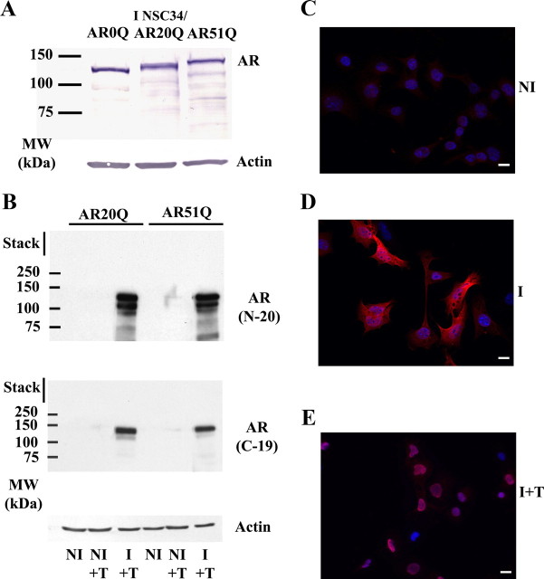
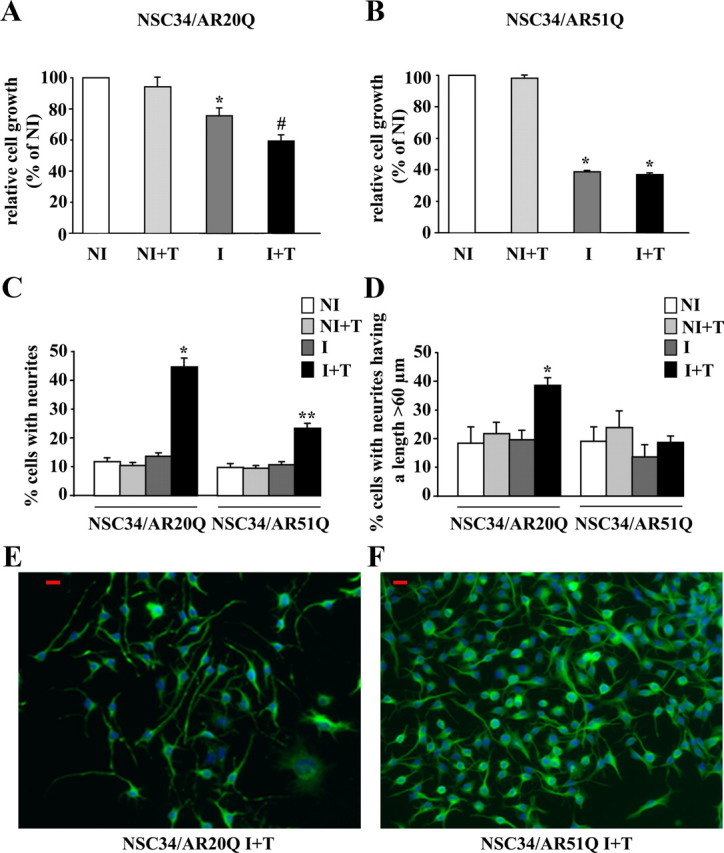
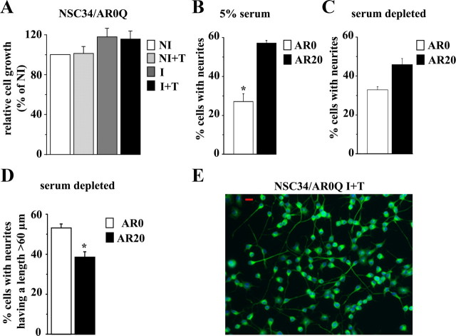
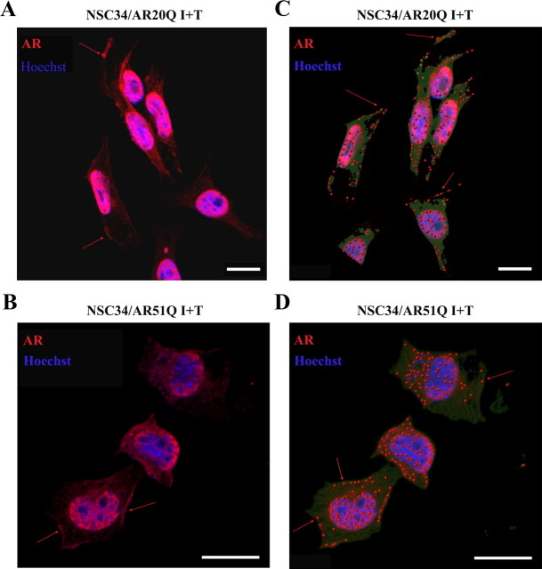
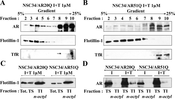
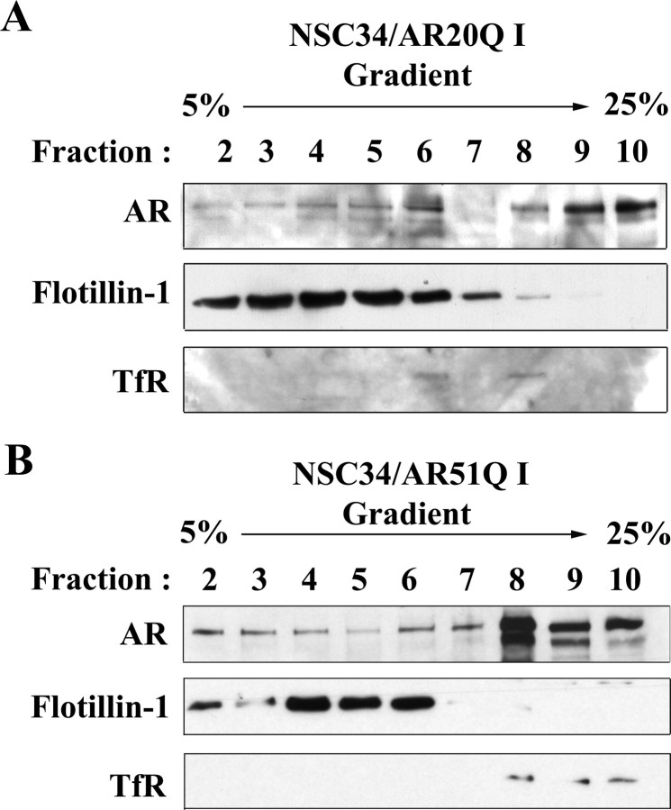
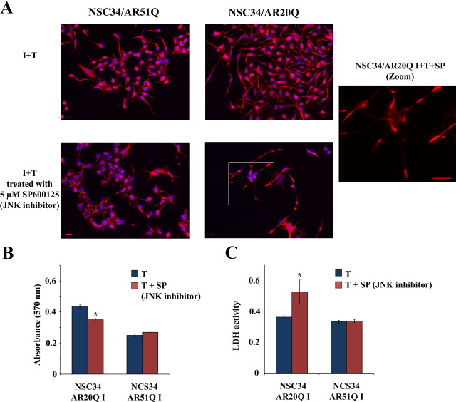
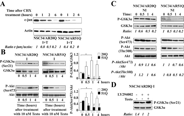
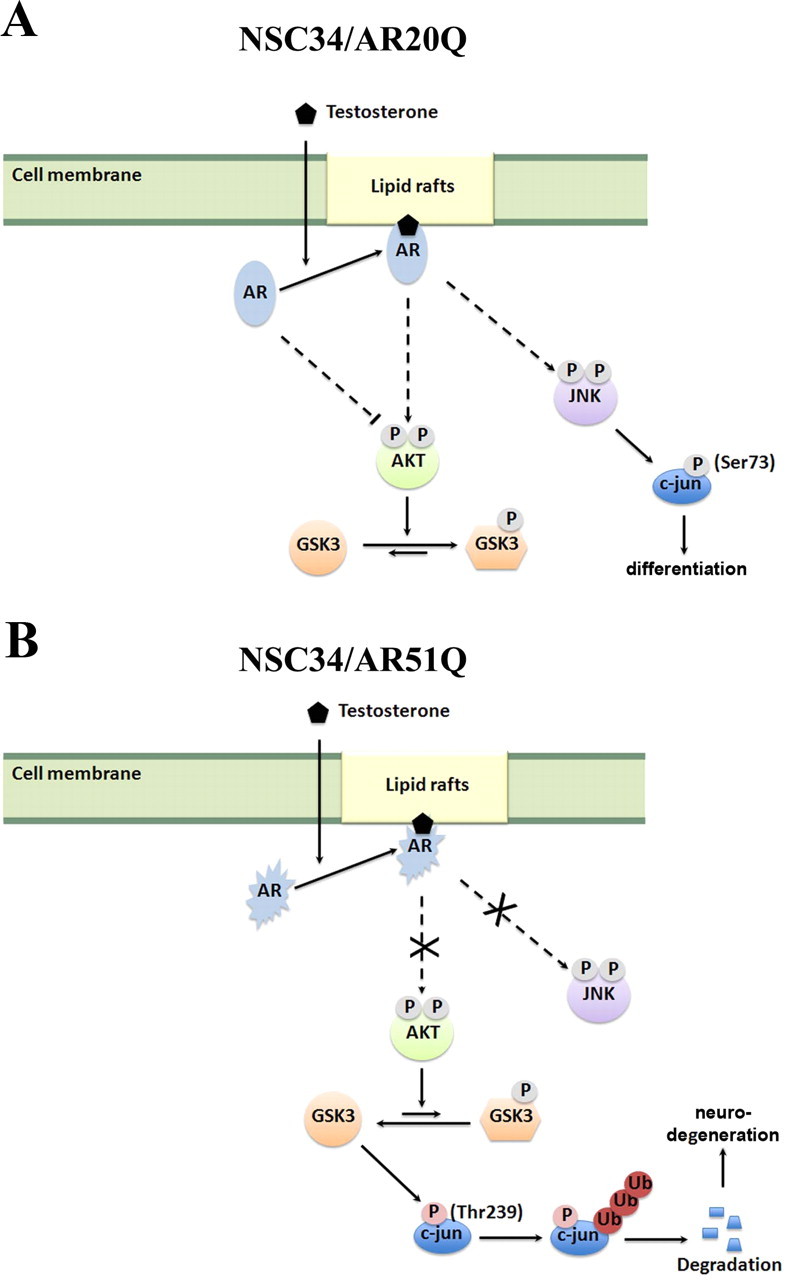
As one of the nine hereditary neurodegenerative polyQ disorders, spinal and bulbar muscular atrophy (SBMA) results from a polyQ tract expansion in androgen receptor (AR). Although protein aggregates are the pathological hallmark of many neurodegenerative diseases, their direct role in the neurodegeneration is more and more questioned. To determine the early molecular mechanisms causing motor neuron degeneration in SBMA, we established an in vitro system based on the tetracycline-inducible expression of normal (AR20Q), the mutated, 51 glutamine-extended (AR51Q), or polyQ-deleted (AR0Q) AR in NSC34, a motor neuron-like cell line lacking endogenous AR. Although no intracellular aggregates were formed, the expression of the AR51Q leads to a loss of function characterized by reduced neurite outgrowth and to a toxic gain of function resulting in decreased cell viability. In this study, we show that both AR20Q and AR51Q are recruited to lipid rafts in response to testosterone stimulation. However, whereas testosterone induces the activation of the c-jun N-terminal kinase/c-jun pathway via membrane-associated AR20Q, it does not so in NSC34 expressing AR51Q. Phosphorylation of c-jun N-terminal kinase plays a crucial role in AR20Q-dependent survival and differentiation of NSC34. Moreover, c-jun protein levels decrease more slowly in AR20Q- than in AR51Q-expressing NSC34 cells. This is due to a rapid and transient inhibition of glycogen synthase kinase 3α occurring in a phosphatidylinositol 3-kinase-independent manner. Our results demonstrate that the deregulation of nongenomic AR signaling may be involved in SBMA establishment, opening new therapeutic perspectives.


Figures

Fig. 1.
Fig. 1.
Characterization of the cellular models. A, Western blot analysis of stably transfected NSC34/AR(n)Q (n = 0, 20, or 51). NSC34/AR(n)Q were treated with 1 μg/ml doxycycline (I) or vehicle (NI) for 48 h and then incubated with 10 nm testosterone (+T) for 24 h. B, Validation of inducible expression of AR(n)Q. Cells were treated with doxycycline or with vehicle for 48 h and then treated with 10 nm testosterone or with vehicle for 24 h. Total proteins (10 μg) were loaded and calibrated against β-actin. Western blots were probed with the AR(N-20) polyclonal antibody or with the AR(C-19) polyclonal antibody. C–E, Expression and localization of AR(51)Q in NSC34 determined by immunofluorescence. Cells were treated with doxycycline (I) or vehicle (NI) for 48 h and then incubated for 24 h with vehicle or 10 nm testosterone (+T). Immunofluorescence were carried out using the AR(N-20) polyclonal antibody (red). Nuclei were stained by Hoechst (blue). Scale bar, 20 μm. MW, Molecular weight.
Fig. 2.
Fig. 2.
Influence of polyQ tract length on cell viability/growth and neurite outgrowth: AR20Q vs. AR51Q. A and B, Effects of polyQ region length on cell viability measured by MTT assay. NSC34/AR(n)Q were incubated for 48 h with doxycycline or with vehicle. Then cells were treated with 10 nm testosterone or vehicle for 242 h. Cell proliferation and viability are expressed as a percentage relative to NI control cells. Results are presented as means ± sem (n = 8). A, Influence of AR20Q expression on NSC34 viability/growth. *, P < 0.05, I vs. NI and NI+T NSC34/AR20Q ; #, P < 0.01, I+T vs. NI and NI+T NSC34/AR20Q, ANOVA test. B, Influence of AR51Q expression on NSC34 growth. *, P < 0.001, NI vs. I+T NSC34/AR51Q, ANOVA test. C and D, Influence of polyQ tract on neurite outgrowth. After 48 h after exposure to doxycycline (I) or vehicle (NI), NSC34/AR(n)Q (n = 20 or 51) were depleted in serum and treated with 10 nm testosterone (+T) or vehicle for 24 h. Results are represented as means ± sem of pooled data from three separate experiments. C, Influence of AR(n)Q expression and testosterone treatment on percentage of neurite-bearing cells. Asterisks indicate significant difference compared with control (NI and NI+T) or induced cells (I without testosterone): *, P < 0.01; **, P < 0.001 (ANOVA test). D, Influence of AR(n)Q expression and testosterone treatment on cell number with a longest neurite longer than 60 μm. *, P < 0.01, I+T NSC34/AR20Q vs. NI, NI+T, and I NSC34/AR20Q (ANOVA test). E and F, Immunofluorescence of serum-depleted I+T NSC34/AR(n)Q (n = 20 or 51) were performed using the peripherin antibody detected with Alexa 488-conjugated secondary antibody. Nuclei are stained with Hoechst. Peripherin, green; nuclei, blue; red scale bars, 20 μm.
Fig. 3.
Fig. 3.
Effects of AR0Q on cell viability and neurite elongation. A, At 48 h after exposure with doxycycline or vehicle, NSC34/AR0Q were treated with 10 nm testosterone or vehicle for 120 h. *, P < 0.001 (ANOVA test). B, Effect of AR0Q expression at 24 h after testosterone treatment (10 nm) on percentage of neurite-bearing cells in the presence of serum (5%). *, P < 0.01 (ANOVA test); *, significant difference compared with I+T NSC34/AR20Q at P < 0.05 (ANOVA test). C–E, After 48 h after exposure to doxycycline (I), NSC34/AR(n)Q (n = 0 or 20) were depleted in serum and treated with 10 nm testosterone (+T) for 24 h. C and D, Percentage of neurite-bearing cells (C) and the percentage of cells with the longest neurite having a length greater than 60 μm (D). *, P < 0.05, ANOVA test. E, Immunofluorescence of serum-depleted I+T NSC34/AR0Q was performed using the peripherin antibody detected with Alexa 488-conjugated secondary antibody. Nuclei are stained with Hoechst. Peripherin, green; nuclei, blue; red scale bar, 20 μm.
Fig. 4.
Fig. 4.
Localization of AR(n)Q within plasma membrane by immunofluorescence. Induction of AR(n)Q expression through treatment with doxycycline for 48 h in NSC34/AR(n)Q was followed by incubation with 10 nm testosterone for 2 h. A and B, Immunofluorescence of AR(n)Q (red). Nuclei (blue) are stained with Hoechst 33258. These images were obtained by confocal microscopy. Discontinuous membrane staining is identified using red arrows. C and D, Pixel quantification using Imaris version 7.0.0 software realized from previous confocal images. Points represent zones where AR(n)Q staining shows the highest intensity.
Fig. 5.
Fig. 5.
Localization of AR(n)Q within lipid rafts. A and B, Analysis of AR(n)Q in density gradient fractions. Both I NSC34/AR20Q and I NSC34/AR51Q were depleted in serum and treated with 1 μm testosterone for 24 h. Ten fractions were collected from the top of the gradient, and 10 μg of proteins of the last nine fractions was resolved by SDS-PAGE. Western blots were probed with anti-AR(C-19), anti-flotillin-1, and anti-TfR antibodies. C and D, Localization of AR(n)Q within lipid rafts by isolation of detergent-resistant membrane. Treatment of NSC34/AR20Q and NSC34/AR51Q with doxycycline for 48 h was followed by exposure with 1 μm testosterone for 24 h. Proteins (10 μg) of the total cell lysate (Tot.) and TS and TIn-octyl fractions were resolved by SDS-PAGE. Western blots were probed with anti-flotillin-1 (C) and anti-AR(N20) (D).
Fig. 6.
Fig. 6.
Density gradient of I NSC34/AR(n)Q. A and B, AR(n)Q localization in Optiprep density gradient fractions of I NSC34/AR(n)Q (without testosterone). Both I NSC34/AR20Q and I NSC34/AR51Q were depleted in serum and treated with 1 μm vehicle for 24 h. Ten fractions were collected from the top of the gradient, and 10 μg of proteins of the last nine fractions was resolved by SDS-PAGE. Western blots were probed with anti-AR(C-19), anti-flotillin-1, and anti-TfR antibodies.
Fig. 7.
Fig. 7.
Influence of testosterone on JNK/c-jun pathway. A and B, Both serum-depleted I NSC34/AR20Q and I NSC34/AR51Q were treated with 10 nm of testosterone at the indicated times. Equal amounts of total proteins (30 μg) per well were resolved by SDS-PAGE, and Western blots were probed with antibodies against phospho-c-jun(Ser73), phospho-SAPK/JNK(Thr183/Tyr185), total c-jun, and total JNK. The c-jun and JNK phosphorylation levels from the autoradiograph were normalized to total c-jun or total JNK. A, Testosterone effects on c-jun(Ser73) phosphorylation levels in I NSC34/AR20Q (left) and I NSC34/AR51Q (right). Histograms represent the mean ratio of phospho-c-jun(Ser73) level to total c-jun level ± sem of three independent experiments at indicated times of exposure with testosterone. #, P < 0.05, t test. B, Testosterone effects on JNK1–3 activity in I NSC34/AR20Q (left) and I NSC34/AR51Q (right). Histograms represent the mean ratio of phospho-SAPK/JNK(Thr183/Tyr185) level to total SAPK/JNK level (n = 3 independent experiments) at indicated times of testosterone treatment. Error bars represent ± sem. #, P < 0.05; *, P < 0.01, t test.
Fig. 8.
Fig. 8.
Influence of JNK inhibition on NSC34/AR(n)Q. A, Influence of JNK inhibition on neurite morphology in NSC34/AR(n)Q. Immunofluorescence of peripherin in serum-depleted I NSC34/AR(n)Q (n = 20 or 51) treated with 10 nm testosterone together with 5 μm SP600125 (I+T+SP) or with vehicle (I+T). Peripherin is in red, and nuclei are stained blue. These images are representative of several images from three different experiments. Red scale bar, 20 μm. B, Influence of JNK inhibition on cell viability in NSC34/AR(n)Q, measured by MTT assay. Serum-depleted I NSC34/AR(n)Q (n = 20 or 51) were treated with 10 nm testosterone for 24 h together with 5 μm SP600125 or with vehicle control. The mean of absorbance (λ = 570 nm) in four wells ± sem is plotted. *, P < 0.05, ANOVA test. C, LDH release measured by absorbance at λ = 490 nm and λ = 655 nm in serum-depleted I NSC34/AR(n)Q (n = 20 or 51) treated with 10 nm testosterone for 24 h together with 5 μm SP600125 or with vehicle control. *, P < 0.05, ANOVA test.
Fig. 9.
Fig. 9.
Influence of testosterone on Akt and GSK3α activity. A, Testosterone effects on total c-jun. Serum-depleted I NSC34/AR20Q and I NSC34/AR51Q were treated with 10 nm testosterone for 2 h and then incubated with cycloheximide (CHX, 100 μg/ml) at various times (1, 2, and 6 h). Total proteins (30 μg/well) were subjected to SDS-PAGE, and Western blots were carried out using anti-total c-jun (60A8) antibody. Levels of total c-jun were normalized to β-actin. B, GSK3α and Akt phosphorylation levels. Both serum-depleted I NSC34/AR20Q and I NSC34/AR51Q were treated with 10 nm testosterone (Testo) at the indicated times. Data of three independent experiments are expressed as the ratio of phospho-GSK3α to total GSK3α and the ratio of phospho-Akt (Ser473) to total Akt. *, P < 0.05; **, P < 0.05; #, P < 0.01, t test. C, GSK3α and Akt phosphorylation levels in noninduced cells. Both serum-depleted NI NSC34/AR20Q and NI NSC34/AR51Q were treated with 10 nm testosterone (Testo) at the indicated times. All data of relative activity are expressed in comparison with untreated cells (time zero). Data are representative of two experiments. D, Inhibitory phosphorylation of GSK3α induced by AR20Q is PI3K independent. Serum-depleted I NSC34/AR20Q cells were treated with vehicle (I) or with 5 μm LY294002 alone or together with 10 nm testosterone. Equal amounts of total proteins (30 μg) per well were resolved by SDS-PAGE (B–D), and Western blots were probed with antibodies against phospho-GSK3α(Ser21), phospho-Akt(Ser473), total GSK3α, and total Akt. Histograms represent quantification of phospho-GSK3α and phospho-Akt(Ser473) in each sample.
Fig. 10.
Fig. 10.
Scheme of nongenomic activation of AR20Q and AR51Q. Solid lines indicate direct action on the target. Dotted lines indicate indirect action. Ub, Ubiquitin.

Similar articles

Cited by

References

    1. Zoghbi HY , Orr HT. 2000. Glutamine repeats and neurodegeneration. Annu Rev Neurosci 23:217–247 - PubMed
    1. Kennedy WR , Alter M , Sung JH. 1968. Progressive proximal spinal and bulbar muscular atrophy of late onset. A sex-linked recessive trait. Neurology 18:671–680 - PubMed
    1. La Spada AR , Wilson EM , Lubahn DB , Harding AE , Fischbeck KH. 1991. Androgen receptor gene mutations in X-linked spinal and bulbar muscular atrophy. Nature 352:77–79 - PubMed
    1. Li M , Miwa S , Kobayashi Y , Merry DE , Yamamoto M , Tanaka F , Doyu M , Hashizume Y , Fischbeck KH , Sobue G. 1998. Nuclear inclusions of the androgen receptor protein in spinal and bulbar muscular atrophy. Ann Neurol 44:249–254 - PubMed
    1. McCampbell A , Taylor JP , Taye AA , Robitschek J , Li M , Walcott J , Merry D , Chai Y , Paulson H , Sobue G , Fischbeck KH. 2000. CREB-binding protein sequestration by expanded polyglutamine. Hum Mol Genet 9:2197–2202 - PubMed

Publication types

MeSH terms