. 2012 Jul;11(7):874-84.
doi: 10.1128/EC.00095-12. Epub 2012 May 4.

The Tlo proteins are stoichiometric components of Candida albicans mediator anchored via the Med3 subunit

Affiliations

The Tlo proteins are stoichiometric components of Candida albicans mediator anchored via the Med3 subunit

Anda Zhang et al. Eukaryot Cell. 2012 Jul.

Abstract

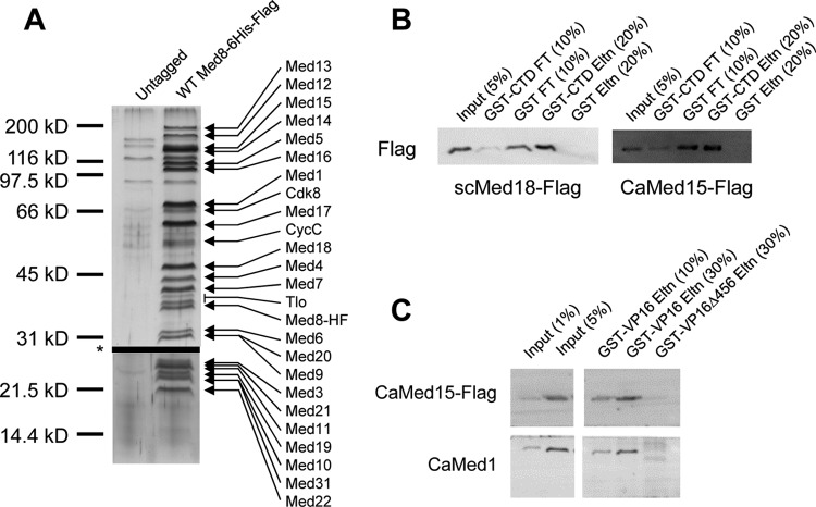
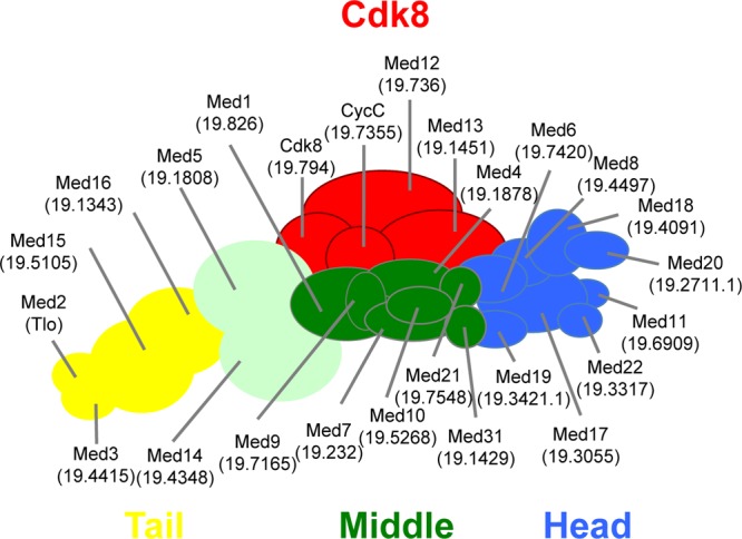
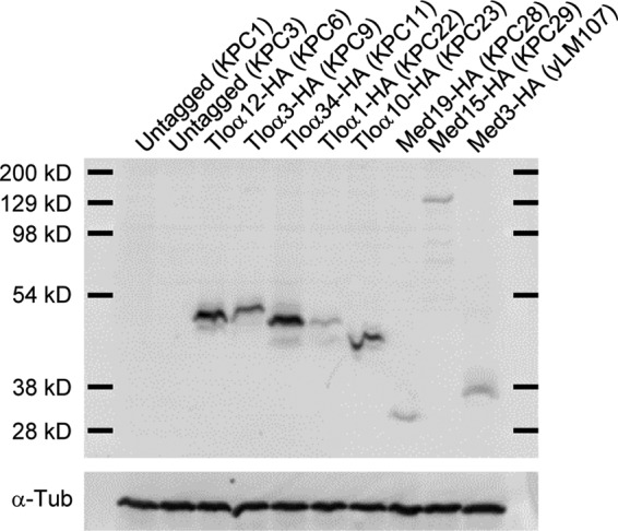
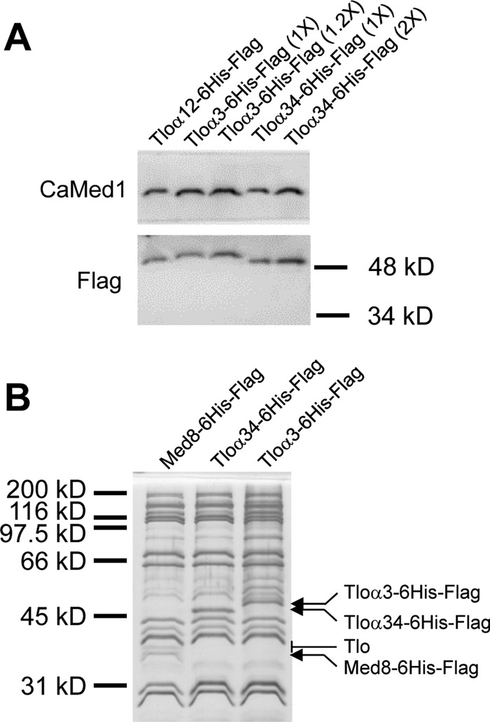
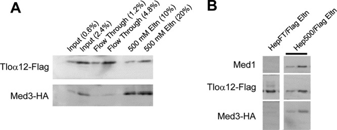
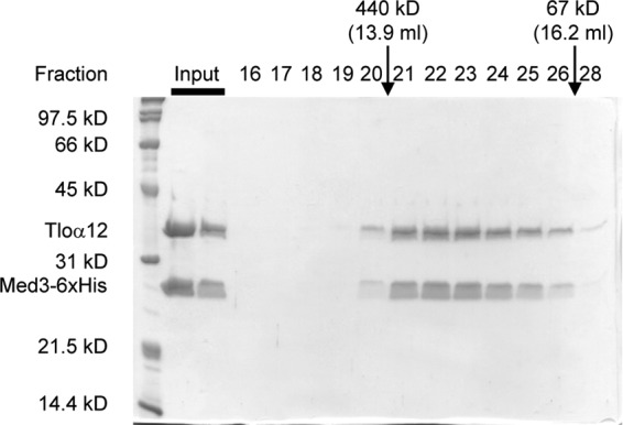
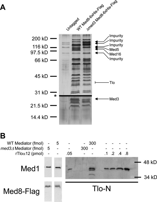
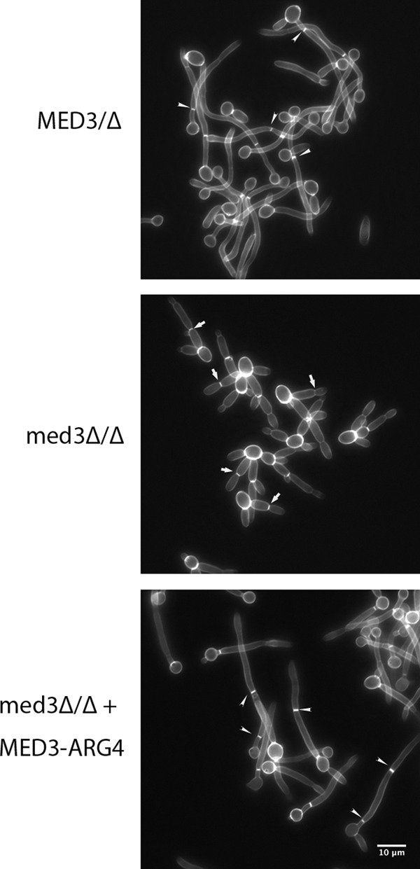
The amplification of the TLO (for telomere-associated) genes in Candida albicans, compared to its less pathogenic, close relative Candida dubliniensis, suggests a role in virulence. Little, however, is known about the function of the Tlo proteins. We have purified the Mediator coactivator complex from C. albicans (caMediator) and found that Tlo proteins are a stoichiometric component of caMediator. Many members of the Tlo family are expressed, and each is a unique member of caMediator. Protein expression analysis of individual Tlo proteins, as well as the purification of tagged Tlo proteins, demonstrate that there is a large free population of Tlo proteins in addition to the Mediator-associated population. Coexpression and copurification of Tloα12 and caMed3 in Escherichia coli established a direct physical interaction between the two proteins. We have also made a C. albicans med3Δ/Δ strain and purified an intact Mediator from this strain. The analysis of the composition of the med3Δ Mediator shows that it lacks a Tlo subunit. Regarding Mediator function, the med3Δ/Δ strain serves as a substitute for the difficult-to-make tloΔ/Δ C. albicans strain. A potential role of the TLO and MED3 genes in virulence is supported by the inability of the med3Δ/Δ strain to form normal germ tubes. This study of caMediator structure provides initial clues to the mechanism of action of the Tlo genes and a platform for further mechanistic studies of caMediator's involvement in gene regulatory patterns that underlie pathogenesis.


Figures

Fig 1
Fig 1
Composition and functional characterization of C. albicans Mediator. (A) Lysates from an untagged and a Med8-6His-Flag-tagged strain were subjected in parallel to multiple chromatographic separations. The elutions from the IMAC (Talon) step were analyzed by 7.5% (results above the asterisk) and 12.5% (below the asterisk) SDS-PAGE. The two gels were run on identical samples to ensure adequate resolution in both the high- and low-molecular-mass ranges. Proteins were revealed by staining with silver. The annotated identity of each band was determined by excising the band from another SDS-PAGE gel run in parallel and using mass spectrometry to analyze the protein content of each band. (B) Purified scMediator and caMediator bind to the CTD of RNA Pol II. Either GST or GST-CTD was prebound to magnetic glutathione beads, and a standardized amount of purified scMediator or caMediator was added (Input). The samples were monitored by Western blotting using the anti-Flag antibody against a Flag tag on scMed18 or caMed15. Percentages represent the percentages of the total sample at each stage that was loaded onto the gel. The flowthrough (FT) shows that GST-CTD, but not GST, depleted both scMediator and caMediator from the input. The bound scMediator and caMediator were recovered in the elution (Eltn) from the GST-CTD beads, while no detectable signal was recovered from the GST beads. (C) Purified caMediator binds to the activation domain of VP16, not an activation-deficient mutant (VP16Δ456). Either GST-VP16 or GST-VP16Δ456 was prebound to magnetic glutathione beads, and a standardized amount of purified caMediator was added (Input). The samples were monitored by Western blotting using the caMed1 antibody and the anti-Flag antibody against a Flag tag on caMed15. The percentages are of the total sample at each stage that was loaded onto the gel. Bound caMediator was recovered in the elution (Eltn) from the GST-VP16 beads, while little detectable signal was recovered from the GST-VP16Δ456 beads.
Fig 2
Fig 2
Assignment of C. albicans Mediator subunits to ORFs and model of its structure. This figure shows a model of the C. albicans Mediator based on an updated version of a model of the modular topology of the S. cerevisiae Mediator (14). We have colored Med5 and Med14 lime green, since they have characteristics of both tail and middle module subunits. The standardized Mediator subunit name is followed by the ORF designation in the C. albicans database in parentheses. The Tlo proteins can serve as orthologs for scMed2. Elsewhere in the manuscript we discuss the particular Tlo proteins that we have found can form a subunit of Mediator.
Fig 3
Fig 3
Expression analysis of Tlo proteins and Mediator subunits in whole-cell extracts. Whole-cell extracts were made from strains that were untagged or had a single copy of a Tlo or other Mediator subunit tagged with the HA epitope. The strain used for each extract is listed in parentheses. (Upper panel) A Western blot is shown that used an anti-HA antibody for detection. The total amount of protein loaded in each well was roughly normalized by cell OD, followed by a precise normalization with an anti-tubulin antibody. (Lower panel) The blot in this figure was reprobed with the anti-tubulin antibody, and each lane was shown to have an equal amount of tubulin.
Fig 4
Fig 4
Tloα3, Tloα12, and Tloα34 are Med2 orthologs of caMediator. Whole-cell lysates from Tloα3-6His-Flag, Tloα12-6His-Flag, and Tloα34-6His-Flag strains were subjected to our Mediator purification protocol and analyzed by Western blotting and silver staining. (A) Western blot of various amounts of protein from the Flag-agarose elutions. One quantity of Tloα12-6His-Flag and two quantities of Tloα3-6His-Flag and Tloα34-6His-Flag elutions were loaded. The number in parentheses is the quantity of a sample relative to the lower quantity. The samples were probed with the caMed1 and Flag antibodies. The ratio of the Flag signal to the caMed1 signal is comparable between all three samples, indicating that each of the three Tlo proteins in the heparin elution was able to pull down a proportional amount of intact Mediator. (B) Comparison of equal amounts of the Med8-6His-Flag, Tloα34-6His-Flag, and Tloα3-6His-Flag Talon elutions by SDS-PAGE and silver staining of a 10% gel. The general pattern of silver stain bands indicates that both Tloα34-6His-Flag- and Tloα3-6His-Flag-purified Mediator are largely identical to the Med8-6His-Flag pure Mediator. Tloα34-6His-Flag and Tloα3-6His-Flag protein appear to be the only detectable Tlo component of each population of purified Mediator. The Tlo band observed in the Med8-6His-Flag Mediator is missing from the Tloα34-6His-Flag and Tloα3-6His-Flag samples, and a new band appeared at a molecular size that corresponds to the Tloα34-6His-Flag and Tloα3-6His-Flag signals detected by Western blotting in the corresponding samples. The molecular mass range occupied by the Tlo band in the Med8-6His-Flag Mediator is also left absent by the decrease in the molecular size of Med8, which is untagged in the Tlo-tagged samples.
Fig 5
Fig 5
Majority of Tloα12 protein is in the heparin unbound protein fraction and is not associated with Mediator subunits. Lysate from a Tloα12-Flag-Med3-HA-tagged strain was separated on heparin Sepharose into unbound and bound fractions. Material from both fractions was purified on Flag-agarose. (A) Western blot analysis (using an anti-Flag and anti-HA antibody) of the input, flowthrough, and 500 mM KOAc elution (Eltn) of the heparin column shows that a majority of Med3 binds but that a majority of Tloα12 flows through. The number in parentheses represents the percentage of 1/1,000 of the total amount of each fraction loaded on the gel. (B) Western blot of the Flag elution from the heparin flowthrough (HepFT/Flag Eltn) and two amounts of the Flag elution from the heparin elution (Hep500/Flag Eltn) probed with the caMed1, Flag, and HA antibodies. The Flag signal from the HepFT/Flag elution sample exceeds that from the Hep500/Flag elution samples, yet the HepFT/Flag elution contains no detectable Mediator as monitored by the caMed1 and HA (Med3) antibodies.
Fig 6
Fig 6
Size-exclusion chromatography shows that recombinant C. albicans Tloα12 and Med3 form a cocomplex. IMAC quantitatively depletes coexpressed Tloα12 and Med3-6His from E. coli lysates. The IMAC elution was separated by ion exchange followed by size exclusion. Input and fractions from the size-exclusion column (Superose 6) were analyzed by SDS-PAGE on a 12% gel, followed by staining with Coomassie blue. The relative migrations of two molecular size standards are marked with respect to the fractions collected.
Fig 7
Fig 7
caMediator purified from a med3 null strain lacks Tlo proteins and other tail module subunits. (A) Lysate from an untagged, Med8-6His-Flag-tagged WT strain and a Med8-6His-Flag-tagged med3 null strain were subjected, in parallel, to multiple chromatographic separations. The elutions from the IMAC (Talon) step were analyzed by 7.5% (results above the asterisk) and 12.5% (below the asterisk) SDS-PAGE. The two gels were run on identical samples to ensure adequate resolution in both the high- and low-molecular-mass ranges. Proteins were revealed by staining with silver. The positions of bands identified in Fig. 1, which differ in the med3Δ Mediator, are marked with arrows. (B) Western blotting comparing various amounts of purified WT and med3Δ Mediator and recombinant Tloα12/Med3-6His complex. The amount of WT Mediator was estimated by comparison of band intensity on a Coomassie gel to a known standard (data not shown). caMed1 and Flag antibodies were used to demonstrate a normalized amount of Med1, and Med8-6His-Flag was present in both the purified WT and med3Δ Mediator. The pan-Tlo-N antibody was used to detect Tlo proteins in normalized amounts of the WT and med3Δ Mediator. The sensitivity of the pan-Tlo-N antibody was calibrated by adding known amounts of our recombinant Tloα12/Med3-6His complex. These standards show that the amount of Tlo protein in the WT Mediator is roughly equimolar to the total amount of Mediator loaded, and that the depletion of Tlo protein in the med3Δ Mediator at least exceeds ∼85%.
Fig 8
Fig 8
Morphology of MED3/Δ (yLM93), med3Δ/Δ (yLM96), and med3Δ/Δ plus MED3-ARG4 (AZC53-3) cells undergoing the blastospore-to-filament transition. All strains were grown under non-filament-inducing conditions and were diluted into prewarmed YPD medium at 37°C in the presence of 2.5 mM GlcNAc. Cells were removed at various time points and stained with Blankophor. Each panel shows a separate strain after 2.5 h of induction. Arrows point to the location of septa formed along the filament. A complete set of strains from an entire time course is shown in Fig. S2 in the supplemental material. Bar at bottom left, 10 μm.

Similar articles

Cited by

References

    1. Baidoobonso SM, Guidi BW, Myers LC. 2007. Med19(Rox3) regulates intermodule interactions in the Saccharomyces cerevisiae mediator complex. J. Biol. Chem. 282:5551–5559 - PubMed
    1. Béve J, et al. 2005. The structural and functional role of Med5 in the yeast Mediator tail module. J. Biol. Chem. 280:41366–41372 - PubMed
    1. Bjorklund S, Gustafsson CM. 2005. The yeast Mediator complex and its regulation. Trends Biochem. Sci. 30:240–244 - PubMed
    1. Booth LN, Tuch BB, Johnson AD. 2010. Intercalation of a new tier of transcription regulation into an ancient circuit. Nature 468:959–963 - PMC - PubMed
    1. Borggrefe T, Davis R, Erdjument-Bromage H, Tempst P, Kornberg RD. 2002. A complex of the Srb8, -9, -10 and -11 transcriptional regulatory proteins from yeast. J. Biol. Chem. 277:44202–44207 - PubMed

Publication types

LinkOut - more resources