. 2012 Sep-Oct;6(4):391-9.
doi: 10.4161/pri.20199. Epub 2012 May 7.

Prion formation by a yeast GLFG nucleoporin

Affiliations

Prion formation by a yeast GLFG nucleoporin

Randal Halfmann et al. Prion. 2012 Sep-Oct.

Abstract

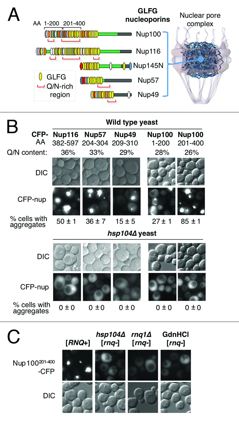
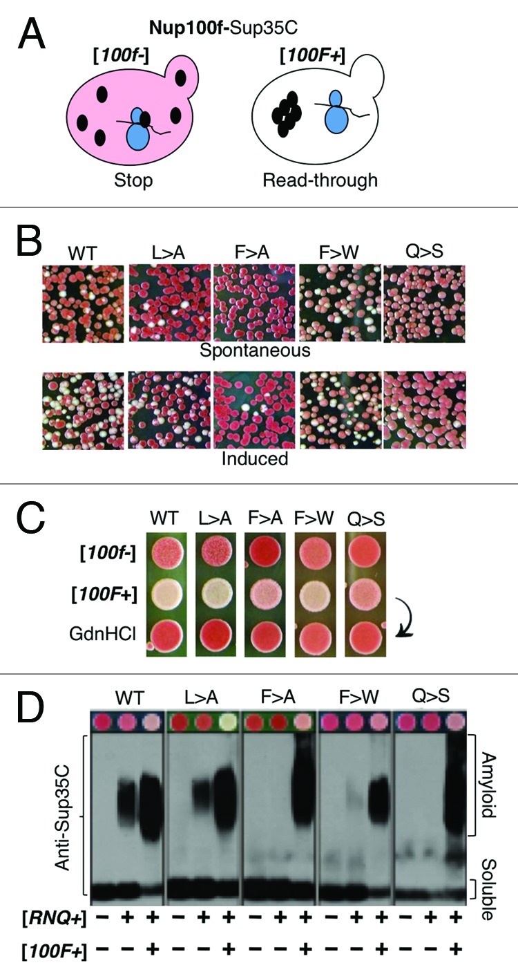
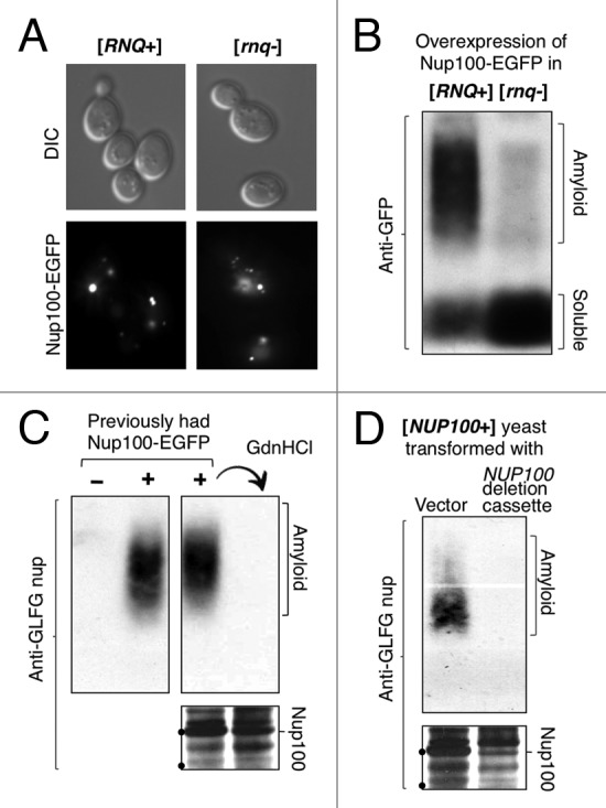
The self-assembly of proteins into higher order structures is both central to normal biology and a dominant force in disease. Certain glutamine/asparagine (Q/N)-rich proteins in the budding yeast Saccharomyces cerevisiae assemble into self-replicating amyloid-like protein polymers, or prions, that act as genetic elements in an entirely protein-based system of inheritance. The nuclear pore complex (NPC) contains multiple Q/N-rich proteins whose self-assembly has also been proposed to underlie structural and functional properties of the NPC. Here we show that an essential sequence feature of these proteins--repeating GLFG motifs--strongly promotes their self-assembly into amyloids with characteristics of prions. Furthermore, we demonstrate that Nup100 can form bona fide prions, thus establishing a previously undiscovered ability of yeast GLFG nucleoporins to adopt this conformational state in vivo.


Figures

None
Figure 1. GLFG nucleoporins form prion-like aggregates. (A) Diagram of the NPC and of intrinsically-disordered GLFG nups populating its conduit. Each GLFG nup is shown as a purple rectangle (N-terminus at left) and the location of FG motifs is indicated by vertical ovals. GLFG motifs are yellow, FxFG red, SPFG dark green, FxFx light gray, SAFG dark blue, PSFG bright green, NxFG light blue, SLFG orange, xxFG white, and FxxFG lime green. The red brackets below each nup highlight the center of Q/N rich regions larger than 100 AA featuring ≥ 30% Q/N content within 80 consecutive AAs. The horizontal gray rectangle in each nup marks its known or presumed NPC anchor domain. (B) Intracellular aggregation of Q/N-rich regions of GLFG nups. Q/N-rich regions of Nup116, Nup57, Nup49 and Nup100 were overexpressed as CFP fusions in WT and hsp104Δ yeast from a constitutive ADH1 promoter. The percentage of cells (n > 400) with fluorescent Nup-CFP aggregates is indicated; standard deviation is from two independent experiments. (C) [RNQ+]-dependence of Nup100201–400 aggregation. Nup100201–400-CFP was overexpressed as in (B) in [RNQ+] cells and in cells converted to [rnq-] by deletion of HSP104 (hsp104Δ); by deletion of RNQ1 (rnq1Δ); or by treatment with 5 mM GdnHCl.
None
Figure 2. The prion-like region of Nup100 forms insoluble aggregates that sequester endogenous GLFG nups. (A) Mislocalization of endogenous nups by overexpressed Nup100201–400. Yeast containing a chromosomal fusion of Nup100, Nup116, Nup49 or Nup2 with GFP were transformed with a plasmid that overexpresses Nup100201–400. The percent of cells with cytoplasmic nup aggregates (n > 400) is shown below the pictures. Standard deviation (± ) is from two independent experiments. The arrowheads point to cytoplasmic nup aggregates in cells, next to the more typical nuclear rim fluorescence pattern of the nup (B) Aggregation of endogenous nups induced by overexpressed Nup100201–400. [rnq-] and [RNQ+] cells overexpressing Nup100201–400-CFP were lysed and cleared of unbroken cells by centrifugation at 2,000 x g. The low speed supernatant fraction (T) was fractionated further at 12,000 x g into medium speed supernatant (MSS) and pellet (MSP) fractions. Proteins in each fraction were resolved by SDS-PAGE, and the presence of Nup100201–400-CFP, Gsp1 and endogenous GLFG nups were detected by western blotting with anti-GFP, anti-Gsp1, or anti-GLFG nup antibodies. The black dots on the gel frames mark the position of 116, 97 and 68 kDa molecular weight markers.
None
Figure 3. Nup100f forms amyloids under physiological conditions. (A) AA sequence of Nup100f. FG motifs are highlighted in bold text; Q/N residues are underlined. (B) Kinetics of Nup100f amyloid formation in vitro. Left panel: Nup100f-Trp-7xHis (WT) was diluted from denaturant to 20 μM in assembly buffer. The reaction was incubated at 30°C with agitation, in the absence or presence of 5% pre-formed aggregate seed. Amyloid assembly was monitored by ThT fluorescence. Data represent means ± SEM from three reactions. Right panel: The indicated Nup100f-Trp-7xHis variants were assembled in the absence of pre-formed fiber seeds. (C) Ultrastructure of WT Nup100f amyloids. Seeded amyloids formed as in (B) were negatively stained with uranyl acetate and examined by transmission electron microscope. Scale bar, 200 nm.
None
Figure 4. Nup100f is a prion-forming domain. (A) Schematic of the phenotypic reporter used to detect Nup100PrD-Sup35C prions. When WT Sup35 forms prion aggregates, it is sequestered away from its role in translation termination, causing a stop codon read-through phenotype that converts cells from red [psi-] to white [PSI+] (not shown). The Sup35PrD can be substituted for the Nup100PrD, resulting in a fully functional chimeric protein that recapitulates both the red ([100f-]) and white ([100F+]) states (shown). (B) Frequency of spontaneous and induced appearance of [100F+] in cells. WT or variant [100f-] [RNQ+] cells containing a galactose-inducible version of the respective Nup100f-EYFP were grown overnight in either glucose- or galactose-containing media, followed by plating to YPD to assess the appearance of white or pink [100F+] colonies. Red colonies derive from cells that remain [100f-]. (C) Hsp104-dependence of [100F+] variants. WT and variant [100f-] strains were spotted onto YPD (top row). The corresponding [100F+] strains before and after GdnHCl treatment were spotted below. (D) Detection of SDS-resistant aggregates of Nup100f-Sup35C in lysates of prion-containing cells. Variant [100f-] and [100F+] cells in both [rnq-] and [RNQ+] strains were analyzed by SDD-AGE. The 100f-Sup35C fusion proteins were detected with anti-Sup35C antibodies. The color phenotype of the corresponding strains grown on YPD is shown above the SDD-AGE blots.
None
Figure 5. Prion-formation by full-length Nup100. (A) Overexpressed full-length Nup100-EGFP forms foci in both [rnq-] and [RNQ+] yeast. Cells containing NUP100-EGFP on a high copy galactose-inducible plasmid were grown overnight in galactose media, and then analyzed by fluorescence microscopy. (B) Detection of Nup100-EGFP amyloids in [rnq-] and [RNQ+] cell extracts. [rnq-] or [RNQ+] yeast expressing Nup100-EGFP as in (A) were analyzed by SDD-AGE. The blot was probed with anti-GFP, revealing SDS-resistant aggregates of Nup100-EGFP in [RNQ+] cells. (C) Prion formation by an endogenous GLFG nup. Cells that either had or had not overexpressed Nup100-EGFP, but no longer contained the Nup100-EGFP overexpression plasmid, were analyzed by SDD-AGE. Note how the transient overexpression of Nup100-EGFP induced persistent amyloids of endogenous GLFG nups, and these were eliminated by passage on GdnHCl. (D) GLFG nup prion amyloids require Nup100. [NUP100+] cells were transformed with either empty vector or a linearized NUP100 gene-deletion cassette (targeted to create Δnup100). Verified transformants were analyzed by SDD-AGE as in (C). The black dots on the gel frames mark the position of 100 and 75 kDa molecular weight markers.

Similar articles

Cited by

References

    1. Prusiner SB. Novel proteinaceous infectious particles cause scrapie. Science. 1982;216:136–44. doi: 10.1126/science.6801762. - DOI - PubMed
    1. Halfmann R, Lindquist S. Epigenetics in the extreme: prions and the inheritance of environmentally acquired traits. Science. 2010;330:629–32. doi: 10.1126/science.1191081. - DOI - PubMed
    1. Hou F, Sun L, Zheng H, Skaug B, Jiang QX, Chen ZJ. MAVS forms functional prion-like aggregates to activate and propagate antiviral innate immune response. Cell. 2011;146:448–61. doi: 10.1016/j.cell.2011.06.041. - DOI - PMC - PubMed
    1. Majumdar A, Cesario WC, White-Grindley E, Jiang H, Ren F, Khan MR, et al. Critical role of amyloid-like oligomers of Drosophila Orb2 in the persistence of memory. Cell. 2012;148:515–29. doi: 10.1016/j.cell.2012.01.004. - DOI - PubMed
    1. Alberti S, Halfmann R, King O, Kapila A, Lindquist S. A systematic survey identifies prions and illuminates sequence features of prionogenic proteins. Cell. 2009;137:146–58. doi: 10.1016/j.cell.2009.02.044. - DOI - PMC - PubMed

Publication types

MeSH terms

LinkOut - more resources