. 2012 Aug;86(15):8296-308.
doi: 10.1128/JVI.00222-12. Epub 2012 May 2.

Functional cooperation between human adenovirus type 5 early region 4, open reading frame 6 protein, and cellular homeobox protein HoxB7

Affiliations

Functional cooperation between human adenovirus type 5 early region 4, open reading frame 6 protein, and cellular homeobox protein HoxB7

Daniela Müller et al. J Virol. 2012 Aug.

Abstract

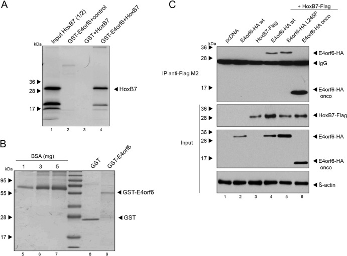
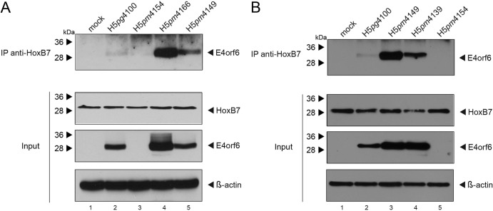
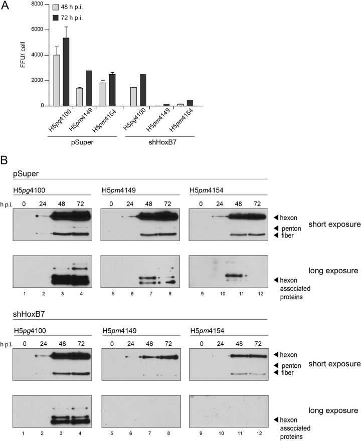
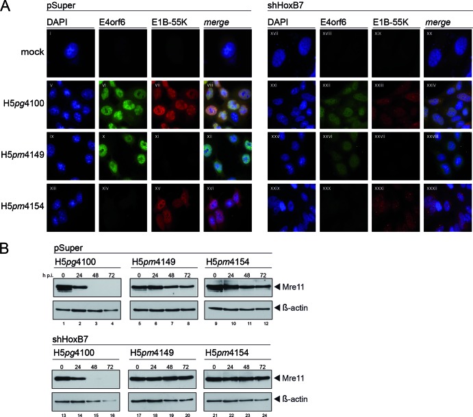
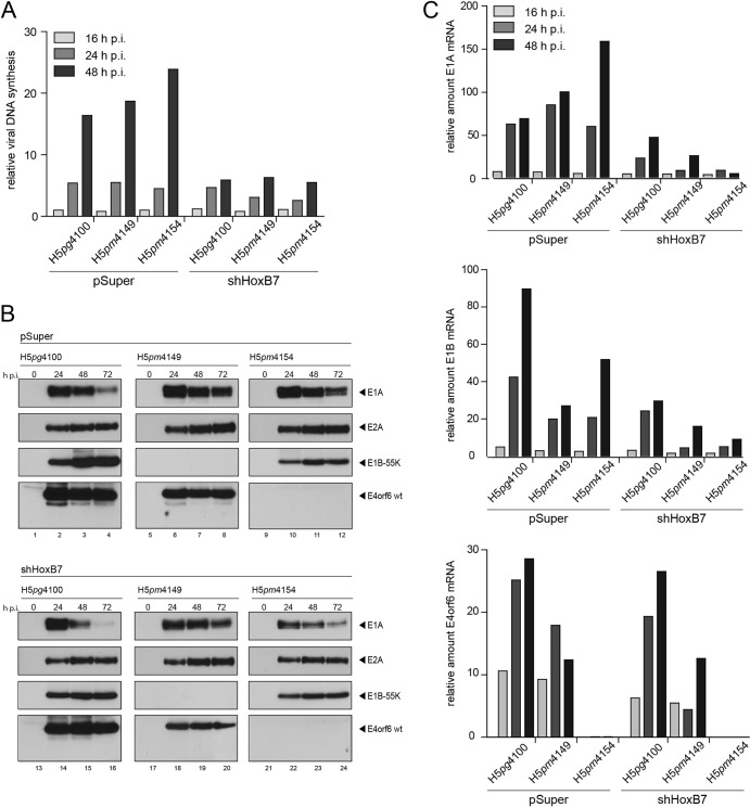
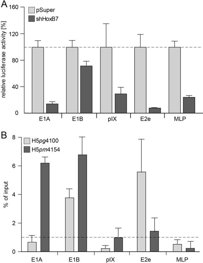
Human adenovirus type 5 (HAdV5) E4orf6 (early region 4 open reading frame 6 protein) is a multifunctional early viral protein promoting efficient replication and progeny production. E4orf6 complexes with E1B-55K to assemble cellular proteins into a functional E3 ubiquitin ligase complex that not only mediates proteasomal degradation of host cell substrates but also facilitates export of viral late mRNA to promote efficient viral protein expression and host cell shutoff. Recent findings defined the role of E4orf6 in RNA splicing independent of E1B-55K binding. To reveal further functions of the early viral protein in infected cells, we used a yeast two-hybrid system and identified the homeobox transcription factor HoxB7 as a novel E4orf6-associated protein. Using a HoxB7 knockdown cell line, we observed a positive role of HoxB7 in adenoviral replication. Our experiments demonstrate that the absence of HoxB7 leads to inefficient viral progeny production, as HAdV5 gene expression is highly regulated by HoxB7-mediated activation of various adenoviral promoters. We have thus identified a novel role of E4orf6 in HAdV5 gene transcription via regulation of homeobox protein-dependent modulation of viral promoter activity.


Figures

Fig 1
Fig 1
E4orf6 interacts with HoxB7 in vitro and in human cells. (A) In vitro-translated [35S]methionine-labeled HoxB7 was incubated with GST-E4orf6 or GST alone, and as a control, GST-E4orf6 was incubated without HoxB7. After precipitation, proteins were separated by SDS-PAGE and visualized by autoradiography. Half the amount of labeled HoxB7 was loaded as input. (B) GST inputs (GST and GST-E4orf6) used for panel A were separated by SDS-PAGE and stained with Coomassie blue. Three concentrations of bovine serum albumin (BSA) were loaded as controls, and molecular mass markers served to position the kDa sizes indicated. (C) H1299 cells were transfected with plasmids encoding E4orf6-HA and HoxB7-FLAG as indicated and harvested at 48 h posttransfection. Total cell extracts were prepared and immunoprecipitated (IP) with mouse monoclonal anti-FLAG M2 antibody (top panel). Proteins from the IP and input samples separated by SDS-PAGE were subjected to immunoblotting using rat anti-HA MAb, anti-FLAG M2, or β-actin antibody as indicated on the right; β-actin served as a loading control.
Fig 2
Fig 2
E4orf6 interacts with HoxB7 independently of E1B-55K and the E3 ubiquitin ligase complex. (A) H1299 cells were infected with wild-type virus (H5pg4100) and mutant viruses (E4orf6-null H5pm4154, E4orf4-null H5pm4166, and E1B-55K-null H5pm4149) at a multiplicity of infection of 50 focus-forming units (FFU) per cell and harvested at 24 h postinfection. Total cell extracts were prepared and immunoprecipitated (IP) with mouse anti-HoxB7 MAb (top panel). IP and input proteins separated by SDS-PAGE were subjected to immunoblotting using rabbit polyclonal E4orf6 (1807) and mouse monoclonal HoxB7 antibodies as indicated on the right. β-Actin was immunoblotted as a loading control. (B) H1299 cells were infected with wild-type virus (H5pg4100) and mutant viruses (H5pm4149, H5pm4154, and H5pm4139, with a mutated E4orf6 protein) at a multiplicity of infection of 50 FFU per cell and harvested at 24 h postinfection. Total cell extracts were prepared and immunoprecipitated (IP) with mouse anti-HoxB7 MAb (top panel). IP and input proteins separated by SDS-PAGE were subjected to immunoblotting using rabbit polyclonal E4orf6 (1807) and mouse monoclonal HoxB7 antibodies as indicated on the right. β-Actin was immunoblotted as a loading control.
Fig 3
Fig 3
Reduction of HoxB7 mRNA and proteins in shRNA-transduced shHoxB7 cell lines. (A) Total RNA was extracted from control (pSuper) and different shHoxB7 knockdown cells, reverse transcribed, and quantified by RT-PCR using primers specific for HoxB7 (Table 1). The data were normalized to 18S rRNA levels. (B) Total cell extracts were prepared from control (pSuper) and shHoxB7 knockdown cells, and proteins were separated by SDS-PAGE and subjected to immunoblotting using a mouse HoxB7 MAb. β-Actin served as a loading control. (C) Control (pSuper) and shHoxB7 knockdown cells were seeded at 2 × 10⁴ cells. At different time points, cells were harvested and viable cells were counted after trypan blue staining. (D) Light microscopy of control (pSuper) and shHoxB7 knockdown cells. (E) Control (pSuper) and shHoxB7-2 knockdown cells were infected with wild-type virus (H5pg4100) at a multiplicity of infection of 25 FFU per cell and harvested at 2 h postinfection. Total cell extracts were prepared and treated with proteinase K. Quantitative PCR was performed using E1B-55K-specific primers. The results represent the averages for three independent experiments. Error bars indicate the standard errors of the means.
Fig 4
Fig 4
HoxB7 depletion decreases HAdV5 progeny production and late viral protein synthesis. (A) Control (pSuper) and shHoxB7-2 knockdown cells were infected with wild-type virus (H5pg4100) and mutant viruses (E1B-55K-null H5pm4149 and E4orf6-null H5pm4154) at a multiplicity of infection of 10 FFU per cell. Viral particles were harvested at 48 and 72 h postinfection, and virus yields were determined by quantitative E2A-72K immunofluorescence staining of W162-infected cells. (B) Control (pSuper) (top panels) and shHoxB7-2 knockdown cells (bottom panels) were infected with wild-type virus (H5pg4100) and mutant viruses (H5pm4154 and H5pm4149) at a multiplicity of infection of 25 FFU per cell and harvested at 24, 48, and 72 h postinfection. Total cell extracts were prepared, and proteins were separated by SDS-PAGE and subjected to immunoblotting using rabbit antiserum to HAdV5 capsid L133, which detects the viral proteins indicated on the right. For the highly expressed, slower-migrating proteins, short exposures are displayed, and for the more weakly expressed, fast-migrating proteins, long exposures are displayed.
Fig 5
Fig 5
HoxB7 depletion exhibits no effects on subcellular localization of E1B-55K or E4orf6 or on E3 ubiquitin ligase activity. (A) Control (pSuper) and shHoxB7-2 knockdown cells were left uninfected (mock) or infected with wild-type virus (H5pg4100) or mutant viruses (E4orf6-null H5pm4154 and E1B-55K-null H5pm4149) at a multiplicity of infection of 25 FFU per cell, fixed at 24 h postinfection, and stained with mouse monoclonal 2A6 (E1B-55K) and rabbit polyclonal 1807 (E4orf6) antibodies. Nuclei are shown in blue, labeled with DAPI. E4orf6 protein localization in wild-type H5pg4100- and E1B-55K-null H5pm4149-infected cells is exclusively nuclear, while E4orf6 is not visible in mock- and E4orf6-null H5pm4154-infected cells. Localization of E1B-55K in wild-type H5pg4100 and E4orf6-null H5pm4154 virus-infected cells is nuclear, with distinct cytoplasmic aggregates. In the absence of HoxB7, the cytoplasmic accumulation of E1B-55K, especially in the H5pm4154 mutant, is enhanced. No E1B-55K protein was detected in mock- and H5pm4149-infected cells. (B) Control (pSuper) and shHoxB7-2 knockdown cells were infected with wild-type virus (H5pg4100) and mutant viruses (H5pm4149 and H5pm4154) at a multiplicity of infection of 25 FFU per cell and harvested at 24, 48, and 72 h postinfection. Total cell extracts were prepared, and proteins separated by SDS-PAGE were subjected to immunoblotting using a rabbit polyclonal Mre11 antibody. β-Actin served as a loading control.
Fig 6
Fig 6
HoxB7 depletion represses HAdV5 DNA, early protein, and mRNA synthesis. (A) Control (pSuper) and shHoxB7-2 knockdown cells were infected with wild-type virus (H5pg4100) and mutant viruses (E1B-55K-null H5pm4149 and E4orf6-null H5pm4154) at a multiplicity of infection of 25 FFU per cell and harvested at 16, 24, and 48 h postinfection. Total cell extracts were prepared and treated with proteinase K. Semiquantitative PCR was performed using E1B-55K-specific primers. (B) Control (pSuper) and shHoxB7 knockdown cells were infected with wild-type virus (H5pg4100) and mutant viruses (H5pm4149 and H5pm4154) at a multiplicity of infection of 25 FFU per cell and harvested at 24, 48, and 72 h postinfection. Total cell extracts were prepared, and proteins were separated by SDS-PAGE and subjected to immunoblotting using mouse MAbs M73 (E1A), B6-8 (E2A), and 2A6 (E1B-55K) and the rabbit polyclonal antibody 1807 (E4orf6). (C) Control (pSuper) and shHoxB7-2 knockdown cells were infected with wild-type virus (H5pg4100) and mutant viruses (H5pm4149 and H5pm4154) at a multiplicity of infection of 25 FFU per cell and harvested at 16, 24, and 48 h postinfection. Total RNA was extracted, reverse transcribed, and quantified by RT-PCR analysis using primers specific for E1A, E1B, and E4orf6 (Table 1). The data were normalized to 18S rRNA levels.
Fig 7
Fig 7
HoxB7-activating function of viral transcription by binding to HAdV5 promoters is affected by E4orf6. (A) Control (pSuper) and shHoxB7-2 knockdown cells were transfected with plasmids encoding firefly luciferase under the control of the E1A, E1B, pIX, E2e, and major late (MLP) promoters. Forty-eight hours after transfection, samples were lysed, absolute luciferase activity was measured, and activity of the HAdV5 promoter in pSuper cells was normalized to 100%. Data are means and standard deviations for three independent experiments. (B) H1299 cells were infected with wild-type virus (H5pg4100) and E4orf6-negative virus (H5pm4154) at a multiplicity of infection of 10 FFU. Cells were fixed with formaldehyde, and ChIP analysis was performed with a HoxB7 mouse MAb as described in Materials and Methods. The average CT value was determined from triplicate reactions and normalized against nonspecific IgG controls by use of a standard curve for each primer pair. Error bars indicate the standard errors of the means. The identities of the products obtained were confirmed by melting curve analysis. The y axis indicates the percentage of immunoprecipitated signal from the input (100%). The dotted line highlights values above 1% of input, commonly stated as significant chromatin/protein binding.

Similar articles

Cited by

References

    1. Abate-Shen C. 2002. Deregulated homeobox gene expression in cancer: cause or consequence? Nat. Rev. Cancer 2:777–785 - PubMed
    1. Araujo FD, Stracker TH, Carson CT, Lee DV, Weitzman MD. 2005. Adenovirus type 5 E4orf3 protein targets the Mre11 complex to cytoplasmic aggresomes. J. Virol. 79:11382–11391 - PMC - PubMed
    1. Babich A, Feldman LT, Nevins, Darnell JE, Jr, Weinberger C. 1983. Effect of adenovirus on metabolism of specific host mRNAs: transport control and specific translational discrimination. Mol. Cell. Biol. 3:1212–1221 - PMC - PubMed
    1. Baker A, Rohleder KJ, Hanakahi LA, Ketner G. 2007. Adenovirus E4 34k and E1b 55k oncoproteins target host DNA ligase IV for proteasomal degradation. J. Virol. 81:7034–7040 - PMC - PubMed
    1. Blackford AN, et al. 2010. Adenovirus 12 E4orf6 inhibits ATR activation by promoting TOPBP1 degradation. Proc. Natl. Acad. Sci. U. S. A. 107:12251–12256 - PMC - PubMed

Publication types

MeSH terms