. 2012 May 2;32(18):6351-63.
doi: 10.1523/JNEUROSCI.4479-11.2012.

PI3-kinase/Akt pathway-regulated membrane insertion of acid-sensing ion channel 1a underlies BDNF-induced pain hypersensitivity

Affiliations

PI3-kinase/Akt pathway-regulated membrane insertion of acid-sensing ion channel 1a underlies BDNF-induced pain hypersensitivity

Bo Duan et al. J Neurosci. .

Abstract

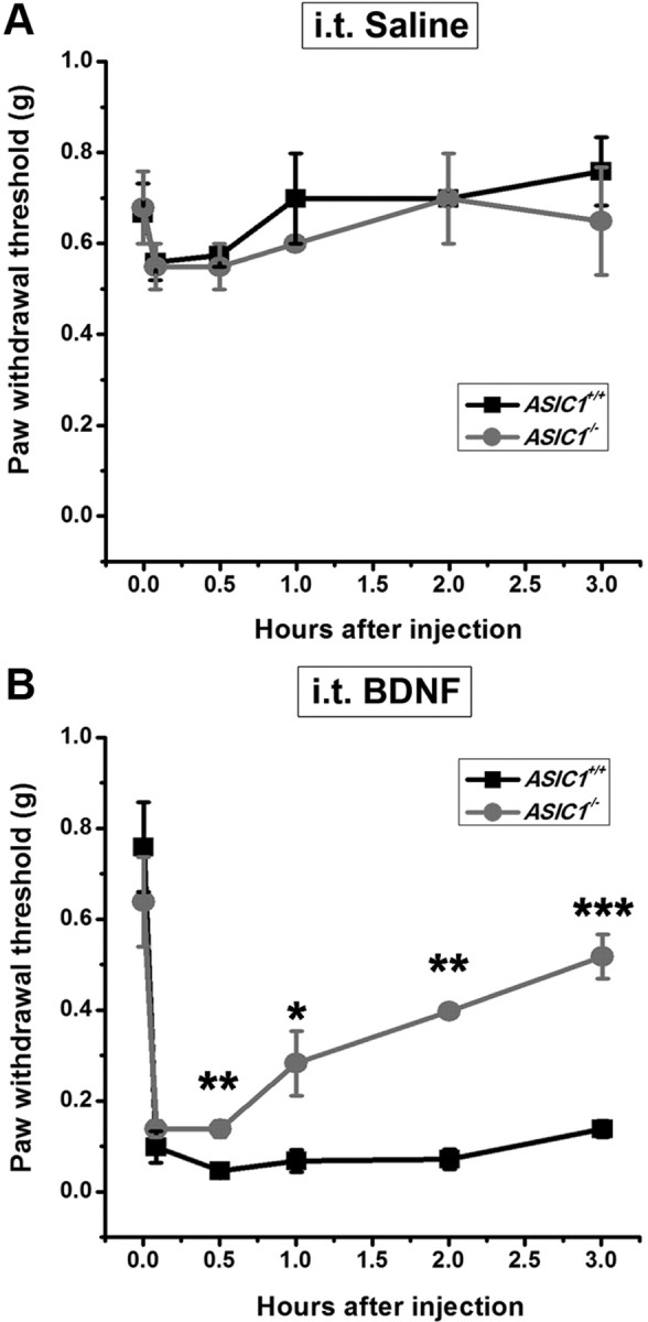
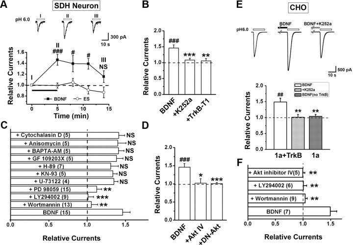
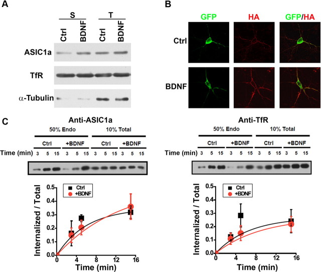
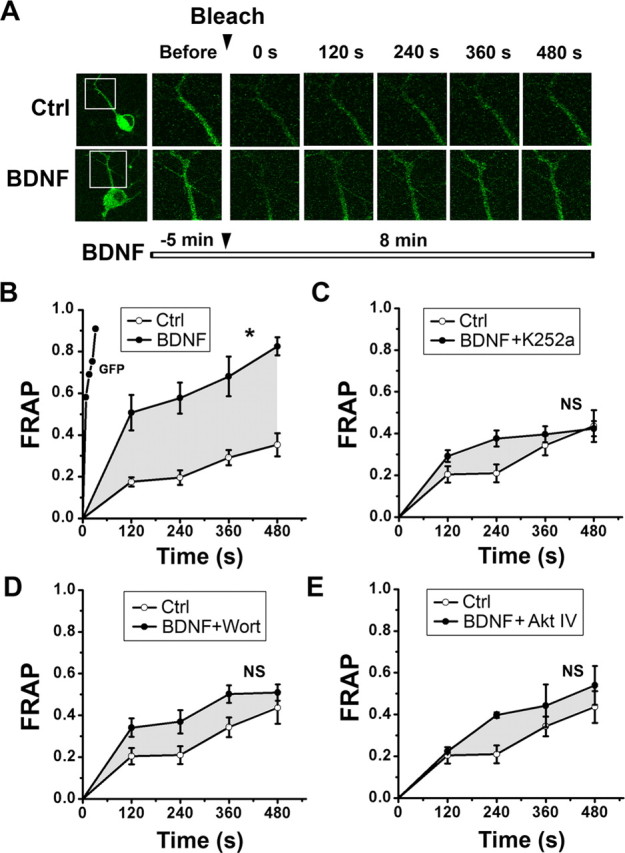
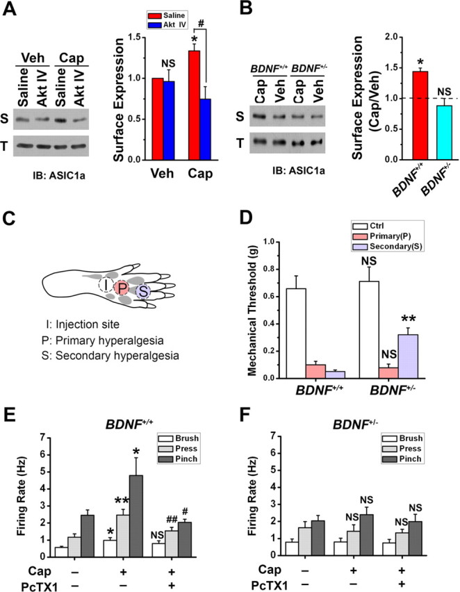
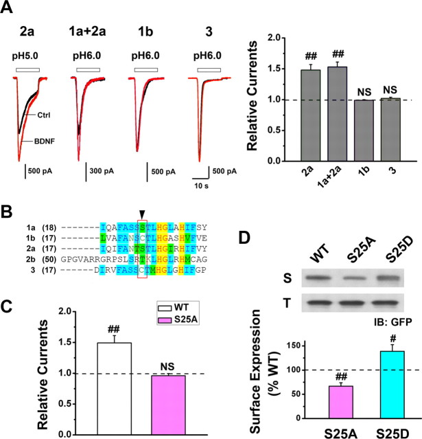
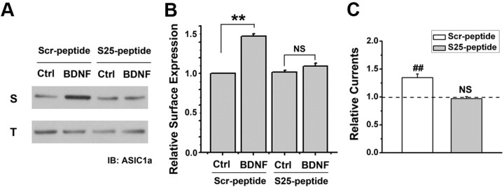
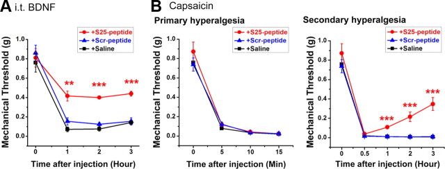
Central neural plasticity plays a key role in pain hypersensitivity. This process is modulated by brain-derived neurotrophic factor (BDNF) and also involves the type 1a acid-sensing ion channel (ASIC1a). However, the interactions between the BDNF receptor, tropomyosin-related kinase B (TrkB), and ASIC1a are unclear. Here, we show that deletion of ASIC1 gene suppressed the sustained mechanical hyperalgesia induced by intrathecal BDNF application in mice. In both rat spinal dorsal horn neurons and heterologous cell cultures, the BDNF/TrkB pathway enhanced ASIC1a currents via phosphoinositide 3-kinase (PI3K)-protein kinase B (PKB/Akt) cascade and phosphorylation of cytoplasmic residue Ser-25 of ASIC1a, resulting in enhanced forward trafficking and increased surface expression. Moreover, in both rats and mice, this enhanced ASIC1a activity was required for BDNF-mediated hypersensitivity of spinal dorsal horn nociceptive neurons and central mechanical hyperalgesia, a process that was abolished by intrathecal application of a peptide representing the N-terminal region of ASIC1a encompassing Ser-25. Thus, our results reveal a novel mechanism underlying central sensitization and pain hypersensitivity, and reinforce the critical role of ASIC1a channels in these processes.


Figures

Figure 1.
Figure 1.
Effect of intrathecal injection of BDNF on paw withdrawal threshold to mechanical stimulus in the wild-type (ASIC1+/+) and ASIC1 deletion (ASIC1−/−) mice. A, Time course of mechanical threshold in the intrathecal saline-treated group. B, Time course of intrathecal BDNF-induced mechanical hyperalgesia. Each point represents the mean ± SEM of 5–6 mice. *p < 0.05, **p < 0.01 versus ASIC1+/+ group, by Student's unpaired t test.
Figure 2.
Figure 2.
BDNF enhances ASIC1a-mediated currents through activation of PI3K-Akt pathway. Perforated electrophysiological recordings were performed. A, The amplitude of the ASIC current (I6.0) in cultured mouse SDH neurons was enhanced by bath application of BDNF (20 ng/ml), but not a control standard extracellular solution (ES), for 5 min. Inset, Examples of ASIC current traces recorded from one neuron at different times before and after BDNF treatment. Data were normalized to the mean amplitude of I6.0 before BDNF treatment (dashed line here and in B–F). #p < 0.05, ###p < 0.001, n = 8–15, by Student's paired t test. B, Blockade of TrkB receptor with either K252a (n = 15) or by transfecting TrkB-T1 (n = 6) abolished the enhancement caused by BDNF treatment for 5 min (n = 15). ###p < 0.001, versus the amplitude of I6.0 before BDNF treatment, by Student's paired t test. **p < 0.01, ***p < 0.001, versus BDNF-treated group, by Student's unpaired t test. C, Summary of effects of various pharmacological treatments on I6.0 at 5 min after the onset of BDNF. **p < 0.01, ***p < 0.001 versus BDNF only (n = 4–15, Student's unpaired t test). NS, No significant difference. D, Blockade of Akt activity with either Akt inhibitor IV (n = 4) or by transfecting DN-Akt (n = 6) abolished the enhancement caused by BDNF. ###p < 0.001, versus before BDNF treatment, by Student's paired t test. *p < 0.05, ***p < 0.001, versus BDNF-treated group, by Student's unpaired t test. E, Sample traces and summary data showing that bath application of BDNF for 5 min (n = 7) enhanced ASIC1a current in CHO cells co-transfected with TrkB and ASIC1a, which was abolished by K252a (n = 7) and not seen if TrkB was omitted in the transfection (n = 6). ##p < 0.01, versus before BDNF treatment, by Student's paired t test. **p < 0.01, versus ASIC1a + TrkB without K252a, by Student's unpaired t test. F, Summary of effects of various pharmacological treatments on I6.0 at 5 min after the onset of BDNF in CHO cells that coexpressed ASIC1a and TrkB (same as in B). n = 5–9, **p < 0.01, versus BDNF only, by Student's unpaired t test.
Figure 3.
Figure 3.
BDNF increases ASIC1a trafficking but not internalization in vitro. A, Surface biotinylation of cultured SDH neurons treated with BDNF for 5 min. Endogenous transferrin receptor (TfR) is shown as a surface protein control and was used to normalize ASIC1a expression level in each group. Endogenous α-tubulin is shown as a cytoplasmic protein control. S, Surface; T, total. BDNF treatment for 5 min increased surface expression of ASIC1a to 150.0 ± 15.6% of the untreated control (Ctrl, n = 4, p < 0.05, vs Ctrl, by Student's paired t test). B, Surface expression of ASIC1a with extracellular HA and N-terminal GFP tags (GFP-ASIC1a-HA) transfected in cultured SDH neurons without (Ctrl) and with BDNF treatment for 5 min. Surface GFP-ASIC1a-HA was labeled using anti-HA antibody without permeabilization (surface), whereas total proteins were visualized with GFP fluorescence (total). The estimated ratio of mean fluorescence intensity of surface/total of the expressed GFP-ASIC1a-HA in BDNF-treated neurons was 167.0 ± 17.5% of Ctrl (n = 4, p < 0.05, vs Ctrl, by Student's paired t test). Shown on the right are summary data of relative surface expression normalized to the mean of the control group (n = 17 cells in each group). **p < 0.01, versus Ctrl, by Student's unpaired t test. C, Representative Western blots of internalized and total ASIC1a and Transferrin receptor (as a negative control) in high-density SDH cultures. Five minutes after treatment with control solution or BDNF (20 ng/ml), surface receptors were labeled with biotin and allowed to internalize at 37°C for 3, 5, or 15 min, after which the remaining surface biotin was stripped off. The internalized biotinylated ASIC1a was measured. In these experiments, total ASIC1a levels were not affected by the BDNF treatment (126.9 ± 10.5% of control, n = 3). Quantification of internalized/total ASIC1a and TfR levels shows endocytosis rates in control and BDNF-treated cultures (n = 3). A best-fit single exponential growth curve was used to obtain time constants (τ) for internalization of ASIC1a (6.7 ± 1.4 min for control, 5.3 ± 1.6 min for BDNF-treated).
Figure 4.
Figure 4.
Dendritic transport of ASIC1a is facilitated by BDNF-TrkB and downstream PI3K/Akt signaling. A, Images of FRAP experiments showing that bleaching a defined dendritic segment of a neuron expressing ASIC1a-GFP resulted in a slow fluorescent recovery in 480 s (top row). Application of BDNF before bleaching resulted in faster recovery (bottom row). White rectangles label the region of the neuron that was bleached. B, FRAP was quantified by measuring the intensity of fluorescence in the frame at fixed intervals. GFP alone showed recovery (by diffusion) within 60 s (n = 5). Application of BDNF facilitated the recovery of ASIC1a-GFP (n = 8). *p < 0.05, versus Ctrl (n = 11), by one-way ANOVA with Tukey post hoc tests. C–E, Suppression of BDNF-induced ASIC1a FRAP by K252a (C, n = 6), wortmannin (Wort) (D, n = 8), and Akt IV (E, n = 6). NS, By one-way ANOVA with Tukey post hoc tests. The region between the drug-treatment and no-treatment curves is superimposed as a gray envelope on FRAP graphs for comparison.
Figure 5.
Figure 5.
Membrane insertion of ASIC1a triggered by BDNF-Akt pathway contributes to capsaicin-induced secondary mechanical hyperalgesia. A, Akt IV abolished the increase in surface expression (S) of ASIC1a in capsaicin (Cap)-injected rats, but not in vehicle (Veh)-injected rats. The total protein level (T) of ASIC1a in SDH neurons did not change. Shown on the right is a summary (n = 3 in each group). *p < 0.05, Cap versus Veh, by Student's paired t test. NS, Akt IV versus saline in the Veh groups, and #p < 0.05, Akt IV versus saline in the Cap groups, by Student's unpaired t test. B, Representative Western blots showing that intradermal injection of capsaicin induced an increase in surface expression (S), but no change in total protein level (T), of ASIC1a in SDH of wild-type (BDNF+/+) mice. However, the change in surface expression was not detected in BDNF deletion heterozygotes (BDNF+/−). Shown on the right is a summary (dashed line indicates vehicle only; n = 3 in each group). *p < 0.05, by Student's paired t test. C, Diagram showing the sites of capsaicin injection and behavioral test in the hindpaw. Capsaicin was injected into the plantar surface of the right hindpaw (“I”). Mechanical thresholds to von Frey filament stimuli were measured at site “P” for primary hyperalgesia 15 min after capsaicin injection and at site “S” for secondary hyperalgesia 2 h after capsaicin injection. D, Capsaicin-induced mechanical hyperalgesia in the BDNF+/− (n = 9) and wild-type (WT or BDNF+/+, n = 7) littermates. Control (Ctrl) was the mechanical threshold before capsaicin injection. **p < 0.01, versus the corresponding WT, by Student's unpaired t test. E, F, Summary of in vivo single unit recording of WDR neurons in the SDH before and 2 h after capsaicin injection from the BDNF+/+ (E, n = 5) and BDNF+/− mice (F, n = 4), in response to innocuous (brushing) or noxious (pressing and pinching) mechanical stimulation. PcTX1 (10 μm, 10 μl) was directly administered to the dorsal surface of the spinal cord 15 min before recording. *p < 0.05, **p < 0.01, capsaicin (Cap) versus vehicle (no Cap, no PcTX1). #p < 0.05, ##p < 0.01, PcTX1-plus-Cap versus Cap, by Student's unpaired t test.
Figure 6.
Figure 6.
Ser-25 at ASIC1a N terminus mediates BDNF-induced ASIC1a trafficking. A, Proton-activated currents in CHO cells that coexpressed TrkB with ASIC2a, ASIC1a+2a, ASIC1b, or ASIC3 before (black lines) and after (red lines) BDNF treatment (5 min). Left, Representative traces; right, summary for current amplitude after BDNF normalized to that before BDNF (dashed line; n = 5, 6, 4, and 5 for ASIC2a, ASIC1a+2a, ASIC1b, and ASIC3, respectively). BDNF increased the current mediated by ASIC2a and ASIC1a+2a but not that by ASIC1b and ASIC3. ##p < 0.01, by Student's paired t test. B, Sequence alignment of the ASIC N termini indicating that Ser-25 is present in ASIC1a and 2a but not in other ASIC subunits. C, Mutation of ASIC1a Ser-25 to Ala (S25A, n = 7) abolished BDNF-induced enhancement of ASIC1a current. Data were normalized to the mean amplitude of I6.0 before BDNF treatment (dashed line) for each cell. ##p < 0.01, by Student's paired t test. D, Representative Western blots and summary data showing that the surface expression level of S25A was significantly lower than that of WT ASIC1a (dashed line), whereas that of S25D significantly increased. Data were normalized to the surface expression level of WT ASIC1a (n = 4). #p < 0.05, ##p < 0.01, by Student's paired t test.
Figure 7.
Figure 7.
Effects of a TAT-fusion peptide containing ASIC1a N-terminal sequence encompassing Ser-25 on ASIC expression and activation in SDH neurons. A, B, Representative Western blots and summary showing bath application of TAT-Ser-25 peptide (S25-peptide) abolished BDNF-induced surface expression of ASIC1a. Incubation with the scrambled peptide (Scr-peptide) had no effect. S, Surface; T, total. Data were normalized to the surface expression level of ASIC1a in Ctrl group incubated with Scr-peptide. n = 3, **p < 0.01, by Student's paired t test. C, Bath incubation (2 h) of S25-peptide abolished BDNF-induced enhancement of ASIC current in cultured SDH neurons. Incubation with the Scr-peptide had no effect. Data were normalized to the mean amplitude of I6.0 before BDNF treatment (dashed line) for each neuron, n = 6 for each group. ##p < 0.01, by Student's paired t test.
Figure 8.
Figure 8.
Effects of S25-peptide on BDNF-mediated mechanical hyperalgesia. A, Changes in paw withdrawal threshold of WT mice in response to mechanical stimuli after intrathecal coinjection of BDNF with S25-peptide (n = 5), or Scr-peptide (n = 5), or saline (n = 6). **p < 0.01, ***p < 0.001, S25-peptide versus Scr-peptide, by Student's paired t test. B, Changes in paw withdrawal threshold of mice in response to mechanical stimuli after intradermal injection of capsaicin. Intrathecal injection of S25-peptide (n = 7), but not Scr-peptide (n = 8), or saline (n = 12) 1 h before capsaicin injection significantly attenuated capsaicin-induced secondary mechanical hyperalgesia but not primary mechanical hyperalgesia. ***p < 0.001, S25-peptide versus Scr-peptide, by Student's unpaired t test.
Figure 9.
Figure 9.
Schematic diagram of the effect of BDNF/TrkB and downstream signaling pathways on central sensitization following C-fiber activity. Both BDNF and glutamate are released from presynaptic DRG neurons following C-fiber activity by peripheral capsaicin treatment. Activation of postsynaptic TrkB receptor in the spinal dorsal horn by BDNF induces intracellular PKC activation and enhances NMDAR function via phosphorylation. On the other hand, BDNF also activates intracellular PI3K/Akt pathway, and then induces ASIC1a phosphorylation and forward targeting to neuron surface. ASIC1a channels sense synaptic acidosis and cause membrane depolarization via Na+ and Ca2+ influx, which may facilitate NMDAR activation and induce central sensitization. Blockade of ASIC1a channel trafficking by fusion of S25-peptide to obstruct the phosphorylation of ASIC1a at Ser25 attenuates BDNF-mediated mechanical hyperalgesia (right). Moreover, release of BDNF from spinal microglia following peripheral nerve injury (Coull et al., 2005) may also facilitate forward trafficking of ASIC1a and then induces central sensitization.

Similar articles

Cited by

References

    1. Adelson DW, Wei JY, Kruger L. Warm-sensitive afferent splanchnic C-fiber units in vitro. J Neurophysiol. 1997;77:2989–3002. - PubMed
    1. Baron A, Voilley N, Lazdunski M, Lingueglia E. Acid sensing ion channels in dorsal spinal cord neurons. J Neurosci. 2008;28:1498–1508. - PMC - PubMed
    1. Basbaum AI, Bautista DM, Scherrer G, Julius D. Cellular and molecular mechanisms of pain. Cell. 2009;139:267–284. - PMC - PubMed
    1. Benson CJ, Eckert SP, McCleskey EW. Acid-evoked currents in cardiac sensory neurons: a possible mediator of myocardial ischemic sensation. Circ Res. 1999;84:921–928. - PubMed
    1. Blair LA, Bence-Hanulec KK, Mehta S, Franke T, Kaplan D, Marshall J. Akt-dependent potentiation of L channels by insulin-like growth factor-1 is required for neuronal survival. J Neurosci. 1999;19:1940–1951. - PMC - PubMed

Publication types