Rab20 regulates phagosome maturation in RAW264 macrophages during Fc gamma receptor-mediated phagocytosis
- PMID: 22545127
- PMCID: PMC3335809
- DOI: 10.1371/journal.pone.0035663
Rab20 regulates phagosome maturation in RAW264 macrophages during Fc gamma receptor-mediated phagocytosis
Abstract
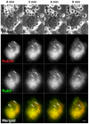
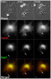
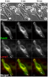
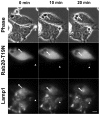
Rab20, a member of the Rab GTPase family, is known to be involved in membrane trafficking, however its implication in FcγR-mediated phagocytosis is unclear. We examined the spatiotemporal localization of Rab20 during phagocytosis of IgG-opsonized erythrocytes (IgG-Es) in RAW264 macrophages. By the live-cell imaging of fluorescent protein-fused Rab20, it was shown that Rab20 was transiently associated with the phagosomal membranes. During the early stage of phagosome formation, Rab20 was not localized on the membranes of phagocytic cups, but was gradually recruited to the newly formed phagosomes. Although Rab20 was colocalized with Rab5 to some extent, the association of Rab20 with the phagosomes persisted even after the loss of Rab5 from the phagosomal membranes. Then, Rab20 was colocalized with Rab7 and Lamp1, late endosomal/lysosomal markers, on the internalized phagosomes. Moreover, our analysis of Rab20 mutant expression revealed that the maturation of phagosomes was significantly delayed in cells expressing the GDP-bound mutant Rab20-T19N. These data suggest that Rab20 is an important component of phagosome and regulates the phagosome maturation during FcγR-mediated phagocytosis.
Conflict of interest statement
Figures


Similar articles
-
Transient recruitment of M-Ras GTPase to phagocytic cups in RAW264 macrophages during FcγR-mediated phagocytosis.Microscopy (Oxf). 2018 Apr 1;67(2):68-74. doi: 10.1093/jmicro/dfx131. Microscopy (Oxf). 2018. PMID: 29340604
-
Rab35 regulates phagosome formation through recruitment of ACAP2 in macrophages during FcγR-mediated phagocytosis.J Cell Sci. 2011 Nov 1;124(Pt 21):3557-67. doi: 10.1242/jcs.083881. Epub 2011 Nov 1. J Cell Sci. 2011. PMID: 22045739
-
Spatiotemporal Localization of Rab20 in Live RAW264 Macrophages during Macropinocytosis.Acta Histochem Cytochem. 2012 Dec 26;45(6):317-23. doi: 10.1267/ahc.12014. Epub 2012 Nov 3. Acta Histochem Cytochem. 2012. PMID: 23378675 Free PMC article.
-
Molecular imaging analysis of Rab GTPases in the regulation of phagocytosis and macropinocytosis.Anat Sci Int. 2016 Jan;91(1):35-42. doi: 10.1007/s12565-015-0313-y. Epub 2015 Nov 3. Anat Sci Int. 2016. PMID: 26530641 Review.
-
The complexity of Rab5 to Rab7 transition guarantees specificity of pathogen subversion mechanisms.Front Cell Infect Microbiol. 2014 Dec 22;4:180. doi: 10.3389/fcimb.2014.00180. eCollection 2014. Front Cell Infect Microbiol. 2014. PMID: 25566515 Free PMC article. Review. No abstract available.
Cited by
-
Interferon-γ-inducible Rab20 regulates endosomal morphology and EGFR degradation in macrophages.Mol Biol Cell. 2015 Sep 1;26(17):3061-70. doi: 10.1091/mbc.E14-11-1547. Epub 2015 Jul 8. Mol Biol Cell. 2015. PMID: 26157167 Free PMC article.
-
Identification of an immune-regulated phagosomal Rab cascade in macrophages.J Cell Sci. 2014 May 1;127(Pt 9):2071-82. doi: 10.1242/jcs.144923. Epub 2014 Feb 25. J Cell Sci. 2014. PMID: 24569883 Free PMC article.
-
Rab GTPases in Immunity and Inflammation.Front Cell Infect Microbiol. 2017 Sep 29;7:435. doi: 10.3389/fcimb.2017.00435. eCollection 2017. Front Cell Infect Microbiol. 2017. PMID: 29034219 Free PMC article. Review.
-
Identification of contributing genes of Huntington's disease by machine learning.BMC Med Genomics. 2020 Nov 23;13(1):176. doi: 10.1186/s12920-020-00822-w. BMC Med Genomics. 2020. PMID: 33228685 Free PMC article.
-
Consequences of Rab GTPase dysfunction in genetic or acquired human diseases.Small GTPases. 2018 Mar 4;9(1-2):158-181. doi: 10.1080/21541248.2017.1397833. Epub 2017 Dec 28. Small GTPases. 2018. PMID: 29239692 Free PMC article. Review.
References
Publication types
MeSH terms
Substances
LinkOut - more resources
Full Text Sources
Miscellaneous

