. 2012 Jun;23(12):2240-52.
doi: 10.1091/mbc.E11-11-0926. Epub 2012 Apr 25.

Cell survival, DNA damage, and oncogenic transformation after a transient and reversible apoptotic response

Affiliations

Cell survival, DNA damage, and oncogenic transformation after a transient and reversible apoptotic response

Ho Lam Tang et al. Mol Biol Cell. 2012 Jun.

Abstract

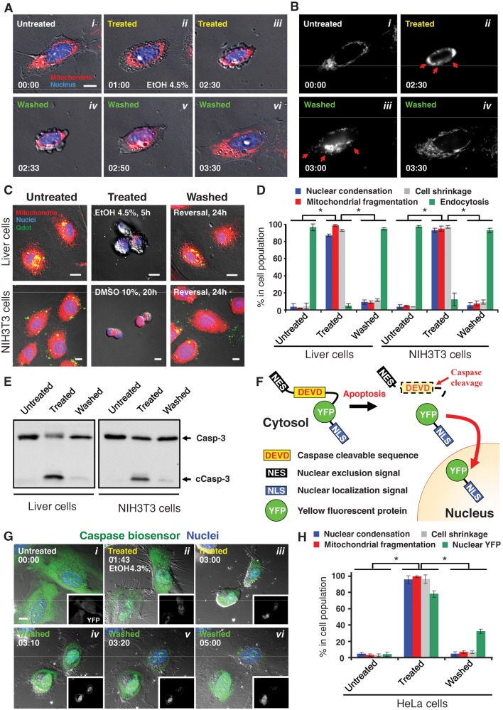
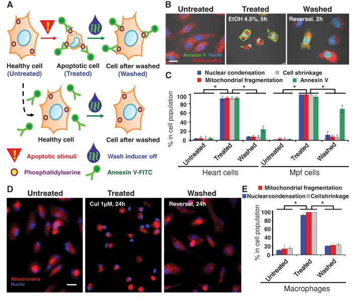
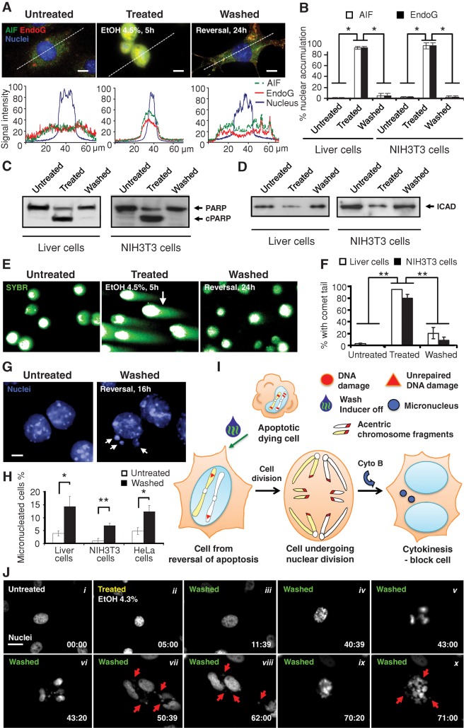
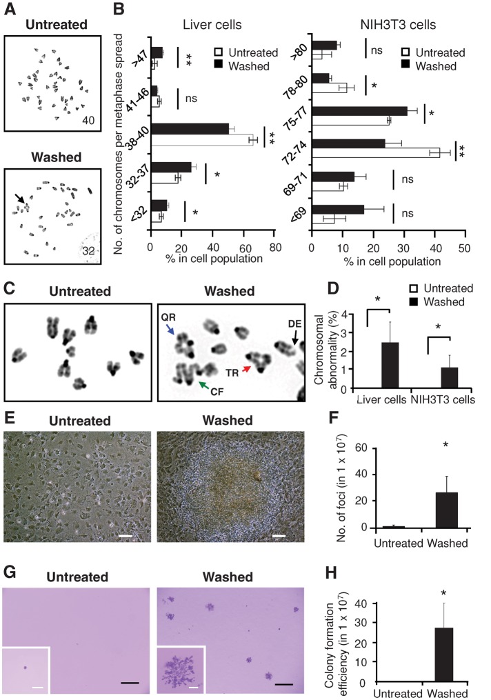
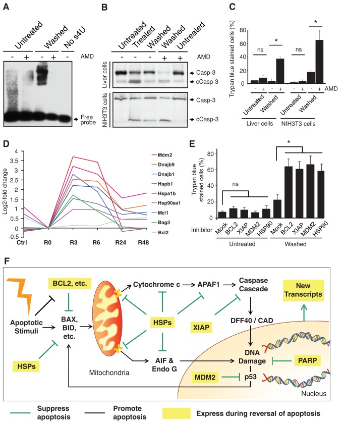
Apoptosis serves as a protective mechanism by eliminating damaged cells through programmed cell death. After apoptotic cells pass critical checkpoints, including mitochondrial fragmentation, executioner caspase activation, and DNA damage, it is assumed that cell death inevitably follows. However, this assumption has not been tested directly. Here we report an unexpected reversal of late-stage apoptosis in primary liver and heart cells, macrophages, NIH 3T3 fibroblasts, cervical cancer HeLa cells, and brain cells. After exposure to an inducer of apoptosis, cells exhibited multiple morphological and biochemical hallmarks of late-stage apoptosis, including mitochondrial fragmentation, caspase-3 activation, and DNA damage. Surprisingly, the vast majority of dying cells arrested the apoptotic process and recovered when the inducer was washed away. Of importance, some cells acquired permanent genetic changes and underwent oncogenic transformation at a higher frequency than controls. Global gene expression analysis identified a molecular signature of the reversal process. We propose that reversal of apoptosis is an unanticipated mechanism to rescue cells from crisis and propose to name this mechanism "anastasis" (Greek for "rising to life"). Whereas carcinogenesis represents a harmful side effect, potential benefits of anastasis could include preservation of cells that are difficult to replace and stress-induced genetic diversity.


Figures

FIGURE 1:
FIGURE 1:
Reversibility of apoptosis in primary mouse liver, NIH 3T3, and HeLa cells. (A) Time-lapse live-cell fluorescence microscopy of a primary liver cell before, during, and after exposure to ethanol. The same cell before ethanol induction (Untreated, i), induced with 4.5% ethanol in culture medium for 2.5 h (Treated, ii and iii), and then washed and further cultured with fresh medium (Washed, iv–vi). Merged images, mitochondria (red) and nuclei (blue) were visualized by fluorescence and cell morphology by DIC. Time presented as h:min. Scale bar, 10 μm. Also see Supplemental Figure S1 and Supplemental Video S1. (B) Monochrome images of mitochondria from A. Mitochondrial fragmentation is indicated by red arrows. (C) Fluorescence and DIC microscopy of healthy liver and NIH 3T3 cells (Untreated), cells that were exposed to apoptotic inducers (liver cells, 4.5% ethanol for 5 h; NIH 3T3 cells, 10% DMSO for 20 h) (Treated), and treated cells that were washed to remove apoptotic inducers and further cultured for 24 h (Washed). Merged images, mitochondria (red), nuclei (blue), and Quantum Dots (Qdots) taken up by endocytosis (green) were visualized by fluorescence and cell morphology by DIC. Scale bar, 10 μm. (D) Quantification of the apoptotic response and its reversal. Morphological signs of apoptosis included nuclear condensation, mitochondrial fragmentation, and cell shrinkage. Uptake of Quantum Dots by endocytosis is characteristic of healthy cells, whereas the other features are characteristic of apoptotic cells. Apoptosis was induced in liver cells with 4.5% ethanol for 5 h and in NIH 3T3 cells with 10% DMSO for 20 h (Treated). Treated cells were then washed and further cultured for 24 h in standard conditions. *p < 0.01; n = 3 independent experiments. Error bars denote SD. (E) Western blot analysis of the total cell lysate of untreated, treated, and washed liver and NIH 3T3 cells for the protein level of caspase-3 (Casp-3). c, cleaved form. (F) Schematic diagram of the caspase biosensor NES-DEVD-YFP-NLS. (G) Real-time live-cell microscopy of HeLa cells expressing the caspase biosensor before (Untreated, i), during (Treated, ii and iii), and after (Washed, iv-vi) exposure to 4.3% ethanol. Merged images, caspase biosensor (YFP, green) and nuclei (blue) were visualized by confocal microscopy and cell morphology by DIC. Corresponding monochromatic YFP image is shown in each panel. Time presented as h:min. Scale bar, 10 μm. Also see Supplemental Figure S4 and Supplemental Videos S2 and S3. (H) Quantification of the caspase biosensor response in HeLa cells. Treated cells were exposed to 4.3% ethanol for 5 h. After washing, cells were cultured for 2 h in standard conditions. *p< 0.01; n = 3 independent experiments. Error bars denote SD.
FIGURE 2:
FIGURE 2:
Reversibility of apoptosis in primary rat heart cells, ferret brain cells, and primary mouse macrophages. (A) Schematic diagram of approach using annexin V-FITC to track cells that reverse apoptosis. (B) Confocal and DIC microscopy of rat primary heart cells that were exposed to 4.5% ethanol for 5 h (Treated) or not (Untreated). Treated cells were then washed to remove apoptotic inducers and further cultured for 2 h (Washed). Merged images, mitochondria (red), nuclei (blue), and annexin V-FITC (annexin V)–labeled exposed phosphatidylserine (PS) (green) were visualized by fluorescence, and cell morphology was by DIC. Scale bar, 10 μm. (C) Quantification of the apoptotic response and its reversal on primary rat heart cells and Mpf brain cells. Percentage of cells showing morphological signs of apoptosis including mitochondrial fragmentation, nuclear condensation, cell shrinkage, and cell surface phosphatidylserine labeled with annexin V-FITC (Annexin V) for control cells (Untreated), cells treated with apoptotic inducer (heart cells with 4.5% ethanol for 5 h, brain cells with 2 μM jasplakinolide for 50 h) (Treated), and treated cells that were washed and further cultured with fresh medium (heart cells for 2 h, brain cells for 3 h) in standard conditions (Washed). *p < 0.01; n = 3 independent experiments. Error bars denote SD. (D) Fluorescence of healthy, untreated macrophages, those that were exposed to 1 μM cucurbitacin I (CuI) for 24 h (Treated), and treated cells that were washed to remove apoptotic inducers and further cultured for 24 h (Washed). Merged images, mitochondria (red) and nuclei (blue). Scale bar, 30 μm. (E) Percentage of the untreated, treated, and washed macrophages that displayed mitochondrial fragmentation, nuclear condensation, and cell shrinkage. *p < 0.01; n = 3 independent experiments. Error bars denote SD.
FIGURE 3:
FIGURE 3:
Damage of DNA in dying cells before reversal of apoptosis. (A) Fluorescence micrographs showing the subcellular localization of AIF (green), EndoG (red), and nuclei (blue) in primary liver cells that were untreated (Untreated) or treated with 4.5% ethanol for 5 h (Treated) and treated cells that were then cultured for 24 h in fresh medium after removal of the ethanol (Washed) from cells. Quantification of the corresponding fluorescence signals of AIF, EndoG, and nucleus along the dotted line as indicated in their respective images. Scale bar, 10 μm. (B) Percentage of liver and NIH 3T3 cells that displayed nuclear accumulation of AIF and EndoG. Treated liver cells were exposed to 4.5% ethanol for 5 h. NIH 3T3 cells were treated with 10% DMSO for 20 h. Treated cells that were then washed to remove apoptotic inducers were further cultured for 24 h (Washed). *p < 0.01; n = 3 independent experiments. Error bars denote SD. (C, D) Western blot analysis of total cell lysates of untreated, treated, and washed liver and NIH 3T3 cells were probed for (C) PARP and (D) ICAD. c, cleaved form. (E) Fluorescence microscopy for the SYBR-stained DNA of untreated, treated, and washed liver cells subjected to the comet assay for DNA damage. Cells embedded in agarose were subjected to electrophoresis. Broken DNA forms comet tails such as the one indicated by an arrow. Intact DNA remains within the nuclear envelop. (F) Percentage of untreated, treated, and washed liver and NIH 3T3 cells that displayed a comet tail. **p < 0.01; n = 3 independent experiments. Error bars denote SD. (G) Fluorescence microscopy on nuclear morphology of untreated as well as treated and washed primary liver cells 16 h after removal of apoptotic inducer. Cytokinesis was blocked with cytochalasin B (Cyto B). Arrows indicate micronuclei in the washed cells. Scale bar, 10 μm. (H) Percentage of untreated, as well as treated and then washed, liver, NIH 3T3, and HeLa cells that displayed micronuclei. Apoptotic inductions for the liver and NIH 3T3 cells were as described in (B) and for HeLa as in (J). See Materials and Methods for detail. *p < 0.05; **p < 0.01; n = 3 independent experiments. Error bars denote SD. (I) Proposed model for the formation of micronuclei in cells that divide after reversal of apoptosis, likely as a result of unrepaired DNA damage. (J) Time-lapse live-cell fluorescence microscopy of HeLa cells before, during, and after exposure to ethanol. The same cell before ethanol induction (Untreated, i), induced with 4.3% ethanol in culture medium for 5 h (Treated, ii), and then washed and further cultured with fresh medium (Washed, iii–x). Monochromatic images of nuclei stained with Hoechst 33342. Arrows indicate some of the micronuclei. Time presented as h:min. Scale bar, 30 μm. See Supplemental Figure S5 and Supplemental Video S4 for images and video with fluorescence microscopy for the corresponding mitochondria and nuclei and phase contrast microscopy for cell morphology of the same cells. Cytochalasin B was not present in this experiment.
FIGURE 4:
FIGURE 4:
Genetic alterations and transformation after reversal of apoptosis. (A) Inverted DAPI-banding image of metaphase spreads of untreated liver cells (Untreated) compared with cells that were treated with 4.5% ethanol for 5 h, washed, and then cultured for 3 d after removal of apoptotic inducer (Washed). The number of metaphase chromosomes is indicated on the corresponding images. An abnormal chromosomal configuration is indicated by an arrow. (B) Percentage of untreated and washed liver cells (3 d after 5-h exposure to 4.5% ethanol) and NIH 3T3 cells (3 d after 20-h exposure to 10% DMSO) displaying the indicated number of chromosomes in metaphase spreads. ns, p > 0.05; *p < 0.05; **p < 0.01; n = 3 independent experiments. Error bars denote SD. (C) Representative inverted DAPI-banding images of the configuration of metaphase chromosomes of untreated (Untreated) as well as treated and washed (Washed) liver cells. Configurations indicated by arrows: triradial (TR, red); quadriradial (QR, blue); complex figures (CF, green); deletion (DE, black). (D) Percentage of the untreated and the washed liver and NIH 3T3 cells displaying at least one abnormality in chromosomal configuration. *p < 0.05; n = 3 independent experiments. Error bars denote SD. (E) Image of untreated NIH 3T3 cells and the foci from the cells after being treated and washed (Washed) after 3 wk of culture. Scale bars: 50 μm. (F) Number of foci in untreated as well as treated and washed NIH 3T3 cells after 3 wk of culture. *p < 0.05; n = 3 independent experiments. Error bars denote SD. (G) Image of crystal violet–stained colonies in soft agar in untreated as well as treated and washed NIH 3T3 cells from the foci after 5 wk of culture. Inset, enlarged image of a colony. Scale bars, 400 μm (white), 2 mm (black). (H) Number of colonies formed in soft agar in untreated as well as treated and washed NIH 3T3 cells after 5 wk of culture. *p < 0.05; n = 3 independent experiments. Error bars denote SD.
FIGURE 5:
FIGURE 5:
Critical contributing factors in reversal of apoptosis. (A) RNA blot for detecting new RNA synthesis in untreated vs. treated and washed (Washed) liver cells 1 h after a 5-h exposure to 4.5% ethanol and with and without transient exposure (1 h, 1μg/ml) to actinomycin D (AMD). No s4U served as negative control for the probe binding to RNA. The detailed procedure is described in Materials and Methods. (B) Western blot analysis of the cleavage of caspase-3 (Casp-3) on the total lysate of untreated, treated (5-h exposure to 4.5% ethanol for liver cells, 20-h exposure to 10% DMSO for NIH 3T3 cells), and washed liver cells and NIH 3T3 cells with and without transient exposure to actinomycin D (AMD) immediately after removal of apoptotic stimuli. The AMD-exposed cells were cultured in fresh medium for 23 h and then subjected to the analysis. c, cleaved form. (C) Percentage of untreated as well as treated and washed liver cells and NIH 3T3 cells with and without transient AMD exposure that displayed plasma membrane permeability in trypan blue exclusion assay. The AMD exposed-cells were cultured in fresh medium for 23 h and then subjected to the assay. ns, p > 0.05; *p < 0.05; n = 3 independent experiments. Error bars denote SD. (D) A time-course microarray study in liver cells, log2-fold change of gene expression comparison between ethanol-induced apoptotic cells (R0), untreated cells (Ctrl), and induced cells that were then washed and further cultured in fresh medium for 3 (R3), 6 (R6), 24 (R24), and 48 h (R48). The log2 signal values from three biological replicates were averaged (geometric mean) for each time point. (E) Percentage of untreated and washed liver cells with and without 24-h exposure of inhibitors of BCL-2 (ABT 263, 1 μM), XIAP (Embelin, 20 μM), MDM2 (Nutlin-3, 10 μM), and HSP90 (17-allylaminogeldanamycin, 0.5 μM) that displayed full plasma membrane permeability in trypan blue exclusion assay. The corresponding cells were exposed to the inhibitors after apoptotic stimuli had been removed from the treated cells and then further cultured for 24 h. ns, p > 0.05; *P < 0.05; n = 3 independent experiments. Error bars denote SD. (F) Proposed model for reversal of apoptosis. Expression of multiple prosurvival factors and new transcripts during reversal of apoptosis promotes cell survival by suppressing the activated apoptotic pathways and repairing the cells.

Similar articles

Cited by

References

    1. Aitken RJ, Findlay JK, Hutt KJ, Kerr JB. Apoptosis in the germ line. Reproduction. 2011;141:139–150. - PubMed
    1. Arama E, Agapite J, Steller H. Caspase activity and a specific cytochrome C are required for sperm differentiation in Drosophila. Dev Cell. 2003;4:687–697. - PubMed
    1. Bloom AD. Induced chromosomal aberrations: biological and clinical significance. J Pediatr. 1972;81:1–8. - PubMed
    1. Boffetta P, Hashibe M. Alcohol and cancer. Lancet Oncol. 2006;7:149–156. - PubMed
    1. Capy P, Gasperi G, Biemont C, Bazin C. Stress and transposable elements: co-evolution or useful parasites? Heredity. 2000;85:101–106. - PubMed

Publication types

MeSH terms