. 2012 Mar 15;125(Pt 6):1556-67.
doi: 10.1242/jcs.095729.

Regulation of nuclear TDP-43 by NR2A-containing NMDA receptors and PTEN

Affiliations

Regulation of nuclear TDP-43 by NR2A-containing NMDA receptors and PTEN

Mei Zheng et al. J Cell Sci. .

Abstract

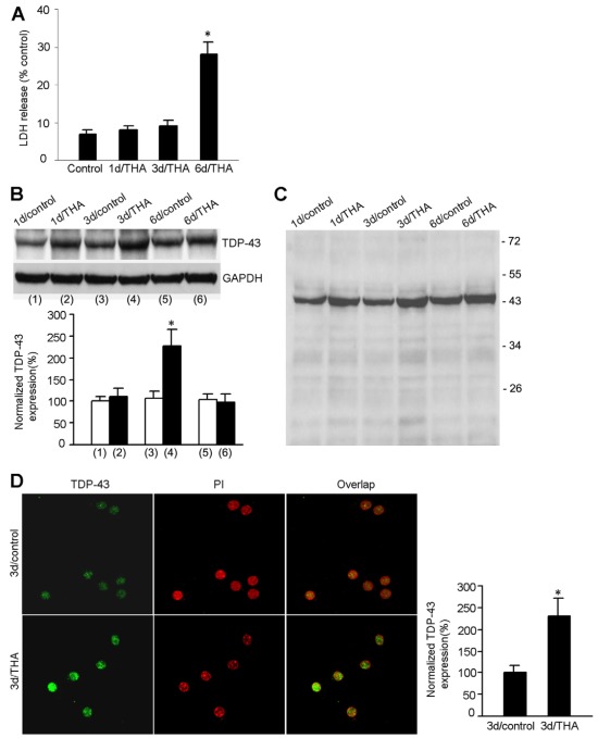
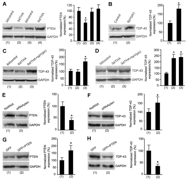
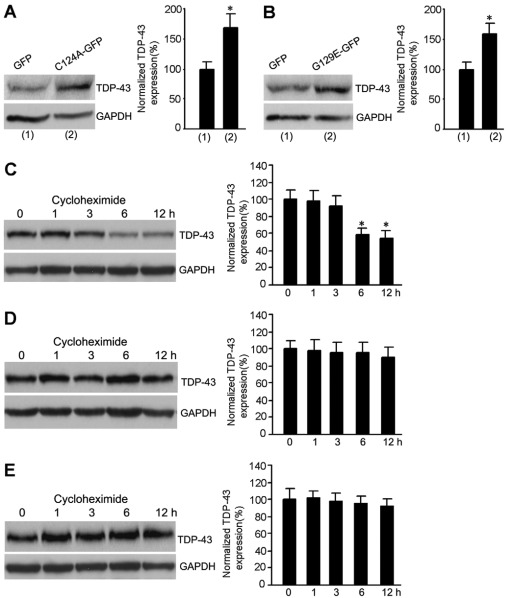
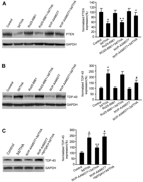
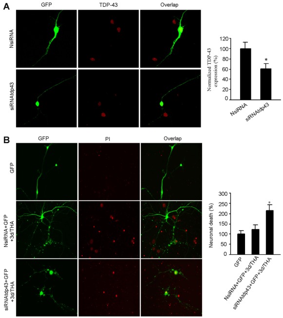
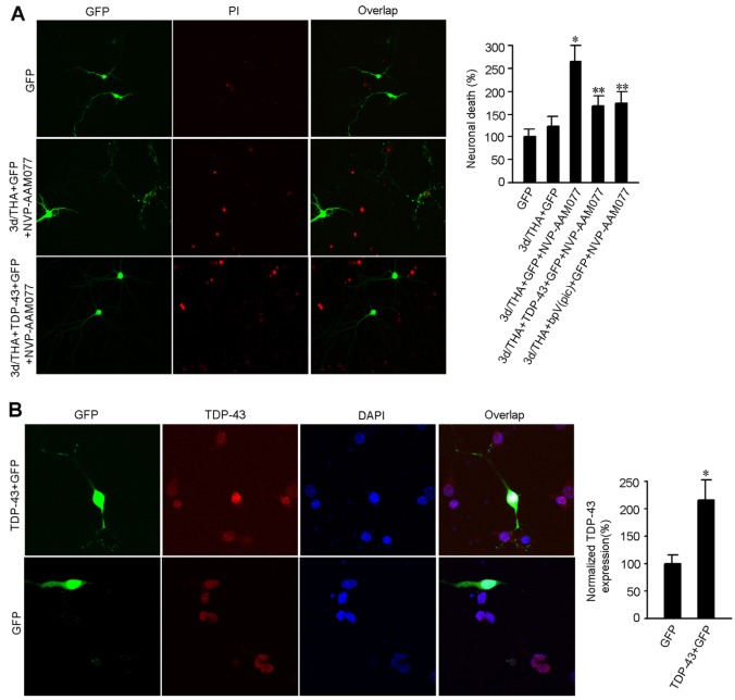
The dysfunction of TAR DNA-binding protein-43 (TDP-43) is implicated in neurodegenerative diseases. However, the function of TDP-43 is not fully elucidated. Here we show that the protein level of endogenous TDP-43 in the nucleus is increased in mouse cortical neurons in the early stages, but return to basal level in the later stages after glutamate accumulation-induced injury. The elevation of TDP-43 results from a downregulation of phosphatase and tensin homolog (PTEN). We further demonstrate that activation of NR2A-containing NMDA receptors (NR2ARs) leads to PTEN downregulation and subsequent reduction of PTEN import from the cytoplasm to the nucleus after glutamate accumulation. The decrease of PTEN in the nucleus contributes to its reduced association with TDP-43, and thereby mediates the elevation of nuclear TDP-43. We provide evidence that the elevation of nuclear TDP-43, mediated by NR2AR activation and PTEN downregulation, confers protection against cortical neuronal death in the late stages after glutamate accumulation. Thus, this study reveals a NR2AR-PTEN-TDP-43 signaling pathway by which nuclear TDP-43 promotes neuronal survival. These results suggest that upregulation of nuclear TDP-43 represents a self-protection mechanism to delay neurodegeneration in the early stages after glutamate accumulation and that prolonging the upregulation process of nuclear TDP-43 might have therapeutic significance.


Figures

Fig. 1.
Fig. 1.
Protein expression of nuclear TDP-43 is increased in mouse cortical neurons in the early stages after THA treatment. (A) Time course of THA-induced neuronal damage. Summarized data indicate that LDH release is remarkably increased at 6 days after 100 μM THA treatment (mean ± s.e.m.; n=6 animals for each group; *P<0.05 vs control). (B) Representative immunoblots (top panel) and summarized data (bottom panel) show that the protein level of TDP-43 is increased at 3 days after 100 μM THA treatment (n=8 animals for each group; *P<0.05 vs 3d/control; data are normalized to 1d/control). (C) A full blot image of the sample TDP-43 blot in B. (D) Sample images (left) and summarized data (right) show that the protein expression of TDP-43 in the nucleus of cultured cortical motor neurons is increased at 3 days after 100 μM THA treatment (for each group, n=30 cells from three independent experiments; *P<0.05 vs 3d/control; data are normalized to 3d/control).
Fig. 2.
Fig. 2.
The increase of TDP-43 is mediated by PTEN downregulation soon after THA treatment. (A) Representative immunoblots (left) and summarized data (right) show that the protein expression of PTEN is decreased at 3 days after 100 μM THA treatment (n=7 for each group; *P<0.05 vs 3d/control; data are normalized to 3d/control). (B) Sample immunoblots (left) and summarized data (right) show that treatment with the PTEN inhibitor bpV(pic) (100 nM) for 24 hours increases the protein expression of TDP-43 in normal cortical neurons (n=8 for each group; *P<0.05 vs control). (C) Representative immunoblots (left) and summarized data (right) show that treatment with bpV(pic) (100 nM) at 5 days after 100 μM THA insult increases the protein expression of TDP-43 in cortical neurons at 6 days after THA insult (n=6 for each group; *P<0.05 vs 6d/THA; data are normalized to 6d/control). (D) Sample immunoblots (left) and summarized data (right) show that treatment withbpV(pic) (100 nM) at 2 days after 100 μM THA insult has no effect on the protein expression of TDP-43 at 3 days after THA insult (n=6 for each group; *P<0.05 vs 3d/control; #P>0.05 vs 3d/THA). (E) Treatment with PTEN siRNA (siRNApten) but not the non-targeting control siRNA (NsiRNA) in cultured cortical neurons suppresses PTEN expression (n=5 for each group; *P<0.05 vs NsiRNA). (F) Treatment with siRNApten but not NsiRNA increases TDP-43 expression (n=5 for each group; *P<0.05 vs NsiRNA). (G) Transfection of PTEN cDNA increases PTEN expression in cultured cortical neurons (n=4 for each group; *P<0.05 vs GFP). (H) PTEN overexpression reduces TDP-43 expression in cultured cortical neurons (n=4 for each group. *P<0.05 vs. GFP). All bar graphs show means ± s.e.m.
Fig. 3.
Fig. 3.
The lipid phosphatase activity of PTEN mediates the regulation of TDP-43 by PTEN. (A) Transfection of C124A induces increased expression of TDP-43 in cultured cortical neurons (n=4 for each group; *P<0.05 vs GFP). (B) Transfection of G129E increases TDP-43 expression in cultured cortical neurons (n=4 for each group; *P<0.05 vs GFP). (C) The level of TDP-43 is decreased in control neurons at 6 hours after cycloheximide treatment in cultured cortical neurons (n=6 for each group; *P<0.05 vs 0 h). (D) The protein level of TDP-43 is not altered following treatment for 12 hours with cycloheximide in THA-treated neurons. (E) The protein level of TDP-43 remains unchanged following treatment with cycloheximide for 12 hours in bpV(pic)-treated neurons. All bar graphs show means ± s.e.m.
Fig. 4.
Fig. 4.
NR2AR activation mediates PTEN downregulation and TDP-43 upregulation in early stages after THA treatment. (A) Sample immunoblots (left) and summarized data (right) show that the NR2AR antagonist NVP-AAM077 (0.4 μM) but not the NR2BR antagonist Ro25-6981 (0.5 μM) reduces THA-induced PTEN downregulation at 3 days after 100 μM THA treatment (n=7 for each group; *P<0.05 vs control; **P>0.05 vs 3d/THA; #P<0.05 vs. 3d/THA; data are normalized to control). (B) Representative immunoblots (left) and summarized data (right) show that NVP-AAM077 (0.4 μM) but not Ro25-6981 (0.5 μM) prevented upregulation of TDP-43 expression at 3 days after 100 μM THA treatment (n=6 for each group; *P<0.05 vs control; #P<0.05 vs 3d/THA; data are normalized to control). (C) Sample immunoblots (left) and summarized data (right) show that bpV(pic) (100 nM) prevented NR2AR-inhibition-induced blockade of TDP-43 upregulation in cortical neurons at 3 days after 100 μM THA treatment (n=6 for each group; *P<0.05 vs control; **P<0.05 vs 3d/THA; #P<0.05 vs NVP-AAM077+3d/THA; data are normalized to control). All bar graphs show means ± s.e.m.
Fig. 5.
Fig. 5.
The association of PTEN with TDP-43 is reduced at the early stage after glutamate accumulation. (A) Sample immunoblots (left) and summarized data (right) from subcellular fractionation assays show that the protein expression of PTEN is decreased in both cytosolic and nuclear fractions at 3 days after 100 μM THA treatment (n=6 for each group; *P<0.05 vs cytoplasm or nucleus control; data were normalized to cytoplasm control). Tubulin, a marker of cytoplasmic fraction; p85, a marker of nucleus fraction. (B) Representative immunoblots from co-immunoprecipitation assays show that TDP-43 is co-immunoprecipitated by an antibody against PTEN. No Ab, no antibody added to the assay. (C) Sample immunoblots from co-immunoprecipitation assays show that PTEN is co-immunoprecipitated by by an antibody against TDP-43. (D) Representative immunoblots (left) and summarized data (right) from co-immunoprecipitation assays show that the level of co-precipitated TDP-43 by anti-PTEN antibody is significantly reduced at 3 days after 100 μM THA treatment compared with that in control cortical neurons, and that the NR2AR antagonist NVP-AAM077 (0.4 μM) blocks the reduction of coprecipitated TDP-43 induced by THA treatment (n=6 for each group; *P<0.05 vs control; #P<0.05 vs 3d/THA; data are normalized to control). (E) Representative immunoblots (left) and summarized data (right) from co-immunoprecipitation assays show that the level of coprecipitated PTEN by anti-TDP-43 antibody is significantly decreased at 3 days after 100 μM THA treatment compared with that in control cortical neurons, and that the NR2AR antagonist NVP-AAM077 (0.4 μM) blocks the reduction of coprecipitated PTEN induced by THA treatment (n=6 for each group; *P<0.05 vs control; #P<0.05 vs 3d/THA; data are normalized to control). (F) In vitro binding assay showing the direct binding of [35S]PTEN to GST–NR1-1aCT but not GST–TDP-43. (G) Representative immunoblot (left) and summarized data (right) show that THA (treatment for 3 days) and bpV(pic) have no significant effect on the serine phosphorylation of TDP-43 (n=3 for each group). (H) Representative immunoblot (left) and summarized data (right) show that THA (treatment for 3 days) and bpV(pic) have no significant effect on the tyrosine phosphorylation of TDP-43 (n=3 for each group). All bar graphs show means ± s.e.m.
Fig. 6.
Fig. 6.
Suppressing nuclear TDP-43 increases the death of cortical neurons at early stages after THA treatment. (A) Sample images (left) and summarized data (right) indicate that TDP-43 siRNA (siRNAtdp43) but not the non-targeting control siRNA (NsiRNA) inhibits TDP-43 expression in the nucleus of normal cortical neurons (means ± s.e.m.; for each group, n=35 cells from three independent experiments; *P<0.05 vs NsiRNAtdp43; data are normalized to NsiRNA). (B) Representative images (left) and summarized data (right) show that knockdown of TDP-43 with siRNAtdp43 promotes cortical neuronal death at 3 days after 100 μM THA treatment compared with the control neurons transfected with NsiRNA (means ± s.e.m.; for each group, n=50 cells from three independent experiments; *P<0.05 vs GFP; data are normalized to GFP). The neurons were transfected with siRNAs at 1 day after THA treatment.
Fig. 7.
Fig. 7.
Upregulation of nuclear TDP-43 protects against cortical neuronal death. (A) Representative images (left) and summarized data (right) show that overexpression of TDP-43 or treatment with bpV(pic) reduces NR2AR inhibition-mediated increase of neuronal death at 3 days after 100 μM THA treatment (means ± s.e.m.; for each group, n=50 cells from three independent experiments; *P<0.05 vs GFP; **P<0.05 vs 3d/THA+GFP+NVP-AAM077; data are normalized to GFP). The neurons were transfected with cDNA encoding TDP-43 at 1 day after THA treatment. (B) Sample images (left) and summarized data (right) indicate that transient transfection of TDP-43 cDNA increases TDP-43 expression in nucleus in normal cortical neurons (means ± s.e.m.; for each group, n=30 cells from three independent experiments; *P<0.05 vs GFP; data are normalized to GFP).

Similar articles

Cited by

References

    1. Alilain W. J., Goshgarian H. G. (2008). Glutamate receptor plasticity and activity-regulated cytoskeletal associated protein regulation in the phrenic motor nucleus may mediate spontaneous recovery of the hemidiaphragm following chronic cervical spinal cord injury. Exp. Neurol. 212, 348-357 - PMC - PubMed
    1. Annis C. M., Vaughn J. E. (1998). Differential vulnerability of autonomic and somatic motor neurons to N-methyl-D-aspartate-induced excitotoxicity. Neuroscience 83, 239-249 - PubMed
    1. Arai T., Hasegawa M., Akiyama H., Ikeda K., Nonaka T., Mori H., Mann D., Tsuchiya K., Yoshida M., Hashizume Y., et al. (2006). TDP-43 is a component of ubiquitin-positive tau-negative inclusions in frontotemporal lobar degeneration and amyotrophic lateral sclerosis. Biochem. Biophys. Res. Commun. 351, 602-611 - PubMed
    1. Arai T., Mackenzie I. R., Hasegawa M., Nonoka T., Niizato K., Tsuchiya K., Iritani S., Onaya M., Akiyama H. (2009). Phosphorylated TDP-43 in Alzheimer's disease and dementia with Lewy bodies. Acta Neuropathol. 117, 125-136 - PubMed
    1. Arundine M., Tymianski M. (2004). Molecular mechanisms of glutamate-dependent neurodegeneration in ischemia and traumatic brain injury. Cell. Mol. Life Sci. 61, 657-668 - PMC - PubMed

Publication types