. 2012 Sep 1;21(17):3739-52.
doi: 10.1093/hmg/dds154. Epub 2012 Apr 20.

Calpain and STriatal-Enriched protein tyrosine phosphatase (STEP) activation contribute to extrasynaptic NMDA receptor localization in a Huntington's disease mouse model

Affiliations

Calpain and STriatal-Enriched protein tyrosine phosphatase (STEP) activation contribute to extrasynaptic NMDA receptor localization in a Huntington's disease mouse model

Clare M Gladding et al. Hum Mol Genet. .

Abstract

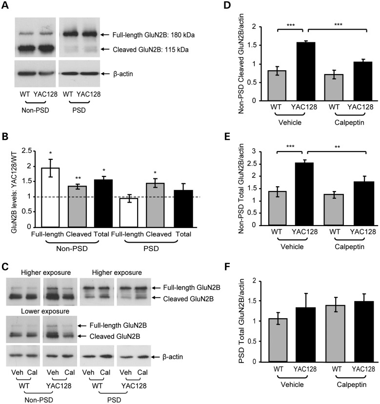
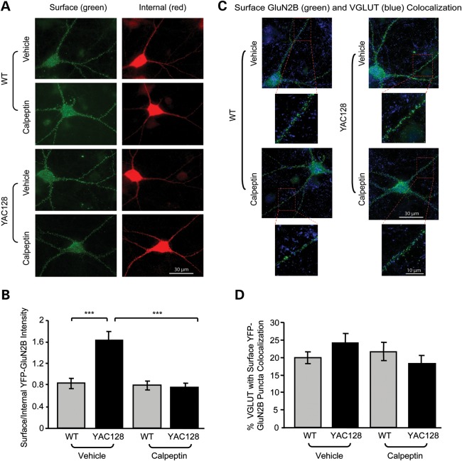
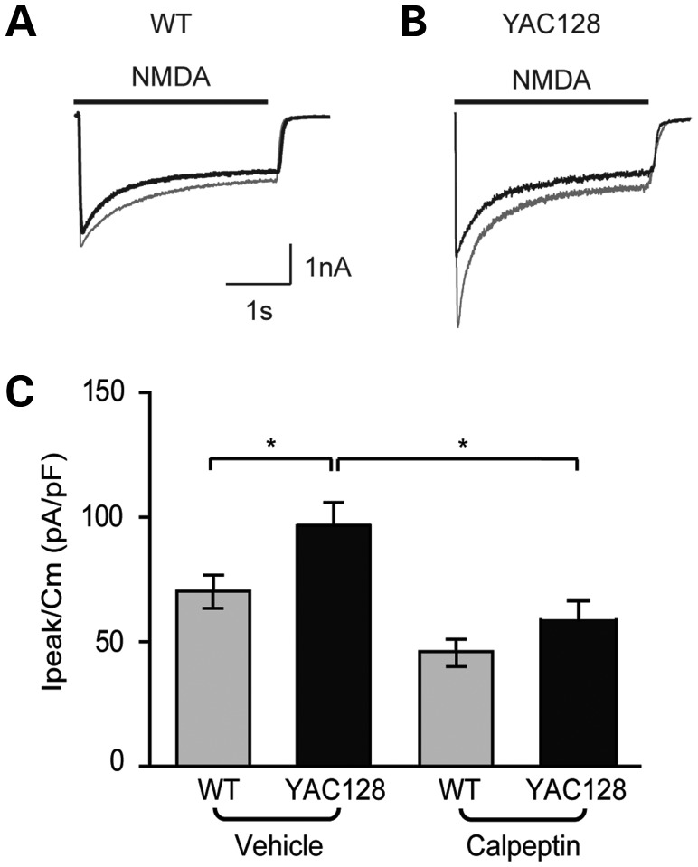
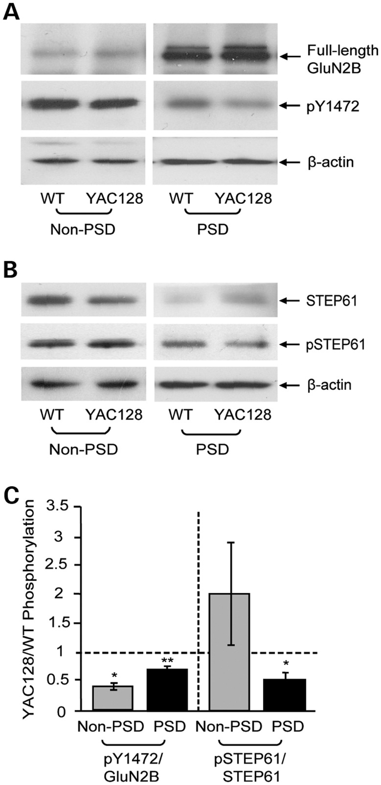
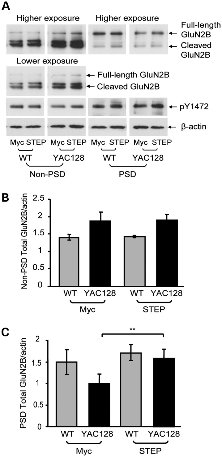
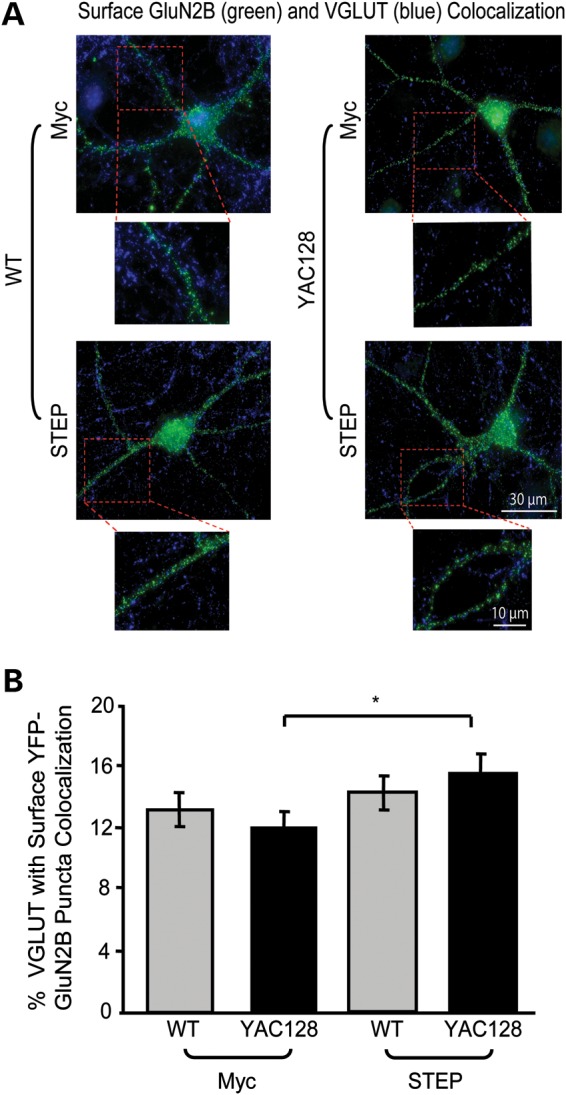
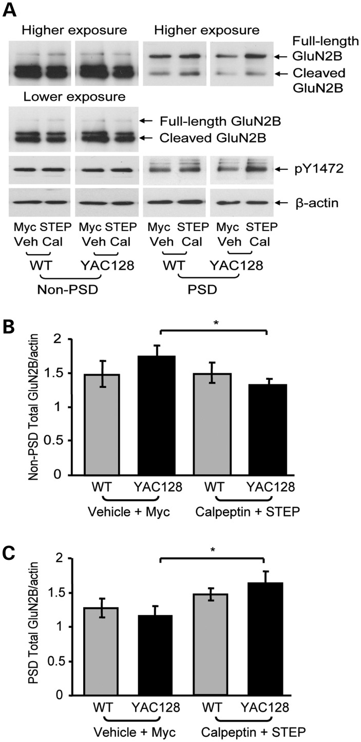
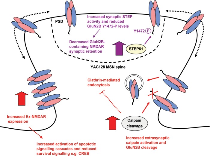
In Huntington's disease (HD), the mutant huntingtin (mhtt) protein is associated with striatal dysfunction and degeneration. Excitotoxicity and early synaptic defects are attributed, in part, to altered NMDA receptor (NMDAR) trafficking and function. Deleterious extrasynaptic NMDAR localization and signalling are increased early in yeast artificial chromosome mice expressing full-length mhtt with 128 polyglutamine repeats (YAC128 mice). NMDAR trafficking at the plasma membrane is regulated by dephosphorylation of the NMDAR subunit GluN2B tyrosine 1472 (Y1472) residue by STriatal-Enriched protein tyrosine Phosphatase (STEP). NMDAR function is also regulated by calpain cleavage of the GluN2B C-terminus. Activation of both STEP and calpain is calcium-dependent, and disruption of calcium homeostasis occurs early in the HD striatum. Here, we show increased calpain cleavage of GluN2B at both synaptic and extrasynaptic sites, and elevated extrasynaptic total GluN2B expression in the YAC128 striatum. Calpain inhibition significantly reduced extrasynaptic GluN2B expression in the YAC128 but not wild-type striatum. Furthermore, calpain inhibition reduced whole-cell NMDAR current and the surface/internal GluN2B ratio in co-cultured striatal neurons, without affecting synaptic GluN2B localization. Synaptic STEP activity was also significantly higher in the YAC128 striatum, correlating with decreased GluN2B Y1472 phosphorylation. A substrate-trapping STEP protein (TAT-STEP C-S) significantly increased VGLUT1-GluN2B colocalization, as well as increasing synaptic GluN2B expression and Y1472 phosphorylation. Moreover, combined calpain inhibition and STEP inactivation reduced extrasynaptic, while increasing synaptic GluN2B expression in the YAC128 striatum. These results indicate that increased STEP and calpain activation contribute to altered NMDAR localization in an HD mouse model, suggesting new therapeutic targets for HD.


Figures

Figure 1.
Figure 1.
Synaptic and extrasynaptic full-length and cleaved GluN2B expression and effect of calpain inhibition. (A) Representative immunoblots of WT and YAC128 striatal non-PSD and PSD fractions probed for GluN2B (using an N-terminal antibody) and β-actin. (B) Quantification of non-PSD and PSD full-length, calpain-cleaved and total (full-length plus cleaved) GluN2B in YAC128 mice relative to WT mice. Synaptic (PSD) cleaved GluN2B and non-PSD full-length, cleaved and total GluN2B are elevated in YAC128 mice relative to the WT. Significance tested using one-sample t-tests (n = 8, *P< 0.05, **P< 0.01). (C) Representative immunoblots of striatal non-PSD and PSD fractions from WT or YAC128 parasagittal slices treated with either vehicle (Veh; DMSO) or calpeptin (Cal), and probed for GluN2B and β-actin. For the non-PSD, lower and higher exposures were used for cleaved and full-length GluN2B quantification, respectively. (D and E) In the vehicle condition, YAC128 non-PSD cleaved and total GluN2B expression was significantly higher than WT and was significantly reduced by calpeptin treatment. Quantification of vehicle- and calpeptin-treated WT and YAC128 non-PSD calpain-cleaved (D) and total (E) GluN2B expression relative to the protein loading control, β-actin. A two-way ANOVA revealed significant differences for non-PSD cleaved GluN2B for treatment (F(1,7) = 100.3, ***P < 0.001), genotype (F(1,7) = 14.76, **P < 0.01), interaction (F(1,7) = 49.63, ***P < 0.001), subjects (matching, F(7,7) = 22.11, ***P < 0.001); ***P < 0.001 by Bonferroni's post hoc tests; n = 4 (D). Significant differences were also detected for non-PSD total GluN2B for treatment (F(1,7) = 19.94, **P < 0.01), genotype (F(1,7) = 12.90, **P < 0.01), interaction (F(1,7) = 10.07, *P < 0.05), subjects (matching, F(7,7) = 5.38, *P < 0.05); **P < 0.01, ***P < 0.001 by Bonferroni's post hoc tests; n = 4 (E). (F) Calpeptin treatment had no effect on synaptic total GluN2B expression in either WT or YAC128 mice. Quantification of vehicle- and calpeptin-treated WT and YAC128 PSD total GluN2B expression relative to β-actin; no significance detected for either total (F) or cleaved GluN2B expression (quantification not shown) with a two-way ANOVA (P > 0.05, n = 4).
Figure 2.
Figure 2.
Calpain inhibition reduces the YAC128 surface/internal YFP-GluN2B intensity ratio with no effect on VGLUT1-GluN2B puncta colocalization. (A) Representative embryonic YFP-GluN2B-transfected co-cultured MSNs live stained with a chicken anti-GFP antibody (surface GluN2B, green), fixed, permeabilized and stained again with a rabbit anti-GFP (internal GluN2B, red). Cultures were treated with either vehicle (DMSO) or calpeptin (1 h). (B) In the vehicle condition, the surface/internal YFP-GluN2B intensity ratio was significantly increased in YAC128 MSNs relative to WT and was significantly reduced to WT levels after calpain inhibition. Quantification of vehicle- and calpeptin-treated surface/internal YFP-GluN2B fluorescence intensities for WT and YAC128. With a two-way ANOVA, there was a significant effect of treatment (F(1,40) = 20.79, ***P < 0.001), genotype (F(1,40) = 9.79, **P < 0.01), interaction (F(1,40) = 17.69, ***P < 0.001); ***P < 0.001 by Bonferroni's post hoc tests; cell n = 21 from four cultures. (C) Representative images of vehicle- and calpeptin-treated WT and YAC128 YFP-GluN2B-transfected co-cultured MSNs live stained with an anti-GFP antibody (surface GluN2B, green), fixed, permeabilized and stained with an anti-VGLUT1 antibody (blue). Magnified images are example dendrites with VGLUT1-GluN2B puncta colocalization. (D) Calpain inhibition does not alter VGLUT1-GluN2B colocalization in either WT or YAC128 MSNs. The percentage of VGLUT1 puncta colocalized with surface YFP-GluN2B is quantified. No significance was obtained with either a two-tailed unpaired t-test or a two-way ANOVA.
Figure 3.
Figure 3.
Calpain inhibition decreases the YAC128 NMDAR current. (A and B) Representative NMDAR currents from postnatal YFP-transfected WT (A) or YAC128 (B) co-cultured MSNs evoked by NMDA (solid line) in the vehicle condition (grey trace) or following treatment with calpeptin (black trace). (C) Mean peak current density is not changed by calpain inhibition in WT MSNs (vehicle n = 14 cells, calpeptin n = 9). In the vehicle condition, YAC128 MSNs (n = 9) show a greater peak current density than WT MSNs (*P < 0.05), which is decreased by calpeptin treatment (n = 6) to WT levels. A minimum of three independent cultures was used per condition. Differences between groups were tested by a one-way ANOVA followed by Bonferroni's post hoc tests.
Figure 4.
Figure 4.
GluN2B Y1472 and STEP phosphorylation levels. (A and B) Representative immunoblots of WT and YAC128 striatal non-PSD and PSD samples probed with anti-GluN2B (N-terminal), pY1472 and β-actin antibodies (A) or with anti-STEP, pSTEP and β-actin antibodies (B). (C) YAC128 non-PSD and PSD GluN2B-pY1472 is significantly reduced compared with WT, and YAC128 PSD pSTEP61 is also significantly lower than WT levels. YAC128 pY1472/full-length GluN2B and pSTEP61/STEP61 levels are normalized to WT for both PSD and non-PSD fractions. Significance tested using one-sample t-tests [n = 4 (non-PSD) and n = 5 (PSD), *P < 0.05, **P < 0.01].
Figure 5.
Figure 5.
STEP inactivation increases YAC128 synaptic GluN2B and pY1472 levels. (A) Representative immunoblots of striatal non-PSD and PSD fractions isolated from parasagittal slices treated with either TAT-myc or TAT-STEP C-S fusion proteins (1 h). Samples were probed with anti-GluN2B (N-terminal), pY1472 and β-actin antibodies. Low and high exposure blots were used for cleaved and full-length GluN2B quantification, respectively. (B) STEP inactivation has no effect on either WT or YAC128 non-PSD total GluN2B expression levels. Quantification of TAT-myc and TAT-STEP C-S-treated WT and YAC128 non-PSD total GluN2B expression relative to β-actin; no significance detected with a two-way ANOVA (P > 0.05, n = 4). (C) STEP inactivation significantly increases YAC128 but not WT total GluN2B expression in the PSD. The effect of TAT-myc and TAT-STEP C-S on total GluN2B levels is quantified for both WT and YAC128 in the PSD fraction. A two-way ANOVA revealed significant differences in synaptic total GluN2B for treatment (F(1,8) = 15.14, **P < 0.01) and subjects (matching, F(8,8) = 8.72, **P < 0.01); **P < 0.01 by Bonferroni's post hoc tests; n = 5. As indicated in the immunoblots (A), this was associated with increased Y1472 phosphorylation in the PSD of YAC128 but not WT striata (quantification not shown).
Figure 6.
Figure 6.
Inactivating STEP increases YAC128 VGLUT1-GluN2B colocalization. (A) Representative images of TAT-myc and TAT-STEP C-S treated (1 h) YFP-GluN2B-transfected MSNs, live stained with an anti-GFP antibody (surface GluN2B, green), fixed, permeabilized and stained with anti-VGLUT1 antibody (blue). Magnified images are example dendrites of VGLUT1-GluN2B puncta colocalization. (B) STEP inactivation significantly increases YAC128 but not WT VGLUT1-GluN2B colocalization. The percentage of VGLUT1-GluN2B puncta colocalization is quantified for TAT-myc and TAT-STEP C-S-treated WT and YAC128 MSNs. Significance obtained for treatment (F(1,58) = 4.14, *P < 0.05) with both a two-way ANOVA and a two-tailed unpaired t-test (*P < 0.05; cell n = 30 from four different cultures).
Figure 7.
Figure 7.
Combined calpain inhibition and STEP inactivation normalizes YAC128 NMDAR expression. (A) Representative immunoblots of striatal non-PSD and PSD fractions isolated from parasagittal slices treated with either vehicle (Veh; DMSO) and TAT-myc or calpeptin and TAT-STEP C-S (1 h). Samples were probed with anti-GluN2B (N-terminal), pY1472 and β-actin antibodies. Low and high exposure blots were used for cleaved and full-length GluN2B quantification, respectively. (B and C) Combined calpain inhibition and STEP inactivation significantly decreases non-PSD YAC128 total GluN2B expression (B), while significantly increasing YAC128 PSD total GluN2B levels (C). The effect of the vehicle plus TAT-myc and calpeptin plus TAT-STEP C-S on non-PSD (B) and PSD (C) total GluN2B levels is quantified relative to β-actin. The decrease in YAC128 non-PSD GluN2B after combined calpain inhibition and STEP inactivation was significant with a two-tailed unpaired t-test [*P < 0.05, n = 4 (B)]. A two-way ANOVA revealed significant differences in synaptic total GluN2B for treatment (F(1,8) = 10.60, *P < 0.05); *P < 0.05 by Bonferroni's post hoc tests; n = 5 (C). As indicated in the immunoblots (A), increased YAC128 synaptic total GluN2B expression was accompanied by increased YAC128 pY1472 after treatment with both calpeptin and TAT-STEP C-S (quantification not shown).
Figure 8.
Figure 8.
Model summarizing consequences of elevated calpain and STEP activation for GluN2B expression in YAC128 MSNs. Elevated calpain activity preferentially promotes YAC128 Ex-NMDAR expression, as calpain inhibition significantly reduced both surface and extrasynaptic GluN2B-containing NMDAR expression and function. Increased STEP activation in the YAC128 PSD could mediate reduced synaptic NMDAR retention. Since receptors laterally diffuse on the plasma membrane, enhanced STEP61-mediated GluN2B Y1472 dephosphorylation may facilitate increased NMDAR lateral diffusion from the PSD to extrasynaptic sites. Alternatively, STEP activation could trigger internalization of synaptic NMDARs followed by rapid recycling to the extrasynaptic membrane (not shown in model). GluN2B-containing NMDARs may then be more stable extrasynaptically because of enhanced calpain-mediated cleavage of the AP-2 α and β2 subunits and reduced clathrin-mediated endocytosis. NMDAR mislocalization could contribute to altered activation of cell survival and apoptotic signalling cascades in HD.

Similar articles

Cited by

References

    1. Bliss T.V., Collingridge G.L. A synaptic model of memory: long-term potentiation in the hippocampus. Nature. 1993;361:31–39. doi:10.1038/361031a0. - DOI - PubMed
    1. Arundine M., Tymianski M. Molecular mechanisms of calcium-dependent neurodegeneration in excitotoxicity. Cell Calcium. 2003;34:325–337. doi:10.1016/S0143-4160(03)00141-6. - DOI - PubMed
    1. Gladding C.M., Raymond L.A. Mechanisms underlying NMDA receptor synaptic/extrasynaptic distribution and function. Mol. Cell Neurosci. 2011;48:308–320. - PubMed
    1. Lau C.G., Zukin R.S. NMDA receptor trafficking in synaptic plasticity and neuropsychiatric disorders. Nat. Rev. Neurosci. 2007;8:413–426. - PubMed
    1. Graveland G.A., Williams R.S., DiFiglia M. Evidence for degenerative and regenerative changes in neostriatal spiny neurons in Huntington's disease. Science. 1985;227:770–773. doi:10.1126/science.3155875. - DOI - PubMed

Publication types

MeSH terms