DICER-LIKE3 activity in Physcomitrella patens DICER-LIKE4 mutants causes severe developmental dysfunction and sterility
- PMID: 22511605
- PMCID: PMC3506028
- DOI: 10.1093/mp/sss036
DICER-LIKE3 activity in Physcomitrella patens DICER-LIKE4 mutants causes severe developmental dysfunction and sterility
Abstract
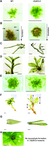
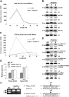
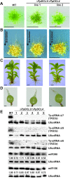
Trans-acting small interfering RNAs (ta-siRNAs) are plant-specific siRNAs released from TAS precursor transcripts after microRNA-dependent cleavage, conversion into double-stranded RNA, and Dicer-dependent phased processing. Like microRNAs (miRNAs), ta-siRNAs direct site-specific cleavage of target RNAs at sites of extensive complementarity. Here, we show that the DICER-LIKE 4 protein of Physcomitrella patens (PpDCL4) is essential for the biogenesis of 21 nucleotide (nt) ta-siRNAs. In ΔPpDCL4 mutants, off-sized 23 and 24-nt ta-siRNAs accumulated as the result of PpDCL3 activity. ΔPpDCL4 mutants display severe abnormalities throughout Physcomitrella development, including sterility, that were fully reversed in ΔPpDCL3/ΔPpDCL4 double-mutant plants. Therefore, PpDCL3 activity, not loss of PpDCL4 function per se, is the cause of the ΔPpDCL4 phenotypes. Additionally, we describe several new Physcomitrella trans-acting siRNA loci, three of which belong to a new family, TAS6. TAS6 loci are typified by sliced miR156 and miR529 target sites and are in close proximity to PpTAS3 loci.
Figures


Similar articles
-
Role of RNA interference (RNAi) in the Moss Physcomitrella patens.Int J Mol Sci. 2013 Jan 14;14(1):1516-40. doi: 10.3390/ijms14011516. Int J Mol Sci. 2013. PMID: 23344055 Free PMC article. Review.
-
Physcomitrella patens DCL3 is required for 22-24 nt siRNA accumulation, suppression of retrotransposon-derived transcripts, and normal development.PLoS Genet. 2008 Dec;4(12):e1000314. doi: 10.1371/journal.pgen.1000314. Epub 2008 Dec 19. PLoS Genet. 2008. PMID: 19096705 Free PMC article.
-
Comprehensive Annotation of Physcomitrella patens Small RNA Loci Reveals That the Heterochromatic Short Interfering RNA Pathway Is Largely Conserved in Land Plants.Plant Cell. 2015 Aug;27(8):2148-62. doi: 10.1105/tpc.15.00228. Epub 2015 Jul 24. Plant Cell. 2015. PMID: 26209555 Free PMC article.
-
Identification of trans-acting siRNAs in moss and an RNA-dependent RNA polymerase required for their biogenesis.Plant J. 2006 Nov;48(4):511-21. doi: 10.1111/j.1365-313X.2006.02895.x. Plant J. 2006. PMID: 17076803
-
miRNAs in the biogenesis of trans-acting siRNAs in higher plants.Semin Cell Dev Biol. 2010 Oct;21(8):798-804. doi: 10.1016/j.semcdb.2010.03.008. Epub 2010 Mar 30. Semin Cell Dev Biol. 2010. PMID: 20359543 Review.
Cited by
-
Novel DICER-LIKE1 siRNAs Bypass the Requirement for DICER-LIKE4 in Maize Development.Plant Cell. 2015 Aug;27(8):2163-77. doi: 10.1105/tpc.15.00194. Epub 2015 Jul 24. Plant Cell. 2015. PMID: 26209554 Free PMC article.
-
Role of RNA interference (RNAi) in the Moss Physcomitrella patens.Int J Mol Sci. 2013 Jan 14;14(1):1516-40. doi: 10.3390/ijms14011516. Int J Mol Sci. 2013. PMID: 23344055 Free PMC article. Review.
-
Sporophyte Formation and Life Cycle Completion in Moss Requires Heterotrimeric G-Proteins.Plant Physiol. 2016 Oct;172(2):1154-1166. doi: 10.1104/pp.16.01088. Epub 2016 Aug 22. Plant Physiol. 2016. PMID: 27550997 Free PMC article.
-
Plant Noncoding RNAs: Hidden Players in Development and Stress Responses.Annu Rev Cell Dev Biol. 2019 Oct 6;35:407-431. doi: 10.1146/annurev-cellbio-100818-125218. Epub 2019 Aug 12. Annu Rev Cell Dev Biol. 2019. PMID: 31403819 Free PMC article. Review.
-
Phylogenetic analyses of seven protein families refine the evolution of small RNA pathways in green plants.Plant Physiol. 2023 May 31;192(2):1183-1203. doi: 10.1093/plphys/kiad141. Plant Physiol. 2023. PMID: 36869858 Free PMC article.
References
-
- Adenot X, et al. DRB4-dependent TAS3 trans-acting siRNAs control leaf morphology through AGO7. Curr. Biol. 2006;16:927–932. - PubMed
-
- Allen E, Xie Z, Gustafson AM, Carrington JC. microRNA-directed phasing during trans-acting siRNA biogenesis in plants. Cell. 2005;121:207–221. - PubMed
-
- Axtell MJ, Jan C, Rajagopalan R, Bartel DP. A two-hit trigger for siRNA biogenesis in plants. Cell. 2006;127:565–577. - PubMed
Publication types
MeSH terms
Substances
Grants and funding
LinkOut - more resources
Full Text Sources
Other Literature Sources
Molecular Biology Databases
Research Materials

