. 2012:2012:723419.
doi: 10.1155/2012/723419. Epub 2012 Feb 29.

Prostaglandins in cancer cell adhesion, migration, and invasion

Affiliations

Prostaglandins in cancer cell adhesion, migration, and invasion

David G Menter et al. Int J Cell Biol. 2012.

Abstract

Prostaglandins exert a profound influence over the adhesive, migratory, and invasive behavior of cells during the development and progression of cancer. Cyclooxygenase-2 (COX-2) and microsomal prostaglandin E(2) synthase-1 (mPGES-1) are upregulated in inflammation and cancer. This results in the production of prostaglandin E(2) (PGE(2)), which binds to and activates G-protein-coupled prostaglandin E(1-4) receptors (EP(1-4)). Selectively targeting the COX-2/mPGES-1/PGE(2)/EP(1-4) axis of the prostaglandin pathway can reduce the adhesion, migration, invasion, and angiogenesis. Once stimulated by prostaglandins, cadherin adhesive connections between epithelial or endothelial cells are lost. This enables cells to invade through the underlying basement membrane and extracellular matrix (ECM). Interactions with the ECM are mediated by cell surface integrins by "outside-in signaling" through Src and focal adhesion kinase (FAK) and/or "inside-out signaling" through talins and kindlins. Combining the use of COX-2/mPGES-1/PGE(2)/EP(1-4) axis-targeted molecules with those targeting cell surface adhesion receptors or their downstream signaling molecules may enhance cancer therapy.


Figures

Figure 1
Figure 1
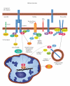
Eicosanoid metabolism. Arachidonic acid (AA) is an essential dietary fatty acid that is transported into cells and stored in membrane phospholipids. First AA is coupled to acyl-CoA by acyl-coenzyme A synthetases (ACLS). Fatty acyltransferases (FACT) then insert AA into membrane phospholipids. Cytoplasmic phospholipase A2 (cPLA2) releases AA from membrane phospholipids after agonist stimulation. In turn, free AA is converted to prostaglandin G2 (PGG2) and then prostaglandin H2 (PGH2) by cyclooxygenases (COXs). PGH2 then becomes a substrate for a variety of PG synthases. These PG synthases are identified by the specific prostaglandin each one produces, namely, PGD2 synthases (PGDSs), PGE2 synthases (PGESs), (PGF2α) synthase (PGFS), PGI2 synthase (PGIS), or TxA2 synthase (TXS). Both COX-2 and microsomal PGE synthase-1 (mPGES-1) are elevated in tumors. Export involves multidrug resistance-associated protein 4 (MRP4). In the extracellular milieu, PGs bind to G-protein-coupled receptors identified as DP1, DP2, EP1-4, FP, IP, and TP. Among these, EP receptors interact with G-stimulatory (Gs) or G-inhibitory (Gi) proteins stimulating downstream signals such as cAMP, Ca2+, inositol phosphates or IP3/Ca2+, and Rho. Catabolism involves uptake by PG transporter (PGT) and inactivation by NAD+ dependent 15-hydroxyprostaglandin dehydrogenase (15-PGDH).
Figure 2
Figure 2
Dynamic adherens junctions. Prostaglandins influence the assembly, stabilization, and disassembly of cell-cell junctions. E-cadherins form Ca2+-dependent transmembrane adhesion complexes between adjacent cells (Figure 2). Cytoplasmic regulatory proteins include α-catenin, β-catenin, IQGAPs scaffold proteins that interact with Rho GTPases to alter morphology and migration. Alternate interactions involve δ-catenin, p190, and RhoA influencing actin assembly. Together, Src and p140Cap influence C-terminal Src kinase (Csk) activity stabilizing cell-cell interactions as well as similar activity by receptor protein tyrosine phosphatase mu (PTPμ). Nectins-afadin complexes also cooperate with cadherins and integrins to regulate cell-cell adhesion. Disassembly of cadherin complexes involves either caveolin- or clathrin-mediated endocytosis and phagosome formation. Inside-out vesicles contain cadherin on the inside and β-catenin and Src exposed to the cytoplasm. When these vesicles interact with Ras-related protein A (RalA), cadherins are recycled. Whereas, interactions with Ras-proximate-1/Ras-related protein-1-(Rap1-)GTPase and E3 ubiquitin ligase followed by ubiquitinization result in proteosomal degradation that prepares cells for migration. The loss of E-cadherin in conjunction with elevations in COX-2 occurs during the transformation and adenoma formation in the presence of Apc mutations causing aberrant β-catenin signaling. Subsequent interactions with T-cell factor/lymphoid-enhancer-factor-(TCF/LEF-) can cause increases in COX-2 expression.
Figure 3
Figure 3
Basement membrane. The basement membrane underlies the typical cellular epithelium or vascular endothelium and consists of two thin structural layers. One layer is the basal lamina made by epithelial or endothelial cells. The second layer is the reticular lamina made by fibroblasts. Electron microscope data show that the basal lamina consists of a clear lamina lucida next to epithelial cells and an opaque lamina densa. The lamina lucida contains integrins, laminins (1, 5, 6 and 10), and collagen XVII, as well as type IV collagen, and dystroglycans. The lamina densa contains type IV collagen fibers, entactin/nidogen-1, perlecan, and heparan sulfate proteoglycans. The reticular lamina contains collagens I, III, and V and various proteoglycans. Invasion through the basement membrane requires the expression of many different cell surface adhesion receptors and matrix degrading enzymes.
Figure 4
Figure 4
Integrins. Integrins are transmembrane glycoprotein adhesion receptor complexes consisting of α and β subunits. The α subunit contains a seven-bladed β-propeller head domain connected to a leg support structure made of a thigh, a calf-1, a calf-2, a transmembrane, and a cytoplasmic domain that mediates ligand specificity. The β subunit contains an N-terminal plexin-semaphorin-integrin (PSI) domain, a hybrid domain, a β-I domain, four cysteine-rich epidermal growth factor (EGF) repeats, a transmembrane, and a cytoplasmic domain that interacts with the cell cytoskeleton. The N-terminal β-I domain of a β subunit inserts into the β-propeller domain of an α subunit forming a headpiece complex. The formation of integrin receptor complexes depends on divalent cation (i.e., Ca2+, Mn2+, Mg2+) that bind to metal-ion-dependent adhesion site (MIDAS) motifs in the α subunits and adjacent to MIDAS (ADMIDAS) motifs in β subunits. Three conformation states exist for α and β subunit complexes. (1) The unliganded conformation has a closed headpiece and a bent receptor structure with the EGF domains of the β-subunit touching the calf-1-calf-2 domains of the α-subunit. (2) The headpiece remains closed, but structural changes in the β-subunit EGF domains cause a separation from the calf-1-calf-2 domains of the α-subunits causing an extended structure. (3) Conformational changes in the β 6-α 7 loops expose the ligand-binding site along with a complete separation of the β-subunit from the calf-1-calf-2 domains in the α-subunit. These conformational changes engage the specific integrin headpiece with its ligand.
Figure 5
Figure 5
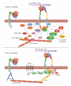
“Outside-in” and “Inside-out” signaling. The “outside-in” binding of ECM ligands to cell surface integrins stimulates conformational changes that activate focal adhesion kinase (FAK). FAK then is autophosphorylated on Tyrosine 397 near the catalytic domain, which binds Src. FAK contains a central kinase domain bordered by FERM (protein 4.1, ezrin, radixin, and moesin homology) domain at the N-terminus and a focal adhesion targeting (FAT) sequence at the C-terminus. Activated Src interacts with human enhancer of filamentation1 (HEF1) and p130 CRK-associated substrate (p130CAS) scaffold proteins that help to positively regulate Src-FAK-Crk interactions with Rac. FAK also activates (PKL/Git2)-β-Pix complexes and β-pix then serves either as an exchange factor for Cdc42 or a scaffold protein to promote signaling via Rac and p21-activated protein kinases (PAK). FAK also interacts with actin-related proteins (ARP2 and ARP3) which is regulated by the Wiskott-Aldrich Syndrome Protein (WASP). ARP2/ARP3 initiates the polymerization of new actin filaments. FAK also influences actin contraction and polarization through another GTPase protein, Rho. The regulation of Rho GTPase hydrolysis of GTP (active) to GDP (inactive) form occurs through the opposing activities of guanine nucleotide exchange factor (GEFs). GTPase regulator associated with FAK (GRAF) and p190RhoGAP blocks actin cytoskeleton changes. In contrast, PDZRhoGEF and p190RhoGEF both serve to activate Rho. “Outside-in signaling” transfers integrin-mediated external signals to the inside of cells.“Inside-out signaling” depends on talin and kindlin. Both talin and kindlin contain FERM (4.1/ezrin/radixin/moesin) domains and a highly conserved C-terminal F3 domains. Talins bind β integrin, actin through the C-terminus, and also vinculin. Kindlins bind integrins, the cell membrane, and various actin adaptor proteins like migfilin, or integrin-linked kinase (ILK). Talin activation occurs through G-protein-coupled receptors that increases cytoplasmic Ca2+ and diacylglycerol. This activates GEF function in conjunction with Ras-proximate-1/Ras-related-protein-1-(Rap1-) GTPase. Rap1 then binds to Rap1-GTP-interacting adaptor molecule (RIAM). RIAM recruits talin to the membrane and the α and β integrin cytoplasmic domains. Kindlin interacts with β integrin cytoplasmic domain stabilizing the activated state of the integrin complex. “Inside-out signaling” strengthens adhesive contacts and the appropriate force necessary for integrin-mediated cell migration, invasion, ECM remodeling, and matrix assembly.
Figure 6
Figure 6
Linking PGE2 to adhesion, migration, and invasion. Prostaglandin E2 elicits profound changes in tumor cells that result in the disassociation of cadherin-mediated cell connections. This is accompanied by the establishment/turnover of integrin-mediated interactions with extracellular matrix during adhesion and subsequent migration and invasion. Stimulation of EP2 or 4 receptors leads to the activation of adenylate cyclase and results in the production of cyclic adenosine monophosphate (cAMP) from adenosine triphosphate (ATP). The accumulation of cAMP in the cell cytoplasm activates protein kinase A (PKA) and the phosphorylation of downstream targets. This accumulation of cAMP can also activate exchange protein activated by cAMP (Epac). The activation of Epac may involve the interactions with Rap1 and subsequent downstream signals that influence adhesion, migration, and invasion. The activation of EP1 and EP3 leads to Ca2+ influx and the activation of Rho-mediated signal transduction that influences cadherin function during the disassociation of cadherin-based adhesive contacts or integrin interactions with the extracellular matrix contacts.

Similar articles

Cited by

References

    1. Wang D, Dubois RN. Eicosanoids and cancer. Nature Reviews Cancer. 2010;10(3):181–193. - PMC - PubMed
    1. Shindou H, Hishikawa D, Harayama T, Yuki K, Shimizu T. Recent progress on acyl CoA: lysophospholipid acyltransferase research. Journal of Lipid Research. 2009;50:S46–51. - PMC - PubMed
    1. Shindou H, Shimizu T. Acyl-CoA: lysophospholipid acyltransferases. The Journal of Biological Chemistry. 2009;284(1):1–5. - PubMed
    1. Ellis JM, Frahm JL, Li LO, Coleman RA. Acyl-coenzyme A synthetases in metabolic control. Current Opinion in Lipidology. 2010;21(3):212–217. - PMC - PubMed
    1. Leslie CC. Regulation of the specific release of arachidonic acid by cytosolic phospholipase A2 . Prostaglandins Leukotrienes and Essential Fatty Acids. 2004;70(4):373–376. - PubMed