. 2012 Jun;86(12):6875-88.
doi: 10.1128/JVI.06310-11. Epub 2012 Apr 11.

The US16 gene of human cytomegalovirus is required for efficient viral infection of endothelial and epithelial cells

Affiliations

The US16 gene of human cytomegalovirus is required for efficient viral infection of endothelial and epithelial cells

Matteo Bronzini et al. J Virol. 2012 Jun.

Abstract

The human cytomegalovirus (HCMV) US12 gene family comprises a set of 10 contiguous genes (US12 to US21), each encoding a predicted seven-transmembrane protein and whose specific functions have yet to be ascertained. While inactivation of individual US12 family members in laboratory strains of HCMV has not been found to affect viral replication in fibroblasts, inactivation of US16 was reported to increase replication in microvascular endothelial cells. Here, we investigate the properties of US16 further by ascertaining the expression pattern of its product. A recombinant HCMV encoding a tagged version of the US16 protein expressed a 33-kDa polypeptide that accumulated with late kinetics in the cytoplasmic virion assembly compartment. To elucidate the function(s) of pUS16, we generated US16-deficient mutants in the TR clinical strain of HCMV. According to previous studies, inactivation of US16 had no effect on viral replication in fibroblasts. In contrast, the US16-deficient viruses exhibited a major growth defect in both microvascular endothelial cells and retinal pigment epithelial cells. The expression of representative IE, E, and L viral proteins was impaired in endothelial cells infected with a US16 mutant virus, suggesting a defect in the replication cycle that occurs prior to IE gene expression. This defect must be due to an inefficient entry and/or postentry event, since pp65 and viral DNA did not move to the nucleus in US16 mutant-infected cells. Taken together, these data indicate that the US16 gene encodes a novel virus tropism factor that regulates, in a cell-specific manner, a pre-immediate-early phase of the HCMV replication cycle.


Figures

Fig 1
Fig 1
HCMV genome and structure of the US16 mutant viruses. (A) Schematic representation of the HCMV US16 gene region and the modifications that were introduced into the US16 ORF. In TRΔUS16, the US16 ORF was replaced with the galactose kinase marker (galK). In TRUS16stop, a single-nucleotide change was introduced into codons 9, 10, and 11 of the US16 ORF. These changes created a stop codon in the 10th codon, as well as a unique restriction site for XbaI. In TRUS16-REV, the changes introduced into codons 9, 10, and 11 of TRUS16stop were reversed to the wt sequence, thus repairing the whole US16 ORF. TRUS16-HA was generated from TRΔUS16 by reintroducing the US16 ORF fused with the coding sequence for an HA epitope tag at its C terminus. Recombinant BACs were examined for the desired mutation by PCR, restriction digestion analysis, and sequencing. (B) PCR and restriction digestion analysis of viral DNA from infectious RV. Viral DNA was genome purified from RVTRwt (lanes 1, 5, 9, and 13), TRΔUS16 (lanes 2, 6, and 10), TRUS16stop (lanes 3, 7, 11, and 14), TRUS16-HA (lanes 4, 8, 12, and 15), and TRUS16-REV (lanes 16, 17, 18, and 19). PCR was performed using the US16-F primer set (lanes 1, 2, 3, 4, and 16), the US16-ORF primer set (lanes 5, 6, 7, 8, and 17), or the galK primer set (lanes 9, 10, 11, 12, and 18) (Table 1). The sizes of the PCR products were as follows: 1,228 bp for the galK primer set; 924 bp for the US16-ORF primer set; 1,249 bp for the US16-F primer set from RVTRwt, RVTRUS16stop, and RVTRUS16-REV; and 1,309 bp for the US16-F primer set from RVTRUS16-HA. To confirm the successful introduction of the desired stop codon in TRUS16stop, PCR fragments from RVTRwt, RVTRUS16stop, RVTRUS16-HA, and RVTRUS16-REV amplified using the US16-F primer set were digested with the XbaI restriction enzyme (lanes 13, 14, 15, and 19, respectively). Lanes M, molecular markers.
Fig 2
Fig 2
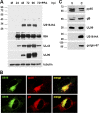
The US16 gene of HCMV encodes a late cytoplasmic protein. (A) Kinetics of pUS16-HA protein expression in infected cells. HELFs were grown to subconfluence and then infected with HCMV RVTRUS16-HA (MOI, 1 PFU/cell). At the indicated times p.i., total protein cell extracts were prepared, fractionated by SDS-PAGE (50 μg protein/lane), and analyzed by immunoblotting with the anti-HA, anti-IEA, anti-UL44, or anti-UL99 MAb, as described in Materials and Methods. The immunodetection of tubulin with a MAb was performed as an internal control. Cell extracts were isolated from mock-infected cells; cells infected for 24, 48, 72, and 96 h; or cells infected and treated with PFA (200 μg/ml) for 72 h. (B) Localization of US16-HA protein in the cytoplasmic assembly compartment of HCMV-infected HELFs. HELFs were grown to subconfluence and then infected with HCMV RVTRUS16-HA (MOI, 0.1 PFU/cell). At 96 h p.i., the cells were fixed, permeabilized, and stained for pUS16-HA (green), pUL99 (red), or gB (red). Immunofluorescence experiments were repeated three times, and representative results are presented. (C) pUS16-HA is not present in extracellular virus particles. RVTRUS16-HA particles were partially purified from clarified culture supernatants by centrifugation through a 20% sorbitol cushion. Protein extracts from virions (V) and infected cells (C) were then fractionated by SDS-PAGE and analyzed by immunoblotting with the anti-HA, anti-pp65, anti-gB, or anti-UL99 MAb. Immunodetection of golgin-97, a commonly used marker for the TGN, was performed as a control.
Fig 3
Fig 3
Growth kinetics of ΔUS16, US16stop, and US16-REV viruses in fibroblasts and in endothelial cells. HELFs or HMVECs were infected with the parental RVTRwt, RVTRΔUS16 (clone 5.3), RVTRUS16stop, or RVTRUS16-REV (MOI, 0.1 PFU/cell). The extent of virus replication was then assessed by titrating the infectivity of supernatants of cell suspensions on HELFs using the IE antigen indirect immunoperoxidase staining technique (15). The data shown are the averages of three experiments ± SD.
Fig 4
Fig 4
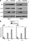
The replicative cycle of US16-deficient viruses in endothelial cells is blocked at a stage prior to the expression of IE genes. (A) Expression of representative IE, E, and L proteins in endothelial cells infected with TR or TRΔUS16 viruses. HMVECs were grown to subconfluence and then mock infected (M) or infected with the parental RVTRwt or RVUS16stop (MOI, 1 PFU/cell). At the indicated times p.i., total cell extracts were prepared and analyzed by immunoblotting with anti-IEA, anti-UL44, or anti-UL99 MAb as described in Materials and Methods. Actin immunodetected with a MAb served as an internal control. (B) The US16 gene product is required for IE gene expression in endothelial cells. HMVECs were infected with RVTRwt or RVUS16stop (MOI, 0.1 PFU/cell). Total RNA was isolated at the indicated time p.i. and reverse transcribed. Real-time RT-PCR was carried out with the appropriate IE1, IE2, and β-actin primers to quantify the expression levels of IE1 and IE2 mRNA. For each time point, IE1 and IE2 mRNA levels were normalized according to the expression of the actin gene. The results were then analyzed using a standard-curve model, and the levels of IE1 and IE2 mRNAs were normalized to the levels of endogenous β-actin mRNA. The value at each time point was normalized to the value observed with cells infected with RVTRwt for 12 h, which was set at 1. Data are shown as means and SD. ***, P ≤ 0.001 versus calibrator sample.
Fig 5
Fig 5
Lack of expression of IE proteins in different types of endothelial cells infected with US16 mutant viruses. HELFs, HMVECs, HUVECs, or LECs were infected with RVTRwt, RVTRΔUS16, RVTRUS16stop, or RVTRUS16-REV at an MOI of 0.1 PFU/cell. At 24 h p.i., cells were fixed, permeabilized, and stained with an anti-IEA (IE1 plus IE2) MAb. Images of ECs infected with US16-deficent viruses were purposely chosen to include positive nuclei to show virus addition, since random fields were on average negative for IEA staining. Immunofluorescence experiments were repeated three times, and representative results are presented (magnification, ×10).
Fig 6
Fig 6
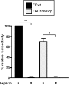
Inactivation of US16 does not impair virion attachment to endothelial cells. HMVECs were infected with equal numbers of [3H]thymidine-labeled RVTR or RVUS16stop virion particles at 4°C in the absence (−) or presence (+) of heparin (30 μg/ml) for 2 h to allow virus adsorption only. The cells were then washed extensively, cell extracts were prepared, and the associated radioactivity was determined by liquid scintillation. The amount of radiolabeled RVTRwt that bound to HMVECs was arbitrarily set to 100 (calibrator sample). Data are shown as means and SD. *, P ≤ 0.05; **, P ≤ 0.01 versus calibrator sample.
Fig 7
Fig 7
The UL83-encoded pp65 tegument protein does not accumulate in the nuclei of endothelial cells infected with US16-deficient viruses. HMVECs were infected with RVTR, RVTRΔUS16, RVTRUS16stop, or RVTRUS16-REV (MOI, 0.1 PFU/cell). At 8 h p.i., the cells were fixed, permeabilized, and stained with an anti-UL83 (pp65) MAb. Immunofluorescence experiments were repeated three times, and representative results are presented (magnification, ×10).
Fig 8
Fig 8
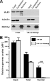
The HCMV ΔUS16 genome does not move to the nucleus in endothelial cells. (A) Separation of nuclear and cytoplasmic fractions. HMVECs were infected with equal numbers of RVTRwt or RVUS16stop virion particles. At 4 h p.i., infected cells were harvested, and nuclear and cytoplasmic extracts were prepared. Equal amounts of protein from the total cell lysate, the nuclear fraction, and the cytoplasmic fraction were then assayed for their pp65 content by immunoblotting. The purity was determined by immunoblotting for the nuclear ribonucleoprotein RNPA2 and tubulin. (B) Viral DNA in the nuclear fraction of HMVECs infected with RVTR or RVUS16stop. HMVECs were infected and fractionated as described for panel A. The amounts of viral DNA present in the nuclear fractions of infected cells were then quantified by real-time PCR using primers specific for the IE1 ORF and normalized to levels of the endogenous 18S gene. The data shown are the averages of three experiments plus SD. ***, P ≤ 0.001 compared to the amount of viral DNA measured in total extracts of cells infected with RVTRwt.
Fig 9
Fig 9
US16-deficient viruses are defective for growth in epithelial cells. (A) Growth kinetics of ΔUS16, US16stop, and US16-REV in ARPE-19 cells. ARPE-19 cells were infected with RVTRwt, RVTRΔUS16 (clone 5.3), RVTRUS16stop, or RVTRUS16-REV (MOI, 0.1 PFU/cell). The extent of virus replication was then assessed by titrating the infectivity of supernatants of cell suspensions by standard plaque assay on HELFs. The data shown are the averages of three experiments ± SD. (B) Lack of IE expression and pp65 nuclear accumulation in epithelial cells infected with US16 mutant viruses. ARPE-19 cells were infected with RVTRwt, RVTRΔUS16, RVTRUS16stop, or RVUS16-REV at an MOI of 0.1 PFU/cell. At 8 h p.i. for pp65 and 24 h p.i. for IEA, cells were fixed, permeabilized, and stained with an anti-IEA (IE1 plus IE2) MAb or an anti-UL83 (pp65) MAb. Immunofluorescence experiments were repeated three times, and representative results are presented (magnification, ×10).

Similar articles

Cited by

References

    1. Adler B, Sinzger C. 2009. Endothelial cells in HCMV infection: one host cell out of many or a crucial target for virus spread? Thromb. Haemost. 102:1057–1063 - PubMed
    1. Bissinger AL, Sinzger C, Kaiserling E, Jahn G. 2002. Human cytomegalovirus as a direct pathogen: correlation of multiorgan involvement and cell distribution with clinical and pathological findings in a case of congenital inclusion disease. J. Med. Virol. 67:200–206 - PubMed
    1. Britt W. 2008. Manifestations of human cytomegalovirus infection: proposed mechanisms of acute and chronic disease. Curr. Top. Microbiol. Immunol. 325:417–470 - PubMed
    1. Brune W, Nevels M, Shenk T. 2003. Murine cytomegalovirus m41 open reading frame encodes a Golgi-localized antiapoptotic protein. J. Virol. 77:11633–11643 - PMC - PubMed
    1. Caposio P, Luganini A, Hahn G, Landolfo S, Gribaudo G. 2007. Activation of the virus-induced IKK/NF-κB signalling axis is critical for the replication of human cytomegalovirus in quiescent cells. Cell. Microbiol. 9:2040–2054 - PubMed

Publication types

MeSH terms

Substances